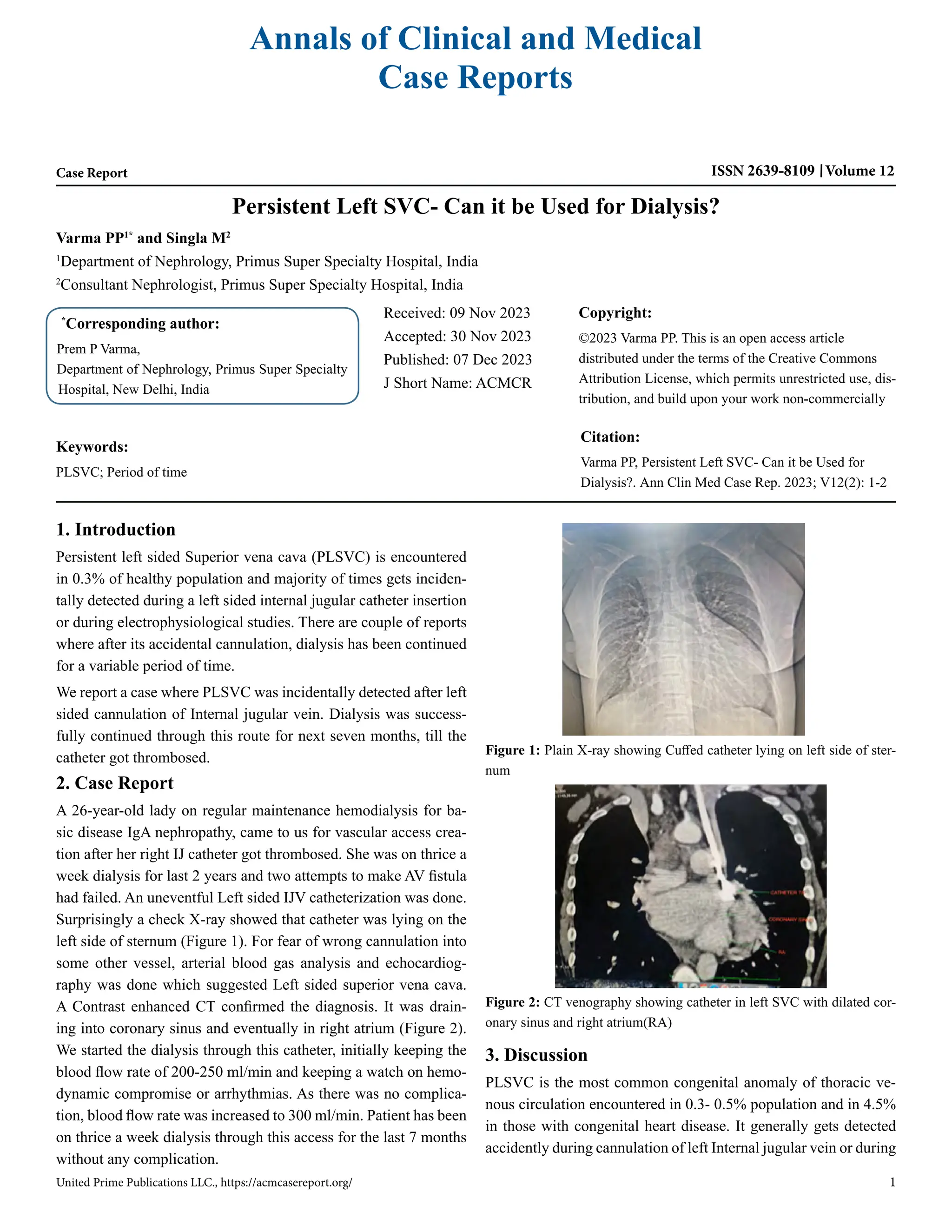
Annals of Clinical and Medical
Case Reports
Case Report ISSN 2639-8109 Volume 12
Varma PP1*
and Singla M2
1
Department of Nephrology, Primus Super Specialty Hospital, India
2
Consultant Nephrologist, Primus Super Specialty Hospital, India
Persistent Left SVC- Can it be Used for Dialysis?
*
Corresponding author:
Prem P Varma,
Department of Nephrology, Primus Super Specialty
Hospital, New Delhi, India
Received: 09 Nov 2023
Accepted: 30 Nov 2023
Published: 07 Dec 2023
J Short Name: ACMCR
Copyright:
©2023 Varma PP. This is an open access article
distributed under the terms of the Creative Commons
Attribution License, which permits unrestricted use, dis-
tribution, and build upon your work non-commercially
Citation:
Varma PP, Persistent Left SVC- Can it be Used for
Dialysis?. Ann Clin Med Case Rep. 2023; V12(2): 1-2
United Prime Publications LLC., https://acmcasereport.org/ 1
Keywords:
PLSVC; Period of time
1. Introduction
Persistent left sided Superior vena cava (PLSVC) is encountered
in 0.3% of healthy population and majority of times gets inciden-
tally detected during a left sided internal jugular catheter insertion
or during electrophysiological studies. There are couple of reports
where after its accidental cannulation, dialysis has been continued
for a variable period of time.
We report a case where PLSVC was incidentally detected after left
sided cannulation of Internal jugular vein. Dialysis was success-
fully continued through this route for next seven months, till the
catheter got thrombosed.
2. Case Report
A 26-year-old lady on regular maintenance hemodialysis for ba-
sic disease IgA nephropathy, came to us for vascular access crea-
tion after her right IJ catheter got thrombosed. She was on thrice a
week dialysis for last 2 years and two attempts to make AV fistula
had failed. An uneventful Left sided IJV catheterization was done.
Surprisingly a check X-ray showed that catheter was lying on the
left side of sternum (Figure 1). For fear of wrong cannulation into
some other vessel, arterial blood gas analysis and echocardiog-
raphy was done which suggested Left sided superior vena cava.
A Contrast enhanced CT confirmed the diagnosis. It was drain-
ing into coronary sinus and eventually in right atrium (Figure 2).
We started the dialysis through this catheter, initially keeping the
blood flow rate of 200-250 ml/min and keeping a watch on hemo-
dynamic compromise or arrhythmias. As there was no complica-
tion, blood flow rate was increased to 300 ml/min. Patient has been
on thrice a week dialysis through this access for the last 7 months
without any complication.
3. Discussion
PLSVC is the most common congenital anomaly of thoracic ve-
nous circulation encountered in 0.3- 0.5% population and in 4.5%
in those with congenital heart disease. It generally gets detected
accidently during cannulation of left Internal jugular vein or during
Figure 1: Plain X-ray showing Cuffed catheter lying on left side of ster-
num
Figure 2: CT venography showing catheter in left SVC with dilated cor-
onary sinus and right atrium(RA)
United Prime Publications LLC., https://acmcasereport.org/ 2
Volume 12 Issue 2 -2023 Case Report
pacemaker placement. Majority of times (92%), it drains through
coronary sinus into right atrium and rarely into left atrium [1-3].
Embryologically human left SVC originates in the third week of
the embryonic period, and then the left anterior cardinal vena cava
gradually atrophies and finally degenerates into the ligament of
Marshall. If the degeneration is not complete, then the remains
of a pipeline structure after birth is PLSVC. Based on anatomy,
Schummer [4] classified SVC into 3 types. Type I, normal anato-
my; type II, only persistent left superior vena cava; type IIIa, right
and left superior vena cava with connection; type IIIb, right and
left superior vena cava without connection.
Based on the drainage characteristic, Zhu [5] classified PLSVC,
into 4 types. Type A- PLSVC draining blood to the right atrium via
coronary sinus, type B- PLSVC draining blood to the right atrium
via coronary sinus with partial right-to-left shunt, type C- PLSVC
drains blood to left atrium directly with right-to-left shunt and type
D- PLSVC is directly connected to left pulmonary vein (coronary
sinus absent)
Our case fits into Schummer’s type III b class and based on drain-
age into Zhu’s type A and this combination is the most common.
Many workers have reported uneventful catheterization like in
our case [1-3]. Most of the times PLSVC drains into right atrium
through coronary sinus. Because of higher blood flow, sinus gets
dilated and enlarged. An enlarged coronary sinus on echocardi-
ography is a hint towards PLSVC. Due to coronary sinus stim-
ulation by catheter, few cases of arrhythmia, angina, stroke and
shock have been reported. If PLSVC drains into left atrium, there
is usually a cardiac septal defect like atrial or ventricular. It can
have dangerous outcome for patient, as there may be cyanosis and
fear of systemic embolization.
Whether dialysis should be continued through PLSVC, is not an
easy answer. If it is draining into Left atrium, dialysis is not pos-
sible. If it is draining into right atrium through coronary sinus,
dialysis can be offered if there is no hemodynamic compromise/
arrhythmia following catheter insertion and dialysis. It is suggest-
ed that to avoid irritation of coronary sinus, the catheter tip (which
is normally placed in right atrium), should be placed at lower end
of PLSVC as it is not possible to place it in right atrium and it is
not advisable to place it in coronary sinus. As cardiac arrhythmias
have been reported even after 2-3 sessions of dialysis, it is prudent
to be aware of this possibility.
A check X-ray showing IJV catheter on left side of sternum, after
left sided IJV cannulation may prompt nephrologist to remove the
catheter fearing cannulation of an artery. However, if fluorosco-
py guided / real time ultrasonography guided cannulation is done,
such fear gets obviated. Arterial blood gas analysis also removes
fear of arterial cannulation. Catheter lying on left side of sternum
should make one suspect of PLSVC and a dilated coronary sinus
on echocardiography gives an additional hint. Echocardiography
generally gives the diagnosis and CT angiography confirms the
diagnosis. Cardiac catheterization is the gold standard for diag-
nosis of PLSVC, however to avoid invasive procedure thoracic
enhanced CTA is an alternative [6]. He et al [7], on compilation
of data, found 28 case reports of PLSVC among dialysis patients,
12 had tunneled cuffed catheter placement and 16 non-cuffed cath-
eters. Dialysis has been performed in some of these patients for
variable a period of time (2-32 months) and few complications like
angina, stroke, arrhythmias etc. have also been reported.
We are reporting the case to bring awareness among nephrologists
(proceduralist) of this rare entity. Dilemma of continuing dialysis
in such cases is not straight forward. We feel PLSVC can be used
as vascular access (like in our case) provided it is providing ade-
quate blood flow and is draining in coronary sinus and into right
atrium. It is prudent to look for any complication like arrhythmias
during initial dialysis sessions.
References
1. Kute VB, Vanikar AV, Gumber MR, Shah PR, Goplani KR, Trivedi
HL. Hemodialysis through persistent left superior vena cava. Indian
J Crit Care Med. 2011; 15(1): 40-2.
2. Anvesh G, Raju SB, Rammurti S, Prasad K. Persistent left superior
vena cava in a hemodialysis patient. Indian J Nephrol. 2018; 28(4):
317-9.
3. Lim TC, H’ng MW. Persistent left superior vena cava: a possible
site for haemodialysis catheter placement. Singapore Med J. 2010;
51(12): e195-7.
4. Schummer W, Schummer C, Fröber R. Persistent left superior vena
cava and central venous catheter position: Clinical impact illustrated
by four cases. Surg Radiol Anat. 2003; 25: 315-21.
5. Zhu X. Basic Illustrations For Cardiac Surgery. 2nd ed. Beijing: Pe-
king union medical college press. 2010; 257-64.
6. Azizova A, Onder O, Arslan S, Ardali S, Hazirolan T. Persistent left
superior vena cava: clinical importance and differential diagnoses.
Insights Imaging. 2020; 11: 110.
7. He H, Li B, Ma Y, Zhang Y, Ye C, Mei C, et al. Catheterization in a
patient with end-stage renal disease through persistent left superior
vena cava: a rare case report and literature review. BMC Nephrol.
2019; 20: 202.

Persistent Left SVC- Can it be Used for Dialysis?

  • 1.
    Annals of Clinicaland Medical Case Reports Case Report ISSN 2639-8109 Volume 12 Varma PP1* and Singla M2 1 Department of Nephrology, Primus Super Specialty Hospital, India 2 Consultant Nephrologist, Primus Super Specialty Hospital, India Persistent Left SVC- Can it be Used for Dialysis? * Corresponding author: Prem P Varma, Department of Nephrology, Primus Super Specialty Hospital, New Delhi, India Received: 09 Nov 2023 Accepted: 30 Nov 2023 Published: 07 Dec 2023 J Short Name: ACMCR Copyright: ©2023 Varma PP. This is an open access article distributed under the terms of the Creative Commons Attribution License, which permits unrestricted use, dis- tribution, and build upon your work non-commercially Citation: Varma PP, Persistent Left SVC- Can it be Used for Dialysis?. Ann Clin Med Case Rep. 2023; V12(2): 1-2 United Prime Publications LLC., https://acmcasereport.org/ 1 Keywords: PLSVC; Period of time 1. Introduction Persistent left sided Superior vena cava (PLSVC) is encountered in 0.3% of healthy population and majority of times gets inciden- tally detected during a left sided internal jugular catheter insertion or during electrophysiological studies. There are couple of reports where after its accidental cannulation, dialysis has been continued for a variable period of time. We report a case where PLSVC was incidentally detected after left sided cannulation of Internal jugular vein. Dialysis was success- fully continued through this route for next seven months, till the catheter got thrombosed. 2. Case Report A 26-year-old lady on regular maintenance hemodialysis for ba- sic disease IgA nephropathy, came to us for vascular access crea- tion after her right IJ catheter got thrombosed. She was on thrice a week dialysis for last 2 years and two attempts to make AV fistula had failed. An uneventful Left sided IJV catheterization was done. Surprisingly a check X-ray showed that catheter was lying on the left side of sternum (Figure 1). For fear of wrong cannulation into some other vessel, arterial blood gas analysis and echocardiog- raphy was done which suggested Left sided superior vena cava. A Contrast enhanced CT confirmed the diagnosis. It was drain- ing into coronary sinus and eventually in right atrium (Figure 2). We started the dialysis through this catheter, initially keeping the blood flow rate of 200-250 ml/min and keeping a watch on hemo- dynamic compromise or arrhythmias. As there was no complica- tion, blood flow rate was increased to 300 ml/min. Patient has been on thrice a week dialysis through this access for the last 7 months without any complication. 3. Discussion PLSVC is the most common congenital anomaly of thoracic ve- nous circulation encountered in 0.3- 0.5% population and in 4.5% in those with congenital heart disease. It generally gets detected accidently during cannulation of left Internal jugular vein or during Figure 1: Plain X-ray showing Cuffed catheter lying on left side of ster- num Figure 2: CT venography showing catheter in left SVC with dilated cor- onary sinus and right atrium(RA)
  • 2.
    United Prime PublicationsLLC., https://acmcasereport.org/ 2 Volume 12 Issue 2 -2023 Case Report pacemaker placement. Majority of times (92%), it drains through coronary sinus into right atrium and rarely into left atrium [1-3]. Embryologically human left SVC originates in the third week of the embryonic period, and then the left anterior cardinal vena cava gradually atrophies and finally degenerates into the ligament of Marshall. If the degeneration is not complete, then the remains of a pipeline structure after birth is PLSVC. Based on anatomy, Schummer [4] classified SVC into 3 types. Type I, normal anato- my; type II, only persistent left superior vena cava; type IIIa, right and left superior vena cava with connection; type IIIb, right and left superior vena cava without connection. Based on the drainage characteristic, Zhu [5] classified PLSVC, into 4 types. Type A- PLSVC draining blood to the right atrium via coronary sinus, type B- PLSVC draining blood to the right atrium via coronary sinus with partial right-to-left shunt, type C- PLSVC drains blood to left atrium directly with right-to-left shunt and type D- PLSVC is directly connected to left pulmonary vein (coronary sinus absent) Our case fits into Schummer’s type III b class and based on drain- age into Zhu’s type A and this combination is the most common. Many workers have reported uneventful catheterization like in our case [1-3]. Most of the times PLSVC drains into right atrium through coronary sinus. Because of higher blood flow, sinus gets dilated and enlarged. An enlarged coronary sinus on echocardi- ography is a hint towards PLSVC. Due to coronary sinus stim- ulation by catheter, few cases of arrhythmia, angina, stroke and shock have been reported. If PLSVC drains into left atrium, there is usually a cardiac septal defect like atrial or ventricular. It can have dangerous outcome for patient, as there may be cyanosis and fear of systemic embolization. Whether dialysis should be continued through PLSVC, is not an easy answer. If it is draining into Left atrium, dialysis is not pos- sible. If it is draining into right atrium through coronary sinus, dialysis can be offered if there is no hemodynamic compromise/ arrhythmia following catheter insertion and dialysis. It is suggest- ed that to avoid irritation of coronary sinus, the catheter tip (which is normally placed in right atrium), should be placed at lower end of PLSVC as it is not possible to place it in right atrium and it is not advisable to place it in coronary sinus. As cardiac arrhythmias have been reported even after 2-3 sessions of dialysis, it is prudent to be aware of this possibility. A check X-ray showing IJV catheter on left side of sternum, after left sided IJV cannulation may prompt nephrologist to remove the catheter fearing cannulation of an artery. However, if fluorosco- py guided / real time ultrasonography guided cannulation is done, such fear gets obviated. Arterial blood gas analysis also removes fear of arterial cannulation. Catheter lying on left side of sternum should make one suspect of PLSVC and a dilated coronary sinus on echocardiography gives an additional hint. Echocardiography generally gives the diagnosis and CT angiography confirms the diagnosis. Cardiac catheterization is the gold standard for diag- nosis of PLSVC, however to avoid invasive procedure thoracic enhanced CTA is an alternative [6]. He et al [7], on compilation of data, found 28 case reports of PLSVC among dialysis patients, 12 had tunneled cuffed catheter placement and 16 non-cuffed cath- eters. Dialysis has been performed in some of these patients for variable a period of time (2-32 months) and few complications like angina, stroke, arrhythmias etc. have also been reported. We are reporting the case to bring awareness among nephrologists (proceduralist) of this rare entity. Dilemma of continuing dialysis in such cases is not straight forward. We feel PLSVC can be used as vascular access (like in our case) provided it is providing ade- quate blood flow and is draining in coronary sinus and into right atrium. It is prudent to look for any complication like arrhythmias during initial dialysis sessions. References 1. Kute VB, Vanikar AV, Gumber MR, Shah PR, Goplani KR, Trivedi HL. Hemodialysis through persistent left superior vena cava. Indian J Crit Care Med. 2011; 15(1): 40-2. 2. Anvesh G, Raju SB, Rammurti S, Prasad K. Persistent left superior vena cava in a hemodialysis patient. Indian J Nephrol. 2018; 28(4): 317-9. 3. Lim TC, H’ng MW. Persistent left superior vena cava: a possible site for haemodialysis catheter placement. Singapore Med J. 2010; 51(12): e195-7. 4. Schummer W, Schummer C, Fröber R. Persistent left superior vena cava and central venous catheter position: Clinical impact illustrated by four cases. Surg Radiol Anat. 2003; 25: 315-21. 5. Zhu X. Basic Illustrations For Cardiac Surgery. 2nd ed. Beijing: Pe- king union medical college press. 2010; 257-64. 6. Azizova A, Onder O, Arslan S, Ardali S, Hazirolan T. Persistent left superior vena cava: clinical importance and differential diagnoses. Insights Imaging. 2020; 11: 110. 7. He H, Li B, Ma Y, Zhang Y, Ye C, Mei C, et al. Catheterization in a patient with end-stage renal disease through persistent left superior vena cava: a rare case report and literature review. BMC Nephrol. 2019; 20: 202.