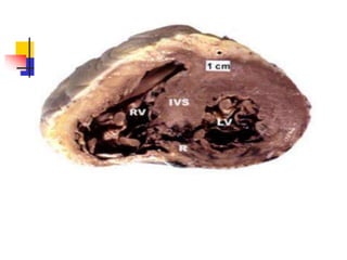
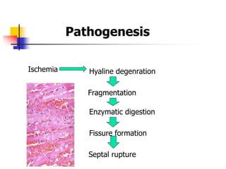
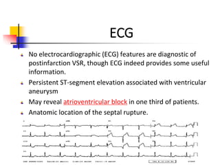
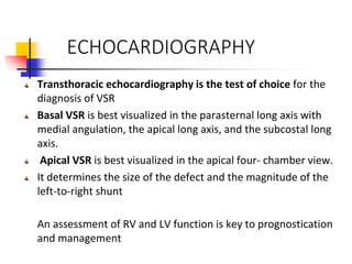
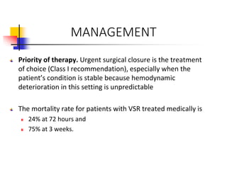
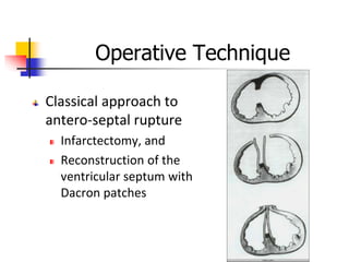
Post MI Ventricular Septal
Rupture
Dr. Asma Iqbal
Dr. Nadish
Dr. Aneera
Dr.Nimra
Ventricular septal rupture (VSR): Tear or Laceration in the
Interventricular septum creating a Left to Right Shunt.
• It is a rare but lethal complication of myocardial
infarction (MI).
• Bimodal peak
• Range: few hours  2 weeks
• Average time to rupture
2-8 days
Time course may be accelerated by thrombolysis, possible
related to intramyocardial hemorrhage
INTRODUCTION
PATHOPHYSIOLOGY
Ischemia Hyaline degenration
Fragmentation
Enzymatic digestion
Fissure formation
Septal rupture
Pathogenesis
Associated Factors
Predictors of VSD
Advanced age,
Anterior location of infarction,
Female gender,
CKD
Thrombolysis after 12 hours also suggested as a
predisposing factor.
Decreased Likelihood
 H/o Smoking
 HTN,
 DM,
 Chronic Angina, previous MI
EPIDEMIOLOGY
Epidemiology
• Uncommon complication of MI.
• Autopsy studies reveal
• It occurs at a rate of approximately 1-2% without
reperfusion therapy
• 0.2% to 0.34% with fibrinolytic therapy
• 3.9% among patients with cardiogenic shock
Relative Improvement in survival due to
Earlier diagnosis
Earlier flow restoration
More aggressive surgical intervention
More aggressive BP control post MI
Septal blood Supply
The septal blood supply comes from branches of
• the left anterior descending coronary artery,
• the posterior descending branch of the right
coronary artery, or the circumflex artery when it
is dominant
ANATOMY OF VSR
Two types of post MI VSR
Simple: through and through single large defect usually
located anteriorly
Complex: serpentiginous dissection tract remote from the
primary septal defect- most commonly an inferior VSD
Apical VSR with anterior MI(due to LAD occlusion)
Basal VSR with inferior MI (due to RCA/LCX occlusion)
CLINICAL PRESENTATION
Patient may remain relatively comfortable early in the disease
course.
Recurrence of angina, pulmonary edema, hypotension, and
shock may develop abruptly later in the course.
Abrupt onset of hemodynamic compromise characterized by
hypotension, biventricular failure, and a new murmur may be
the initial manifestation.
PHYSICAL FINDINGS
The diagnosis should be suspected when a new pansystolic
murmur develops, especially in the setting of worsening
hemodynamics
INVESTIGATIONS
 ECG
 IMAGING MODALITIES
 Catheterization and Pressure Measurement
ECG
No electrocardiographic (ECG) features are diagnostic of
postinfarction VSR, though ECG indeed provides some useful
information.
Persistent ST-segment elevation associated with ventricular
aneurysm
May reveal atrioventricular block in one third of patients.
Anatomic location of the septal rupture.
RADIOGRAPHS
On plain chest radiography, 82% of patients
with postinfarction ventricular septal
rupture (VSR) demonstrate left ventricular
enlargement,
78% have pulmonary edema,
and 64% have a pleural effusion
ECHOCARDIOGRAPHY
Transthoracic echocardiography is the test of choice for the
diagnosis of VSR
Basal VSR is best visualized in the parasternal long axis with
medial angulation, the apical long axis, and the subcostal long
axis.
Apical VSR is best visualized in the apical four- chamber view.
It determines the size of the defect and the magnitude of the
left-to-right shunt
An assessment of RV and LV function is key to prognostication
and management
ECHO AND COLOUR FLOW
DOPPLER VIDEO
Color Flow
Doppler
100% sensitive and
specific in
differentiating VSR
from acute MR
CATHETERIZATION AND PRESSURE
MEASUREMENT
Left-heart catheterization with coronary
angiography is recommended in all stable
patients
An important diagnostic test for differentiating
VSR from mitral valve insufficiency is
catheterization of the right heart with a Swan-
Ganz catheter.
Left- and right-side pressure measurements help
estimate the degree of biventricular failure
Need for cardiac catheterization
2/3 of the patients have multivessel
coronary artery disease
Decreased operative mortality and
improved late survival has been shown in
patients with multivessel disease
Cardiogenic shock not a deterrent to Cath
=> Coronary angiography
should be performed
TREATMENT MODALITIES
Surgical Repair
Percutaneous Device closure
Medical Therapy
MANAGEMENT
Priority of therapy. Urgent surgical closure is the treatment
of choice (Class I recommendation), especially when the
patient’s condition is stable because hemodynamic
deterioration in this setting is unpredictable
The mortality rate for patients with VSR treated medically is
24% at 72 hours and
75% at 3 weeks.
Pre-Operative Management
Hemodynamic stabilization so as to
minimize peripheral organ compromise
Reduce Systemic vascular resistance, and
thus, the left-to-right shunt
Maintain or improve coronary artery blood
flow
Maintain cardiac output and arterial
pressure to ensure peripheral organ
perfusion
Supportive medical management
Vasodilators can decrease left-to-right shunt
and increase systemic flow by means of
reducing systemic vascular resistance (SVR).
Vasopressors
The profound level of cardiogenic shock in
some patients precludes vasodilator treatment,
often necessitating vasopressor support.
Intra-aortic balloon counterpulsation (IABP)
IABP Counterpulsation
Timing of Surgery
Surgery should be performed soon after
diagnosis in most patients
Patients is cardiogenic shock should be
operated on immediately after
anigography
Hemodynamically stable patients should
have surgery on an urgent basis
GOALs OF SURGERY
Resection of Infarcted Myocardium
Elimination of L to R shunt by patch
Operative Technique
Classical approach to
antero-septal rupture
Infarctectomy, and
Reconstruction of the
ventricular septum with
Dacron patches
Operative Technique
Classical approach
to infro-posterior
rupture
Infarctectomy, and
Reconstruction of
infroposterior VSD,
Reconstruction free wall
with Dacron patches.
Outcome
In patients with cardiogenic shock mortality
reported to be the highest
Posterior VSR (IMI) is another factor strongly
associated with poor surgical outcome due to
Difficulty of exposure, and
Frequent concomitant infarction of the postero-
medial papillary muscle
Percutaneous Device Closure
SCANERIO :
A 55 Year old Hypertensive male, diagnosed with
extensive anterior wall MI and managed with
SK,unremarkable general and systemic examination
On 4 th Day He became suddenly Dyspenic with B.P=
80/60, rales heard at lower half of lungs B/L,Cardiac
examination revealed New pansystolic murmur at left
lower parasternal border. Ionotropic support started but
vitals did not improve, so IABP inserted…
WHAT IS YOUR PROVISIONAL DIAGNOSIS?
HOW Will you proceed ?
THANK YOU

Post MI Ventricular Septal Rupture

  • 1.
    Post MI VentricularSeptal Rupture Dr. Asma Iqbal Dr. Nadish Dr. Aneera Dr.Nimra
  • 2.
    Ventricular septal rupture(VSR): Tear or Laceration in the Interventricular septum creating a Left to Right Shunt. • It is a rare but lethal complication of myocardial infarction (MI). • Bimodal peak • Range: few hours  2 weeks • Average time to rupture 2-8 days Time course may be accelerated by thrombolysis, possible related to intramyocardial hemorrhage INTRODUCTION
  • 4.
  • 5.
    Ischemia Hyaline degenration Fragmentation Enzymaticdigestion Fissure formation Septal rupture Pathogenesis
  • 6.
    Associated Factors Predictors ofVSD Advanced age, Anterior location of infarction, Female gender, CKD Thrombolysis after 12 hours also suggested as a predisposing factor. Decreased Likelihood  H/o Smoking  HTN,  DM,  Chronic Angina, previous MI
  • 7.
  • 8.
    Epidemiology • Uncommon complicationof MI. • Autopsy studies reveal • It occurs at a rate of approximately 1-2% without reperfusion therapy • 0.2% to 0.34% with fibrinolytic therapy • 3.9% among patients with cardiogenic shock
  • 9.
    Relative Improvement insurvival due to Earlier diagnosis Earlier flow restoration More aggressive surgical intervention More aggressive BP control post MI
  • 10.
    Septal blood Supply Theseptal blood supply comes from branches of • the left anterior descending coronary artery, • the posterior descending branch of the right coronary artery, or the circumflex artery when it is dominant
  • 11.
    ANATOMY OF VSR Twotypes of post MI VSR Simple: through and through single large defect usually located anteriorly Complex: serpentiginous dissection tract remote from the primary septal defect- most commonly an inferior VSD Apical VSR with anterior MI(due to LAD occlusion) Basal VSR with inferior MI (due to RCA/LCX occlusion)
  • 12.
    CLINICAL PRESENTATION Patient mayremain relatively comfortable early in the disease course. Recurrence of angina, pulmonary edema, hypotension, and shock may develop abruptly later in the course. Abrupt onset of hemodynamic compromise characterized by hypotension, biventricular failure, and a new murmur may be the initial manifestation.
  • 13.
    PHYSICAL FINDINGS The diagnosisshould be suspected when a new pansystolic murmur develops, especially in the setting of worsening hemodynamics
  • 15.
    INVESTIGATIONS  ECG  IMAGINGMODALITIES  Catheterization and Pressure Measurement
  • 16.
    ECG No electrocardiographic (ECG)features are diagnostic of postinfarction VSR, though ECG indeed provides some useful information. Persistent ST-segment elevation associated with ventricular aneurysm May reveal atrioventricular block in one third of patients. Anatomic location of the septal rupture.
  • 17.
    RADIOGRAPHS On plain chestradiography, 82% of patients with postinfarction ventricular septal rupture (VSR) demonstrate left ventricular enlargement, 78% have pulmonary edema, and 64% have a pleural effusion
  • 18.
    ECHOCARDIOGRAPHY Transthoracic echocardiography isthe test of choice for the diagnosis of VSR Basal VSR is best visualized in the parasternal long axis with medial angulation, the apical long axis, and the subcostal long axis. Apical VSR is best visualized in the apical four- chamber view. It determines the size of the defect and the magnitude of the left-to-right shunt An assessment of RV and LV function is key to prognostication and management
  • 19.
    ECHO AND COLOURFLOW DOPPLER VIDEO
  • 20.
    Color Flow Doppler 100% sensitiveand specific in differentiating VSR from acute MR
  • 21.
    CATHETERIZATION AND PRESSURE MEASUREMENT Left-heartcatheterization with coronary angiography is recommended in all stable patients An important diagnostic test for differentiating VSR from mitral valve insufficiency is catheterization of the right heart with a Swan- Ganz catheter. Left- and right-side pressure measurements help estimate the degree of biventricular failure
  • 23.
    Need for cardiaccatheterization 2/3 of the patients have multivessel coronary artery disease Decreased operative mortality and improved late survival has been shown in patients with multivessel disease Cardiogenic shock not a deterrent to Cath => Coronary angiography should be performed
  • 24.
  • 25.
    MANAGEMENT Priority of therapy.Urgent surgical closure is the treatment of choice (Class I recommendation), especially when the patient’s condition is stable because hemodynamic deterioration in this setting is unpredictable The mortality rate for patients with VSR treated medically is 24% at 72 hours and 75% at 3 weeks.
  • 26.
    Pre-Operative Management Hemodynamic stabilizationso as to minimize peripheral organ compromise Reduce Systemic vascular resistance, and thus, the left-to-right shunt Maintain or improve coronary artery blood flow Maintain cardiac output and arterial pressure to ensure peripheral organ perfusion
  • 27.
    Supportive medical management Vasodilatorscan decrease left-to-right shunt and increase systemic flow by means of reducing systemic vascular resistance (SVR). Vasopressors The profound level of cardiogenic shock in some patients precludes vasodilator treatment, often necessitating vasopressor support. Intra-aortic balloon counterpulsation (IABP)
  • 28.
  • 29.
    Timing of Surgery Surgeryshould be performed soon after diagnosis in most patients Patients is cardiogenic shock should be operated on immediately after anigography Hemodynamically stable patients should have surgery on an urgent basis
  • 30.
    GOALs OF SURGERY Resectionof Infarcted Myocardium Elimination of L to R shunt by patch
  • 31.
    Operative Technique Classical approachto antero-septal rupture Infarctectomy, and Reconstruction of the ventricular septum with Dacron patches
  • 32.
    Operative Technique Classical approach toinfro-posterior rupture Infarctectomy, and Reconstruction of infroposterior VSD, Reconstruction free wall with Dacron patches.
  • 33.
    Outcome In patients withcardiogenic shock mortality reported to be the highest Posterior VSR (IMI) is another factor strongly associated with poor surgical outcome due to Difficulty of exposure, and Frequent concomitant infarction of the postero- medial papillary muscle
  • 34.
  • 35.
    SCANERIO : A 55Year old Hypertensive male, diagnosed with extensive anterior wall MI and managed with SK,unremarkable general and systemic examination On 4 th Day He became suddenly Dyspenic with B.P= 80/60, rales heard at lower half of lungs B/L,Cardiac examination revealed New pansystolic murmur at left lower parasternal border. Ionotropic support started but vitals did not improve, so IABP inserted…
  • 36.
    WHAT IS YOURPROVISIONAL DIAGNOSIS? HOW Will you proceed ?
  • 37.

Editor's Notes