Hypertrophic Obstructive
Cardiomyopathy
DR DHANESH
Definition
Hypertrophic cardiomyopathy (HCM) is a
genetic myocardial disorder characterized by
left and/or right ventricular hypertrophy that
is usually, but not always, asymmetric and is
associated with microscopic evidence of
myocardial fiber disarray and fibrosis.
HISTORICAL NOTE
 Pathologic findings compatible with HOCM were described
by two 19th-century French pathologists, Hallopeau and
Liouiville and an early 20th-century German pathologist,
Schmincke
 To distinguish it from other cardiomyopathies, Goodwin
and colleagues named it hypertrophic obstructive
cardiomyopathy, whereas Braunwald and colleagues called
it idiopathic hypertrophic subaortic stenosis and Wigle and
colleagues, muscularsubaortic stenosis.
 Simple myotomy using an aortic approach was used for a
time by the Toronto group
 Later, they modified the procedure to include excision of
muscle (myectomy) as advocated by Morrow
MORPHOGENESIS AND MORPHOLOGY
HCM is caused by a missense mutation in one
of at least 11 genes that encode the proteins
of the cardiac sarcomere.
These include mutations in the β-myosin
heavy-chain gene (chromosome 14q11-q12),
in cardiac troponin-T(chromosome 1), and in
α-tropomyosin (chromosome15q2).
It is transmitted as an AD trait, although
nonfamilial cases probably occur as well.
MORPHOLOGY
• Muscular hypertrophy present in HCM involves
the interventricular septum and LV, and is
variable in its location and severity.
Ventricular Septum
In classic HOCM, hypertrophy is maximal in the
cephalad portion of the ventricular septum
Point of maximal thickening lies just apical
(caudad) to the free edge of the anterior mitral
leaflet in its open position.
 Dynamic Morphology of Septum and Mitral Valve
• When septal hypertrophy is classic, obstruction is sited in the
LVOT between the hypertrophied ventricular septum and AML.
• In systole the PML closes against the body of the elongated AML
at about the junction of the middle and free-edge thirds (rather
than near the free edge as in the normal heart).
• The free-edge portion of the AML beyond the point of coaptation
hinges (angulates) on the remainder of the leaflet in a cephalad
direction toward the aortic annulus.
 Left Ventricular Free Wall
• In obstructive HCM with ASH, free-wall hypertrophy is more
marked than in nonobstructive forms and is fairly uniform,
particularly in the anterolateral and apical portions.
• There is, however, less thickening of the posterior free wall in
almost all varieties of HCM.
Left Ventricular Cavity
In association with unusual shape of the IVS, the
LV cavity is small, even when HF occurs in later
stages of HOCM, and has an S or sigmoid shape in
systole when viewed in its longitudinal axis.
A sigmoid shape is characteristic of patients
younger than age 40 and is rare in patients older
than 65.
Most patients show an “hourglass” contour of the
cardiac border, with midventricular hypertrophy
and intracavitary gradients
Left ventricular (LV) cineangiogram in RAO projection in HOCM. Note characteristic
deformity of LV cavity, with septal muscle encroaching on anterior margin of outflow
tract and the grossly hypertrophied papillary muscles contributing to virtual
elimination of the mid-LV cavity in systole. A, Diastole. Dashed line crossing LV outflow
area represents free-wall portion of mitral annulus, delineated by contrast medium
trapped behind opened posterior leaflet. B, Systole. Lower dashedline outlines a
radiolucent filling defect caused by contact between mitral leaflets and septum
A B
 Histopathology of Left Ventricle
• Increased wall thickness is mainly caused by increased fibrous
tissue, particularly in the ventricular septum but also in the free
wall.
• Increase in muscle cell diameter and number of cell layers also
contributes, with cell diameters being largest in layers closest to the
cavity, perhaps because this is the site of greatest wall stress.
 Left Atrium
• The left atrium is often dilated and thick walled as a result of
decreased compliance of the LV and presence of mitral
regurgitation.
 In obstructive forms of HCM, the MV is positioned closer to the
IVS than in the normal heart
 MV leaflets are disproportionately elongated and thickened,
particularly the leading edge of the AML
 This is presumably the result of SAM
 The mitral annulus forcefully constricts during systole, and this
pursestring action gathers the mitral leaflets into folds
 A further consequence of SAM is production of MR in mid- or
late systole as the anterior leaflet moves forward
 Studies by Bonow and by Wigle and colleagues indicate a direct
relation between magnitude of the pressure gradient and degree
of MR.
 It is likely that severity of MR, magnitude of the pressure
gradient, and degree of prolongation of LVET are determined by
time of onset and duration of mitral leaflet–septal contact
 MR occurs independently of SAM in about 20% of patients with
HOCM
Mitral Valve
Right Ventricle
 The RV chamber is distorted by the hypertrophied IVS,
which projects into the RVOT
 This hypertrophy may cause an important pressure
gradient in the RVOT and, in long-standing cases,
hypertrophy of the free wall
 RVH may also occur secondary to PAH from long-
standing LVH and elevated LA pressure
 Unverferth and colleagues demonstrated an important
increase in amount of fibrous tissue in the RV free wall
in HCM, as well as an increase in myocyte cell diameter
in the subendocardial layer.
Coronary Arteries
 In HCM, coronary arteries are larger in diameter than normal.
 Important coronary arteriosclerosis is present in about 5% of
patients
 Muscular bridging of the LAD during part of its course is more
common in HCM than in normal hearts. The LAD may become
totally occluded during systole at these sites or may have an
irregular saw tooth appearance
 Maron and colleagues and Waller and colleagues have reported
that transmural MI occurs in HCM in the absence of arteriosclerotic
CAD
 This could be caused by systolic arterial compression or spasm,
narrowing of intramural branches, inadequate capillary density, or
reduction in diastolic coronary flow from impaired ventricular
relaxation.
Associated Lesions
Diffuse lentiginosis
Essential hypertension and severe AS
ASD and RHD
With HOCM, functional impairment of von
Willebrand factor is frequent and is closely and
independently related to the magnitude of
outflow obstruction.
A resting peak gradient of 15 mmHg is sufficient
to impair function of this glycoprotein and may
result in abnormal spontaneous bleeding
CLINICAL FEATURES AND DIAGNOSTIC CRITERIA
 Symptoms:
 Angina
 Effort dyspnea
 Syncope
 Dizziness on exertion
 Palpitations(10%)
 Death
Caused by a complex interaction of
diastolic dysfunction, arrhythmias,
myocardial ischemia, and outflow
gradient
 Sign:
• The three cardinal signs of HOCM are
 (1) Late-onset systolic ejection
murmur between the left sternal
edge and apex. The murmur
increases in intensity after a Valsalva
maneuver (or inhalation of amyl
nitrite), because both increase the
degree of obstruction.
 (2) Bifid (Jerky) arterial pulse (S4)
 (3) Palpable left atrial contraction
• An abnormal jugular a wave is
frequently present, and occasionally,
a short, low-pitched diastolic flow
murmur that is enhanced by
inspiration; both are the result of
vigorous atrial contraction
 LV function
• Initially N/ Incresaed and decreased later on
• Diastolic dysfunction
 ECG
• LV strain pattern, Q waves may be present, and rarely, minimal
changes indicative of LVH despite an important gradient
• Occasionally the ECG shows complete right or LBBB and more often
LAHB
• Giant negative T waves in V4-V6 are typical of isolated apical
hypertrophy
• ECG features of LAE are often noted, but those of RAE less often
Chest Radiography
Chest radiography shows mild to moderate
cardiomegaly more often in HOCM than in
other forms of aortic outflow obstruction
The aorta is typically small
The raised LAP may be reflected in the lung
fields by evidence of PVH or frank interstitial
edema
Echocardiography
A:Diastole B:Early systole C
D:Midsystole E:Mid-late systole F
Exercise Echocardiography
• May identify LV outflow obstruction in
symptomatic patients who would not
otherwise be regarded as candidates for
interventional treatment
Cardiac Catheterization and
Cineangiography
 Right-sided heart catheterization will show any Infundibular
stenosis (occasionally severe) and elevation of PAP, which
may also be substantial because of high LAP
 Retrograde left-sided heart catheterization will quantify
and localize the obstruction
 LVEDP is usually increased, often greatly, because of
transmission of a large left atrial a wave
 The beat following a ventricular ectopic beat shows an
abnormal response—a reduced arterial pulse pressure (and
an exaggerated spike-and-dome contour) secondary to
increased obstruction generated by the ectopic beat
 Obstruction is increased by any maneuver that increases LV
contractility or decreases LV preload or afterload
Left ventricular (LV) and aortic pressure tracings in hypertrophic obstructive cardiomyopathy
(HOCM). Peak systolic gradient is 60 mmHg. Aortic pressure pulse shows typical double
contour (“spike and dome”). LV end-diastolic pressure is elevated in association with
transmission of an a wave from the left atrium (arrow). The beat after an ectopic beat
demonstrates reduced aortic pulse pressure characteristic of HOCM (center of tracing).
Postectopic response
NATURAL HISTORY
• The natural history of HCM is typically variable
• Clinical course is often stable over long periods, adverse events
such heart failure, syncope, sudden cardiac death, and peripheral
embolization can occur
• Sudden onset of heart failure is frequently precipitated by atrial
fibrillation, which may be associated with subsequent embolism
• Annual HCM-related mortality reported from referral centers has
ranged from 4% to 6% in children and 2% to 4% in adults
• Sudden cardiac death is common in patients with HCM.
• Risk factors for sudden death include a basal (resting) peak
instantaneous gradient of at least 30 mmHg, young age, syncope,
family history of malignancy, myocardial ischemia (particularly in
the young), sustained ventricular tachycardia on electrophysiologic
testing, and VT on ambulatory monitoring
INDICATIONS FOR OPERATION
 Septal myectomy should be considered for any patient who remains
symptomatic (NYHA class III or IV) after appropriate medical
therapy (β-adrenergic receptor blockade, calcium antagonists,
disopyramide), septal ablation, or pacemaker therapy.
 Who has an important LV subaortic gradient (≥50 mmHg) at rest or
after physiologically base provocation with exercise, caused by SAM
with septal contact
 Atrial fibrillation is also an indication for operation
 May be extended to include less symptomatic patients with severe
gradients, particularly if there is substantial coexisting MR or a
history of syncope or unexplained cardiac arrest, as well as to
asymptomatic younger patients with gradients greater than 100
mmHg
TECHNIQUE OF OPERATION
Postoperative care
 Marked LVH reduces ventricular compliance to such an
extent that LAP of 16 to 18 mmHg may be required
early postoperatively for adequate preload
 Digitalis glycosides and the β-adrenergic receptor
agonist isoproterenol should be avoided because they
increase myocardial contractility and may increase
residual outflow tract gradient
 Hypovolemia and nitroglycerin, which can reduce LV
volume and exaggerate any residual gradient, should
also be avoided
 Avoid AF
Results
 Early (Hospital) death
 Time related survival
 Modes of death
 Incremental risk factors for premature death
 Myocardial Changes
 Conduction disturbances
 Perioperative MI
 Iatrogenic defects
 Systolic Anterior Motion of the Mitral Valve
 Mitral Regurgitation
 Symptomatic Status
 Left Ventricular Function and Structure
Time related survival
Postoperative pressure gradients
Alternative Therapies
• Percutaneous tranluminal septal myocardial
ablation
• Dual chamber pacing
• Cardioverter-Defibrillator
• Cardiac transplantation
• LV-Aortic conduit
THANK
YOU

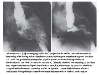
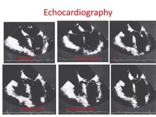
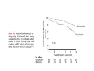
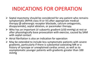
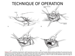
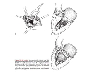
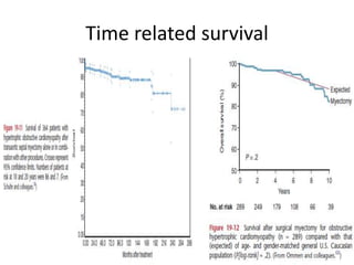
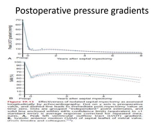
Hypertrophic obstructive cardiomyopathy

  • 1.
  • 2.
    Definition Hypertrophic cardiomyopathy (HCM)is a genetic myocardial disorder characterized by left and/or right ventricular hypertrophy that is usually, but not always, asymmetric and is associated with microscopic evidence of myocardial fiber disarray and fibrosis.
  • 3.
    HISTORICAL NOTE  Pathologicfindings compatible with HOCM were described by two 19th-century French pathologists, Hallopeau and Liouiville and an early 20th-century German pathologist, Schmincke  To distinguish it from other cardiomyopathies, Goodwin and colleagues named it hypertrophic obstructive cardiomyopathy, whereas Braunwald and colleagues called it idiopathic hypertrophic subaortic stenosis and Wigle and colleagues, muscularsubaortic stenosis.  Simple myotomy using an aortic approach was used for a time by the Toronto group  Later, they modified the procedure to include excision of muscle (myectomy) as advocated by Morrow
  • 4.
    MORPHOGENESIS AND MORPHOLOGY HCMis caused by a missense mutation in one of at least 11 genes that encode the proteins of the cardiac sarcomere. These include mutations in the β-myosin heavy-chain gene (chromosome 14q11-q12), in cardiac troponin-T(chromosome 1), and in α-tropomyosin (chromosome15q2). It is transmitted as an AD trait, although nonfamilial cases probably occur as well.
  • 5.
    MORPHOLOGY • Muscular hypertrophypresent in HCM involves the interventricular septum and LV, and is variable in its location and severity. Ventricular Septum In classic HOCM, hypertrophy is maximal in the cephalad portion of the ventricular septum Point of maximal thickening lies just apical (caudad) to the free edge of the anterior mitral leaflet in its open position.
  • 6.
     Dynamic Morphologyof Septum and Mitral Valve • When septal hypertrophy is classic, obstruction is sited in the LVOT between the hypertrophied ventricular septum and AML. • In systole the PML closes against the body of the elongated AML at about the junction of the middle and free-edge thirds (rather than near the free edge as in the normal heart). • The free-edge portion of the AML beyond the point of coaptation hinges (angulates) on the remainder of the leaflet in a cephalad direction toward the aortic annulus.  Left Ventricular Free Wall • In obstructive HCM with ASH, free-wall hypertrophy is more marked than in nonobstructive forms and is fairly uniform, particularly in the anterolateral and apical portions. • There is, however, less thickening of the posterior free wall in almost all varieties of HCM.
  • 7.
    Left Ventricular Cavity Inassociation with unusual shape of the IVS, the LV cavity is small, even when HF occurs in later stages of HOCM, and has an S or sigmoid shape in systole when viewed in its longitudinal axis. A sigmoid shape is characteristic of patients younger than age 40 and is rare in patients older than 65. Most patients show an “hourglass” contour of the cardiac border, with midventricular hypertrophy and intracavitary gradients
  • 8.
    Left ventricular (LV)cineangiogram in RAO projection in HOCM. Note characteristic deformity of LV cavity, with septal muscle encroaching on anterior margin of outflow tract and the grossly hypertrophied papillary muscles contributing to virtual elimination of the mid-LV cavity in systole. A, Diastole. Dashed line crossing LV outflow area represents free-wall portion of mitral annulus, delineated by contrast medium trapped behind opened posterior leaflet. B, Systole. Lower dashedline outlines a radiolucent filling defect caused by contact between mitral leaflets and septum A B
  • 9.
     Histopathology ofLeft Ventricle • Increased wall thickness is mainly caused by increased fibrous tissue, particularly in the ventricular septum but also in the free wall. • Increase in muscle cell diameter and number of cell layers also contributes, with cell diameters being largest in layers closest to the cavity, perhaps because this is the site of greatest wall stress.  Left Atrium • The left atrium is often dilated and thick walled as a result of decreased compliance of the LV and presence of mitral regurgitation.
  • 10.
     In obstructiveforms of HCM, the MV is positioned closer to the IVS than in the normal heart  MV leaflets are disproportionately elongated and thickened, particularly the leading edge of the AML  This is presumably the result of SAM  The mitral annulus forcefully constricts during systole, and this pursestring action gathers the mitral leaflets into folds  A further consequence of SAM is production of MR in mid- or late systole as the anterior leaflet moves forward  Studies by Bonow and by Wigle and colleagues indicate a direct relation between magnitude of the pressure gradient and degree of MR.  It is likely that severity of MR, magnitude of the pressure gradient, and degree of prolongation of LVET are determined by time of onset and duration of mitral leaflet–septal contact  MR occurs independently of SAM in about 20% of patients with HOCM Mitral Valve
  • 11.
    Right Ventricle  TheRV chamber is distorted by the hypertrophied IVS, which projects into the RVOT  This hypertrophy may cause an important pressure gradient in the RVOT and, in long-standing cases, hypertrophy of the free wall  RVH may also occur secondary to PAH from long- standing LVH and elevated LA pressure  Unverferth and colleagues demonstrated an important increase in amount of fibrous tissue in the RV free wall in HCM, as well as an increase in myocyte cell diameter in the subendocardial layer.
  • 12.
    Coronary Arteries  InHCM, coronary arteries are larger in diameter than normal.  Important coronary arteriosclerosis is present in about 5% of patients  Muscular bridging of the LAD during part of its course is more common in HCM than in normal hearts. The LAD may become totally occluded during systole at these sites or may have an irregular saw tooth appearance  Maron and colleagues and Waller and colleagues have reported that transmural MI occurs in HCM in the absence of arteriosclerotic CAD  This could be caused by systolic arterial compression or spasm, narrowing of intramural branches, inadequate capillary density, or reduction in diastolic coronary flow from impaired ventricular relaxation.
  • 13.
    Associated Lesions Diffuse lentiginosis Essentialhypertension and severe AS ASD and RHD With HOCM, functional impairment of von Willebrand factor is frequent and is closely and independently related to the magnitude of outflow obstruction. A resting peak gradient of 15 mmHg is sufficient to impair function of this glycoprotein and may result in abnormal spontaneous bleeding
  • 14.
    CLINICAL FEATURES ANDDIAGNOSTIC CRITERIA  Symptoms:  Angina  Effort dyspnea  Syncope  Dizziness on exertion  Palpitations(10%)  Death Caused by a complex interaction of diastolic dysfunction, arrhythmias, myocardial ischemia, and outflow gradient  Sign: • The three cardinal signs of HOCM are  (1) Late-onset systolic ejection murmur between the left sternal edge and apex. The murmur increases in intensity after a Valsalva maneuver (or inhalation of amyl nitrite), because both increase the degree of obstruction.  (2) Bifid (Jerky) arterial pulse (S4)  (3) Palpable left atrial contraction • An abnormal jugular a wave is frequently present, and occasionally, a short, low-pitched diastolic flow murmur that is enhanced by inspiration; both are the result of vigorous atrial contraction
  • 15.
     LV function •Initially N/ Incresaed and decreased later on • Diastolic dysfunction  ECG • LV strain pattern, Q waves may be present, and rarely, minimal changes indicative of LVH despite an important gradient • Occasionally the ECG shows complete right or LBBB and more often LAHB • Giant negative T waves in V4-V6 are typical of isolated apical hypertrophy • ECG features of LAE are often noted, but those of RAE less often
  • 16.
    Chest Radiography Chest radiographyshows mild to moderate cardiomegaly more often in HOCM than in other forms of aortic outflow obstruction The aorta is typically small The raised LAP may be reflected in the lung fields by evidence of PVH or frank interstitial edema
  • 17.
    Echocardiography A:Diastole B:Early systoleC D:Midsystole E:Mid-late systole F
  • 18.
    Exercise Echocardiography • Mayidentify LV outflow obstruction in symptomatic patients who would not otherwise be regarded as candidates for interventional treatment
  • 19.
    Cardiac Catheterization and Cineangiography Right-sided heart catheterization will show any Infundibular stenosis (occasionally severe) and elevation of PAP, which may also be substantial because of high LAP  Retrograde left-sided heart catheterization will quantify and localize the obstruction  LVEDP is usually increased, often greatly, because of transmission of a large left atrial a wave  The beat following a ventricular ectopic beat shows an abnormal response—a reduced arterial pulse pressure (and an exaggerated spike-and-dome contour) secondary to increased obstruction generated by the ectopic beat  Obstruction is increased by any maneuver that increases LV contractility or decreases LV preload or afterload
  • 20.
    Left ventricular (LV)and aortic pressure tracings in hypertrophic obstructive cardiomyopathy (HOCM). Peak systolic gradient is 60 mmHg. Aortic pressure pulse shows typical double contour (“spike and dome”). LV end-diastolic pressure is elevated in association with transmission of an a wave from the left atrium (arrow). The beat after an ectopic beat demonstrates reduced aortic pulse pressure characteristic of HOCM (center of tracing). Postectopic response
  • 21.
    NATURAL HISTORY • Thenatural history of HCM is typically variable • Clinical course is often stable over long periods, adverse events such heart failure, syncope, sudden cardiac death, and peripheral embolization can occur • Sudden onset of heart failure is frequently precipitated by atrial fibrillation, which may be associated with subsequent embolism • Annual HCM-related mortality reported from referral centers has ranged from 4% to 6% in children and 2% to 4% in adults • Sudden cardiac death is common in patients with HCM. • Risk factors for sudden death include a basal (resting) peak instantaneous gradient of at least 30 mmHg, young age, syncope, family history of malignancy, myocardial ischemia (particularly in the young), sustained ventricular tachycardia on electrophysiologic testing, and VT on ambulatory monitoring
  • 23.
    INDICATIONS FOR OPERATION Septal myectomy should be considered for any patient who remains symptomatic (NYHA class III or IV) after appropriate medical therapy (β-adrenergic receptor blockade, calcium antagonists, disopyramide), septal ablation, or pacemaker therapy.  Who has an important LV subaortic gradient (≥50 mmHg) at rest or after physiologically base provocation with exercise, caused by SAM with septal contact  Atrial fibrillation is also an indication for operation  May be extended to include less symptomatic patients with severe gradients, particularly if there is substantial coexisting MR or a history of syncope or unexplained cardiac arrest, as well as to asymptomatic younger patients with gradients greater than 100 mmHg
  • 24.
  • 26.
    Postoperative care  MarkedLVH reduces ventricular compliance to such an extent that LAP of 16 to 18 mmHg may be required early postoperatively for adequate preload  Digitalis glycosides and the β-adrenergic receptor agonist isoproterenol should be avoided because they increase myocardial contractility and may increase residual outflow tract gradient  Hypovolemia and nitroglycerin, which can reduce LV volume and exaggerate any residual gradient, should also be avoided  Avoid AF
  • 27.
    Results  Early (Hospital)death  Time related survival  Modes of death  Incremental risk factors for premature death  Myocardial Changes  Conduction disturbances  Perioperative MI  Iatrogenic defects  Systolic Anterior Motion of the Mitral Valve  Mitral Regurgitation  Symptomatic Status  Left Ventricular Function and Structure
  • 28.
  • 29.
  • 31.
    Alternative Therapies • Percutaneoustranluminal septal myocardial ablation • Dual chamber pacing • Cardioverter-Defibrillator • Cardiac transplantation • LV-Aortic conduit
  • 33.