
Recommended
More Related Content
What's hot
What's hot (20)
Similar to Spirochetes.pptx
Similar to Spirochetes.pptx (20)
Recently uploaded
Recently uploaded (20)
Spirochetes.pptx
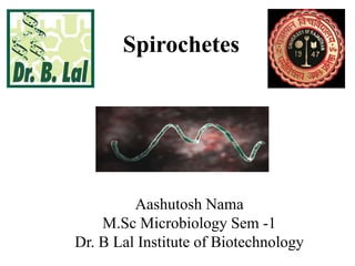
- 1. Spirochetes Aashutosh Nama M.Sc Microbiology Sem -1 Dr. B Lal Institute of Biotechnology
- 2. • Background • Speira = coil Chaite = hair • Gram negative bacteria (differs from other gram negative bacteria due to presence of “endoflagella”). • Thin, flexible, elongated spirally coiled helical bacilli. • Tightly coiled bacteria typically slender and flexuous shpe.
- 3. Endoflagella • Endoflagella do not protrude outside, but present in the periplasmic space between peptidoglycan layer and outer membrane.
- 4. Motility of spirochetes • Endoflagella are responsible for various motility of spirochetes such as: 1. Flexion – extension type 2. Corkscrew type rotatory movement 3. Translatory type motility
- 5. Pathogenes • Most of the spirochetes are saprophytes. Only there genus of them are major humans pathogenes – 1. Borrelia 2. Treponema 3. Leptospira
- 7. Morphological differences between Treponema, Borrelia and Leptospira
- 9. TREPONEMA
- 10. TREPONEMA PALLIDUM • ™ Morphology: extremely thin and delicate with tapering ends 1. ™ Size: 6–14 μm × 0.2 μm 2. ™ Spirals: 6–12 spirals at intervals of 1 μm 3. ™ Motility: flexion extension, translatory and corkscrew motility 4. ™ Endoflagella: About 3–4 flagella - motility & highly antigenic
- 11. PATHOGENESIS OF SYPHILIS • Mode of transmission: - Venereal - Non-venereal - direct contact, blood transfusion or transplacental • ™ Spread: T. pallidum penetrates through mucosa or abraded skin Enter lymphatics and blood systemic primary lesion • ™ Incubation period: Variable (9–90 days) Inversely proportional to the number of organisms inoculated
- 12. Microscopy & Culture • Dark ground or phase contrast microscope • ™ Staining: Do not take up Gram stain - Fluorescence staining - Sliver impregnation methods • ™ Cultivation: Pathogenic treponemes cannot be grown in artificial culture media. Maintained in rabbit testes. E.g, „ Nichols strain • Non-pathogenic Treponemes – grow in Smith Noguchi medium under strict anaerobic conditions. E.g: Reiter’s strain & Noguchi strain
- 13. LABORATORY DIAGNOSIS OF SYPHILIS • Direct Microscopy • Dark Ground Microscopy (DGM) • Direct Fluorescent Antibody Staining • Silver Impregnation Staining
- 14. Serology (Antibody Detection) Cardiolipin antigens test • VDRL test – Venereal Diseases Reference Laboratory • RPR test – Rapid Plasma Reagin Treponemal antigen test • TPHA test (Treponema pallidum haemagglutination test)
- 15. VDRL v/s RPR VDRL RPR RPR Blood, plasma, serum, and CSF can be tested Blood, plasma and serum can be tested but not CSF Rotation of slide is done for 4 mins Rotation of card is done for 8 mins Sensitivity in primary syphilis is 78% Sensitivity in primary syphilis is 86% It is cheaper; one vial of VDRL antigen can be used for 250 tests. It is preferred for field studies and for antenatal screening RPR is expensive than VDRL. It is preferred when sample load is less.
- 16. •Borrelia
- 17. Microscopy • Larger than other spirochaetes • 10-30 µm x 0.2-0.5 µm • Irregular, wide coils (5-8) • Motile, Gram negative, pointed ends • Commensal in mouth or genitals
- 18. Medically important borreliae • B. recurrentis – relapsing fever • B. vicentti – Vincent’s angina • B. burgdoferi – Lyme disease
- 19. Relapsing fever • Relapsing fever is characterized by recurrent episodes of fever and nonspecific symptoms following exposure to insect vector carrying Borrelia species.
- 21. LABORATORY DIAGNOSIS OF SYPHILIS • Serology :– Done for detection of antibodies • ELISA and IFA( indirect fluorescence assay) are available to detect serum antibodies. • Molecular methods :- by RT- PCR (has been developed targeting 16s rRNA and GlpQ genes to identify the various species of borrelia.
- 22. Lyme disease • It is caused by Borrelia burgdoferi. • it is transmitted by tick bite (ixodes tick) • Lyme disease occur in 3 stages:- 1. Early localized infection:- An annular maculopapular lesion developed at the site of the tick bite called “erythema migrans”(after incubation period 3-32 days).
- 23. 2. Early disseminated infection :- B. burgdorferi spreads hematogenously to many site resulting in secondary annular skin lesions, arthralgia , malaise and neurological abnormalities. 3. Late persistent infection :- after the developed arthritis of large joints(e.g. knees).
- 24. Laboratory diganosis • Culture :- B.burgdorferi can be grown on BSK media (barbour- stoener-kelly ) through the CSF, blood, skin lesions. • Molecular methods :- it detects B.burgdorferi flaB, ospA gene(outer surface of lipoprotein) and 16s rRNA. • Serology (by antibody detection) :- it is done by ELISA method (if found positive, it has to be western blot).
- 25. leptospirosis
- 26. • Lepto – fine or thin + spira – coil • obligate aerobe spirochete • Actively motile, delicate, closely wound coils, Characteristic hooked ends • Don’t stain readily, can be seen under DGM(dark ground microscope) • Zoonotic disease – leptospirosis • Largely secreted in urine • Survive many weeks in soil and water
- 27. Classification • Leptospria is antigenically complex; compries two species: a.)L.interrogans b.) L. biflexa
- 28. Transmission • Leptospirosis is zoonotic direct human to human transmission does not occur.it is transmitted by: 1. Direct or indirect contact with urine of infected animal 2. Enter damaged skin which has immersed for a long time in water or mud contaminated with infected urine
- 29. Laboratory diagnosis • Microscopy • Wet films: they may be observed under dark ground microscope or phase contrast microscope. • Staining: they do not stain by ordinary stain, but can be easily stained by sliver impregnation stains. • L. interrogans is 6-12 μm long; tightly and regularly coiled, with hooked ends like umbrella handle.
- 30. Culture • Culture condition : Leptospria is obligate aerobe and slow growing. culture should be incubated at 30⁰C for 4-6 weeks. • Culture media : requires enriched media such as – 1. EMJH liquid (ellinghausen, Mccullough, johnson, harris). 2. Korthof’s media 3. Fletcher’s – semisolid medium.
- 31. Test 1. Serology test – it detect the IgM (appears in 3 or 4 weeks) and IgG (appears later than IgM antibody). • Elisa: it detects IgM and IgG separately. • Immunochromatographic test (ICT): : it detects IgM and IgG antibody separately. 2. Antimicrobial sensitivity test- high doses of penicilin will be effective against L.interrogans. 3. pcr method – it detects the lipL32 genes of L.interrogans.
- 32. Pathogenesis • Two distinct phase 1. Septicemic phase – after entering thorough mucosa or abraded skin L.interrognas spill the bloodsteram and then attack on organ like brain, liver, lung, heart and kidney. 2. Immune phase – as antibodies developed , antigen antibody complexes deposited in various organ. Real colonization – bacilli becomes adherent to the proximal renal tubular brush border and are excreted in urine.
- 33. References • Books 1. Apurba S Sastry, Sandhya Bhat, 2020, Essentials of Medical Microbiology ; Jaypee Brothers Medical Publishers; Third edition. 2. Microbiology: An Introduction by Gerard J. Tortora, Berdell R. Funke , Christine L. Case 3. Prescott L.M., Harley J.P. and Klein D.A., 2007, Microbiology 7th Edition, Mc Grow Hill. • Internet 1. Phylogenetic foundation of spirochetes B J Paster 1, F E Dewhirst https://pubmed.ncbi.nlm.nih.gov/11075904/ 2. Recent discoveries and advancements in research on the Lyme disease spirochete Borrelia burgdorferi https://www.ncbi.nlm.nih.gov/pmc/articles/PMC6545822/ 3. Spirochete Flagella and Motility by Shuichi Nakamura https://www.mdpi.com/2218-273X/10/4/550 4. https://microbeonline.com/spirochetes-morphology-classification-disease/ 5. https://www.slideshare.net/AliaNajiha1/f-43-spirochaetes