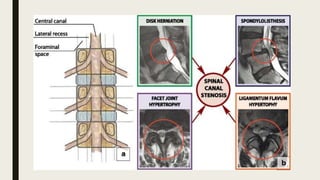
SPINAL CANAL
STENOSIS
• DR.ARPAN CHAUDHARY
GOOD MORNING😊
One of the most common condition in elderly
Defined as narrowing of –Vertebral Canal,
-- Lateral Recess,
-- Intervertebral foramen
First described by “ANTOINE PORTAL 1803”
Spinal Canal is between
-Vertebral Body,Disc,PLL anteriorly,
- Pedicles and lateral part of ligamentum (L)
- Facet joints,lamina and Ligamentum -.
Flavum (dorsally)
Spinal Canal may be Circular,Oval orTrefoil
Trefoil has least crss sectional area, predispose to Lateral. Recess Stenosis (15%)
Circular and Oval has more Space
• Pathogenesis
■ Stenosis can be generalized or Localised to specific
anatomical areas like cervical,thoracic and Lumbar
■ M/C in Lumbar Region,but Cervical also frequent,thoracic
Rare
■ AS Kirkadly-Willis et al. Described pathophysiology for
lumbar spine stenosis,based on considering Spine ,as tripod
of disc and two facet joints
■ At birth annulus and nucleus pulposus has 50% of disc area
■ With time demarcation between both decreases with
increase in collagen
■ As chondroitin sulfate decreases,
■ Ratio of Keratin sulphate/chondroitin sulfate (⬆️ses),that
dehydrate disc over time
■ Annulus contain type 2 collagen (60%),Type 1 (40%)
■ Disc mainly hasType 2 collagen
■ As with timeType 1 Collagen Increase, Hydration of the disc
decrease (type 1 ass. With less water content)
■ Nucleus Pulposus 77-85% & Annulus has 70- 78%Water
■ With Degeneration of disc,water content drops Less than 70
■ Initial stage of degeneration is tearing of annulus leads to
RedialTear
1. Annulus tear+Biochemical change
2. Leads to degeneration of disc and loss disc height
3. Causes bulging of disc and PLL
4. Leads to narrowing of SC and potential Neural Impingement
5. Also can leads to Buckling of Lig.Flavum & subsidence of Facet
Joints
6. Facet joits try to Restabilize And form Osteophytes,causing narrow
canal and compression of neural eliments in foramen
■ Decreased Disc Height,
■ Decreased foramlinal Height
■ Leads to Neural compression
■ ThisVERTICAL/UP-DOWN foraminal stenosis important to
recognise bcz a Posterior Decompression Alone may not
significant to improve the vertcal compression and may result
in persistent symptoms after sx
(1) Central spinal stenosis:
-Area between facet joints (contain dura and its contents)
-pt present with neurological claudication with generalized leg pain
Causes – protrusion of disc,bulging annulus,
-- Osteophytes,thickenes LF
-M/C in Congenital Spinal Stenosis
(2) Lateral Recess (Lees Entrance zone)
-here nerve roots exits dura and extends distally and laterally under Sup.Articular Facet
■ Vent.-Disc and Post.Ligament complex
■ Dorsally-Sup.Articular Facet
■ Laterally-Pedicles
■ Lateral Recess stenosis,
Causes-Facet arthritis(M/C)
-Vertebral Spurring,disc and annular pathology
3.Lees Midzone(Foramlinal Zone)
■ Med.-Lateral Recess
■ Vent.-Post.VertebtalBody and Disc
■ Dors.-Pars and Intertransverse Ligament
■ Lat.-Lateral border of pedicle
Between two Pedicals it is,formed b/w two intervertebral foramen
4.Extraforaminal-Sometimes(Exit Zone)
■ M/C Spinal Stenosis is due to Degenerative Arthritis of Spine
Including Forestier Disease(Characterized by Hyperostosis and
spinal regidity in elderly)
■ Diffuse Idiopathic skeletsl Hyperostosis and hypertrophy of PLL
causes acquired stenosis
■ Congenital is commonly Central
■ Idiopathoic congenital involves AP diameter of cansl secondary
to short pedicles
• Vascular symptoms felt in upper calf,relived by short rest (5mins)
• Neurogenic Claudication improves with trunk flexion,stooping or
lying but may require 20 mins to improve
• SHOPPING CART SIGN – pushing a grocery cart allows
flexion,which enhance endurance and decreases discomfort in pt
with Neurological Claudication
STOOP TEST
• Test is done by asking the pt to walk briskly
• As the pain intensifies,pt complain of Sensory
Symptoms followed by Motor Symptoms
• If pt asked to continue to walk,he or she may assume
Stoop Posture and symptoms maybe eased
• As pt sits in chair And bends forward,same resolution
occurs
■ Diagnostic Imaging:
1. Radiography
-Plain radiograph cannot confirm
-But useful like in short Pedicles, ligament
ossification,narrow foramen, hypertrophy of articular
facet
-
CERVICAL CANAL
• Cervical Canal has AP
Diameter of 17-18 mm
upto C5
• Lower Cervical Canal
Measure 12-14 mm
• Less than 10 Stenotic
THORACIC CANAL
• Also Varies 12-14 mm
• Less than 10 Stenosis
■ MRI:
• Excilent DiagnosticTool
• Provides information for bone and soft tissues
. (Imaging study of choice)
• MRI is conformatory in pt with Neurological
Claudication/radiculopathy
• Axial view provides good view of central spinal Stenosis in
T1 &T2 weighted images
• Far lateral disc obliteration can be identified on axialT1
weighted images by obliteration of normal between the
disc and nerve roots
■ Advantage:
-Provides information
about size and contour of
foramen,and Helps to
assess csf and
Interference and Stenosis
-No radiation exposure
and non-invasive
-Provides images in
axial,coronal and
Saggital planes
■ CT:
-Prior to MRI Ct was the study for choice
-diagnoatic utility can be improved by combining with
myelography
Dye injected in CSF provides good contrast between
the thecal sac and surrounding soft tissues and bony
pathology
Indicated in elder pt with pacemaker,Other metal
implant In which mri not possible,CT Myelography
used
Mid Sagital diameter less than 10 Or less and AP
Diameter less than 3mm suggests Stenosis
Other:
Electromyography:
- Evaluates physiology of nerve roots,whichis useful in delineating disease
Of Lower motor neuron
- It is abnormal in 80% of pt with spinal Stenosis, confirmatory for nerve
involvement
■ NERVE CONDUCTION STUDIES:
-measurea the speed at which nerve impulse travels
-differentiate PERIPHERAL NEUROPATHY from RADICULOPATHY
■ SOMATOSENSORY EVOKED POTENTIAL
Is more useful than EMG to identify neeve root
compression
• Differential Diagnosis:
■ Vascular Claudication – prximal to distal, constant
symptoms,painless extension,weak peripheral pulses
■ Lumbar Spondylosis
■ Lower Extremity Arthritis - Isolated joint pain with ambulation
■ Cord Compression
■ Neurologic Disorders
■ Peripheral Neuropathy - Burning Pain and parasthesia
■ Infectionss,Tremors
• TREATMENT
Non-operative:
■ NSAIDS
■ Medical-like opioids and actaminophen(first drug of choice for
Osteoarthritis
■ Gabapentin anticonvulsant and analgesics useful in neuropathic
pain syndrome
■ Gabapentin found to be more effective with physio and NSAIDS to
increase walking distance
■ Calcitonin hormone found to be analgesic and anti inflammatory,
Increases Beta-Endorphin and neuronal influx of calcium
ions.that in cns,causes Analgesic effect,anti inflammatory by
prostaglandin E2 Release
■ Physical Therapy:
-Improves spine flexibility,pain relief,disabily reduction
-Like in lumbar Stenosis extension exercise
Interventional Therapy:
-Epidural Steroid Injections for inflammatory reactions
and Edema
-can be given caudal,interlaminar, transforaminal
-interlaminar has more effect than caudal
-prior to perform an epidural injection,
anticoagulant,antiplatelets agent,NSAIDS and
Clopidegrol are discontinued for approx 1week
-headche in 25% of treated pts
Surgical Management:
■ Indications:
- Failure of conservation treatment
- Should has Neurological claudication with prolonged walking
- Predominance of low back pain over radicular pain ass. With poor
surgicap outcome
- Selective nerve block is good prognostic indicator of surgical
outcome In whom 50% relief occurred by injection
■ Indicationn for urgent decompression include
progressive neurological deficit and bowel/bladder
dysfunction.
■ LAMINECTOMY-
-Gold Std. Surgical treatment for decompression of spinal canal Stenosis
-Provides space and visibility
“POSTERIOR APPROACH” is used
-care should be taken to to preserve capusle of facet joint
-In posterolateral approach dissection should be extended laterally to
transverse bprocess,to maintain integrith of interatransverse ligament
-decompression carried out from half of spinous process, above and below
compression
-Decompression should begin centrally
-to remove LF curette used,kerrison pouch to remove inf. Aspect of laina
using LF,in above carried out uptu LF ends
■ In lateral Decompression 50% reserve of facet joint and 1mm of pars
interarticularis
■ LAMINECTOMY WITH
Arthrodesis
-when Stenosis with instability
Or spondylitic spine with back pain
-In Stenosis pt decompression and
Arthrodesis vs LAMINECTOMY is good
-currently Arthrodesis with pedicular
Fixation + decompression laminectomy
If instability at involved segment ,revision
Decompression at same level and resection
Of greater than 50% of facet joints
• Laminoplasty
■ Two types (1) Distraction (2)Expansive
■ Distraction for Laminoplasty-useful for central and lateral recess
stenosis with minimal bone resection
■ Allows visual of canal during compression with minimal removal
of posterior bony elements
■ Distraction is Applied across spinal proess, increases 1 cm
interlaminar space
(2) Expensive Laminoplasty- opening of one side lamina using other
side as hinge,bone grafts from excised spinous process are placed in
opened laminae & fixed with braided wire or sutures
It has long term outcome is poor
• Less and minimally invasive techniques
■ The removal of bone and ligament considered when performing
decompression of stenosis,removal of spinal
process,laminae,facets,pars,supraspinous and interspinous
ligament and ligamentum flavum
■ Laminotomy:
-decompression through microdiscectomy like approach
-Targeting stenotic level unilaterally or bilaterally
■ FENESTRATION:
Partial undercutting facetectomy,partial laminectomy maintaining
spinous process and posterior ligaments
Decompression by a 5 mm drill hole in pars interarticularis
immediately below the Superior facet
■ Hole exposes
inferior aspect of
pedicle and root in
nerve canal
■ Using 2mm also can
be used for inferior
aspect of
sup.pedicle is
removed.how this
dorsal nerve root
decompressed as in
lateral and foraminal
stenosis
■ Microendoscopic decopressive
laminotomy
-developed by foley and coworkers for decompression sx that is
endoscopically assisted
Here contralateral lamina is first decompressed by medial
angulation of retractor tube,and drillant. Lamina and
contralateral recesa
-endoscopic kerrison rongeurs used
■ Interspinous devices:
-to distract interspinous
space and block Extension
-X-STOP device waa first used
■ Complications:
■ Dural Tear(4-16%)
■ Nerve root injuries
■ Infection
■ Vascular like DVT,PE,post op hematoloma
■ Meningitis
■ Epidural Hematoma
■ Nonunion,Bony Regowth
Spinal canal stenosis presentation

Spinal canal stenosis presentation

  • 1.
    SPINAL CANAL STENOSIS • DR.ARPANCHAUDHARY GOOD MORNING😊
  • 2.
    One of themost common condition in elderly Defined as narrowing of –Vertebral Canal, -- Lateral Recess, -- Intervertebral foramen First described by “ANTOINE PORTAL 1803” Spinal Canal is between -Vertebral Body,Disc,PLL anteriorly, - Pedicles and lateral part of ligamentum (L) - Facet joints,lamina and Ligamentum -. Flavum (dorsally)
  • 3.
    Spinal Canal maybe Circular,Oval orTrefoil Trefoil has least crss sectional area, predispose to Lateral. Recess Stenosis (15%) Circular and Oval has more Space
  • 4.
    • Pathogenesis ■ Stenosiscan be generalized or Localised to specific anatomical areas like cervical,thoracic and Lumbar ■ M/C in Lumbar Region,but Cervical also frequent,thoracic Rare ■ AS Kirkadly-Willis et al. Described pathophysiology for lumbar spine stenosis,based on considering Spine ,as tripod of disc and two facet joints ■ At birth annulus and nucleus pulposus has 50% of disc area ■ With time demarcation between both decreases with increase in collagen
  • 5.
    ■ As chondroitinsulfate decreases, ■ Ratio of Keratin sulphate/chondroitin sulfate (⬆️ses),that dehydrate disc over time ■ Annulus contain type 2 collagen (60%),Type 1 (40%) ■ Disc mainly hasType 2 collagen ■ As with timeType 1 Collagen Increase, Hydration of the disc decrease (type 1 ass. With less water content) ■ Nucleus Pulposus 77-85% & Annulus has 70- 78%Water ■ With Degeneration of disc,water content drops Less than 70 ■ Initial stage of degeneration is tearing of annulus leads to RedialTear
  • 7.
    1. Annulus tear+Biochemicalchange 2. Leads to degeneration of disc and loss disc height 3. Causes bulging of disc and PLL 4. Leads to narrowing of SC and potential Neural Impingement 5. Also can leads to Buckling of Lig.Flavum & subsidence of Facet Joints 6. Facet joits try to Restabilize And form Osteophytes,causing narrow canal and compression of neural eliments in foramen
  • 8.
    ■ Decreased DiscHeight, ■ Decreased foramlinal Height ■ Leads to Neural compression ■ ThisVERTICAL/UP-DOWN foraminal stenosis important to recognise bcz a Posterior Decompression Alone may not significant to improve the vertcal compression and may result in persistent symptoms after sx
  • 11.
    (1) Central spinalstenosis: -Area between facet joints (contain dura and its contents) -pt present with neurological claudication with generalized leg pain Causes – protrusion of disc,bulging annulus, -- Osteophytes,thickenes LF -M/C in Congenital Spinal Stenosis (2) Lateral Recess (Lees Entrance zone) -here nerve roots exits dura and extends distally and laterally under Sup.Articular Facet ■ Vent.-Disc and Post.Ligament complex ■ Dorsally-Sup.Articular Facet ■ Laterally-Pedicles
  • 15.
    ■ Lateral Recessstenosis, Causes-Facet arthritis(M/C) -Vertebral Spurring,disc and annular pathology 3.Lees Midzone(Foramlinal Zone) ■ Med.-Lateral Recess ■ Vent.-Post.VertebtalBody and Disc ■ Dors.-Pars and Intertransverse Ligament ■ Lat.-Lateral border of pedicle Between two Pedicals it is,formed b/w two intervertebral foramen 4.Extraforaminal-Sometimes(Exit Zone)
  • 16.
    ■ M/C SpinalStenosis is due to Degenerative Arthritis of Spine Including Forestier Disease(Characterized by Hyperostosis and spinal regidity in elderly) ■ Diffuse Idiopathic skeletsl Hyperostosis and hypertrophy of PLL causes acquired stenosis ■ Congenital is commonly Central ■ Idiopathoic congenital involves AP diameter of cansl secondary to short pedicles
  • 21.
    • Vascular symptomsfelt in upper calf,relived by short rest (5mins) • Neurogenic Claudication improves with trunk flexion,stooping or lying but may require 20 mins to improve • SHOPPING CART SIGN – pushing a grocery cart allows flexion,which enhance endurance and decreases discomfort in pt with Neurological Claudication STOOP TEST • Test is done by asking the pt to walk briskly • As the pain intensifies,pt complain of Sensory Symptoms followed by Motor Symptoms • If pt asked to continue to walk,he or she may assume Stoop Posture and symptoms maybe eased • As pt sits in chair And bends forward,same resolution occurs
  • 23.
    ■ Diagnostic Imaging: 1.Radiography -Plain radiograph cannot confirm -But useful like in short Pedicles, ligament ossification,narrow foramen, hypertrophy of articular facet -
  • 24.
    CERVICAL CANAL • CervicalCanal has AP Diameter of 17-18 mm upto C5 • Lower Cervical Canal Measure 12-14 mm • Less than 10 Stenotic THORACIC CANAL • Also Varies 12-14 mm • Less than 10 Stenosis
  • 25.
    ■ MRI: • ExcilentDiagnosticTool • Provides information for bone and soft tissues . (Imaging study of choice) • MRI is conformatory in pt with Neurological Claudication/radiculopathy • Axial view provides good view of central spinal Stenosis in T1 &T2 weighted images • Far lateral disc obliteration can be identified on axialT1 weighted images by obliteration of normal between the disc and nerve roots
  • 26.
    ■ Advantage: -Provides information aboutsize and contour of foramen,and Helps to assess csf and Interference and Stenosis -No radiation exposure and non-invasive -Provides images in axial,coronal and Saggital planes
  • 28.
    ■ CT: -Prior toMRI Ct was the study for choice -diagnoatic utility can be improved by combining with myelography Dye injected in CSF provides good contrast between the thecal sac and surrounding soft tissues and bony pathology Indicated in elder pt with pacemaker,Other metal implant In which mri not possible,CT Myelography used Mid Sagital diameter less than 10 Or less and AP Diameter less than 3mm suggests Stenosis
  • 30.
    Other: Electromyography: - Evaluates physiologyof nerve roots,whichis useful in delineating disease Of Lower motor neuron - It is abnormal in 80% of pt with spinal Stenosis, confirmatory for nerve involvement ■ NERVE CONDUCTION STUDIES: -measurea the speed at which nerve impulse travels -differentiate PERIPHERAL NEUROPATHY from RADICULOPATHY ■ SOMATOSENSORY EVOKED POTENTIAL Is more useful than EMG to identify neeve root compression
  • 31.
    • Differential Diagnosis: ■Vascular Claudication – prximal to distal, constant symptoms,painless extension,weak peripheral pulses ■ Lumbar Spondylosis ■ Lower Extremity Arthritis - Isolated joint pain with ambulation ■ Cord Compression ■ Neurologic Disorders ■ Peripheral Neuropathy - Burning Pain and parasthesia ■ Infectionss,Tremors
  • 32.
    • TREATMENT Non-operative: ■ NSAIDS ■Medical-like opioids and actaminophen(first drug of choice for Osteoarthritis ■ Gabapentin anticonvulsant and analgesics useful in neuropathic pain syndrome ■ Gabapentin found to be more effective with physio and NSAIDS to increase walking distance ■ Calcitonin hormone found to be analgesic and anti inflammatory, Increases Beta-Endorphin and neuronal influx of calcium ions.that in cns,causes Analgesic effect,anti inflammatory by prostaglandin E2 Release
  • 33.
    ■ Physical Therapy: -Improvesspine flexibility,pain relief,disabily reduction -Like in lumbar Stenosis extension exercise Interventional Therapy: -Epidural Steroid Injections for inflammatory reactions and Edema -can be given caudal,interlaminar, transforaminal -interlaminar has more effect than caudal -prior to perform an epidural injection, anticoagulant,antiplatelets agent,NSAIDS and Clopidegrol are discontinued for approx 1week -headche in 25% of treated pts
  • 34.
    Surgical Management: ■ Indications: -Failure of conservation treatment - Should has Neurological claudication with prolonged walking - Predominance of low back pain over radicular pain ass. With poor surgicap outcome - Selective nerve block is good prognostic indicator of surgical outcome In whom 50% relief occurred by injection ■ Indicationn for urgent decompression include progressive neurological deficit and bowel/bladder dysfunction.
  • 35.
    ■ LAMINECTOMY- -Gold Std.Surgical treatment for decompression of spinal canal Stenosis -Provides space and visibility “POSTERIOR APPROACH” is used -care should be taken to to preserve capusle of facet joint -In posterolateral approach dissection should be extended laterally to transverse bprocess,to maintain integrith of interatransverse ligament -decompression carried out from half of spinous process, above and below compression -Decompression should begin centrally -to remove LF curette used,kerrison pouch to remove inf. Aspect of laina using LF,in above carried out uptu LF ends
  • 36.
    ■ In lateralDecompression 50% reserve of facet joint and 1mm of pars interarticularis ■ LAMINECTOMY WITH Arthrodesis -when Stenosis with instability Or spondylitic spine with back pain -In Stenosis pt decompression and Arthrodesis vs LAMINECTOMY is good -currently Arthrodesis with pedicular Fixation + decompression laminectomy If instability at involved segment ,revision Decompression at same level and resection Of greater than 50% of facet joints
  • 37.
    • Laminoplasty ■ Twotypes (1) Distraction (2)Expansive ■ Distraction for Laminoplasty-useful for central and lateral recess stenosis with minimal bone resection ■ Allows visual of canal during compression with minimal removal of posterior bony elements ■ Distraction is Applied across spinal proess, increases 1 cm interlaminar space (2) Expensive Laminoplasty- opening of one side lamina using other side as hinge,bone grafts from excised spinous process are placed in opened laminae & fixed with braided wire or sutures It has long term outcome is poor
  • 38.
    • Less andminimally invasive techniques ■ The removal of bone and ligament considered when performing decompression of stenosis,removal of spinal process,laminae,facets,pars,supraspinous and interspinous ligament and ligamentum flavum ■ Laminotomy: -decompression through microdiscectomy like approach -Targeting stenotic level unilaterally or bilaterally ■ FENESTRATION: Partial undercutting facetectomy,partial laminectomy maintaining spinous process and posterior ligaments Decompression by a 5 mm drill hole in pars interarticularis immediately below the Superior facet
  • 39.
    ■ Hole exposes inferioraspect of pedicle and root in nerve canal ■ Using 2mm also can be used for inferior aspect of sup.pedicle is removed.how this dorsal nerve root decompressed as in lateral and foraminal stenosis
  • 40.
    ■ Microendoscopic decopressive laminotomy -developedby foley and coworkers for decompression sx that is endoscopically assisted Here contralateral lamina is first decompressed by medial angulation of retractor tube,and drillant. Lamina and contralateral recesa -endoscopic kerrison rongeurs used ■ Interspinous devices: -to distract interspinous space and block Extension -X-STOP device waa first used
  • 41.
    ■ Complications: ■ DuralTear(4-16%) ■ Nerve root injuries ■ Infection ■ Vascular like DVT,PE,post op hematoloma ■ Meningitis ■ Epidural Hematoma ■ Nonunion,Bony Regowth