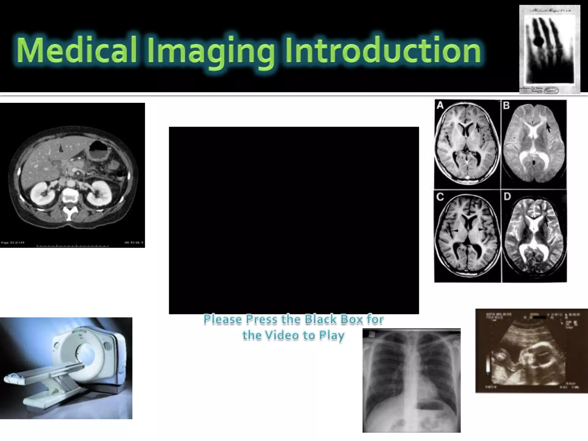
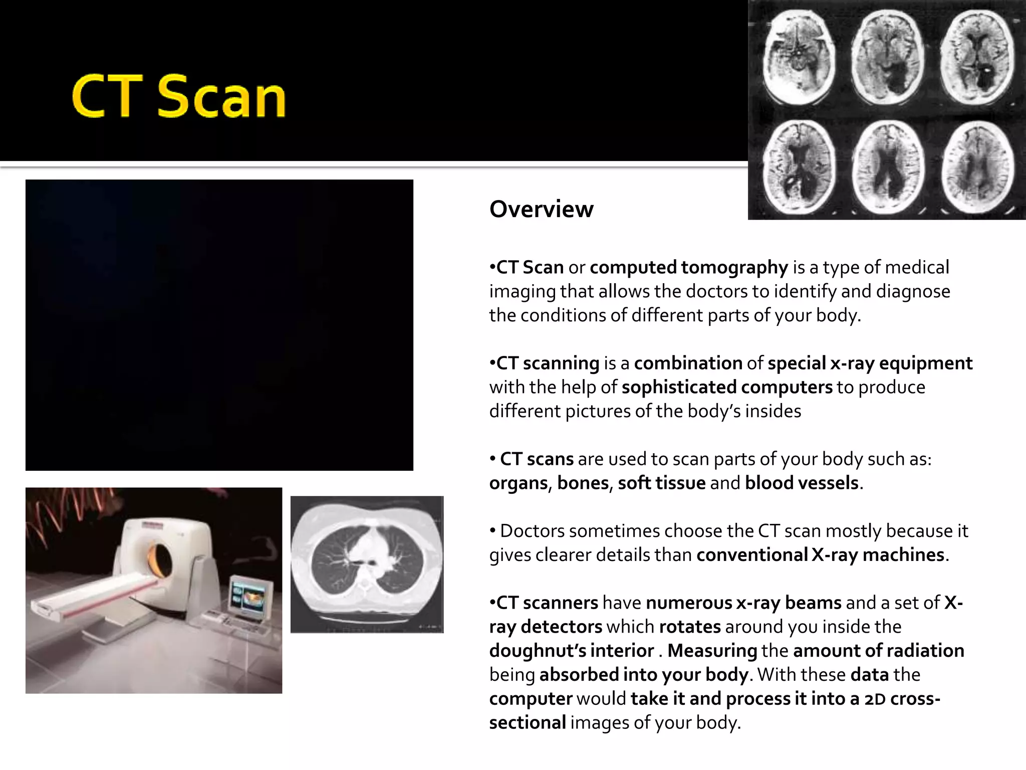
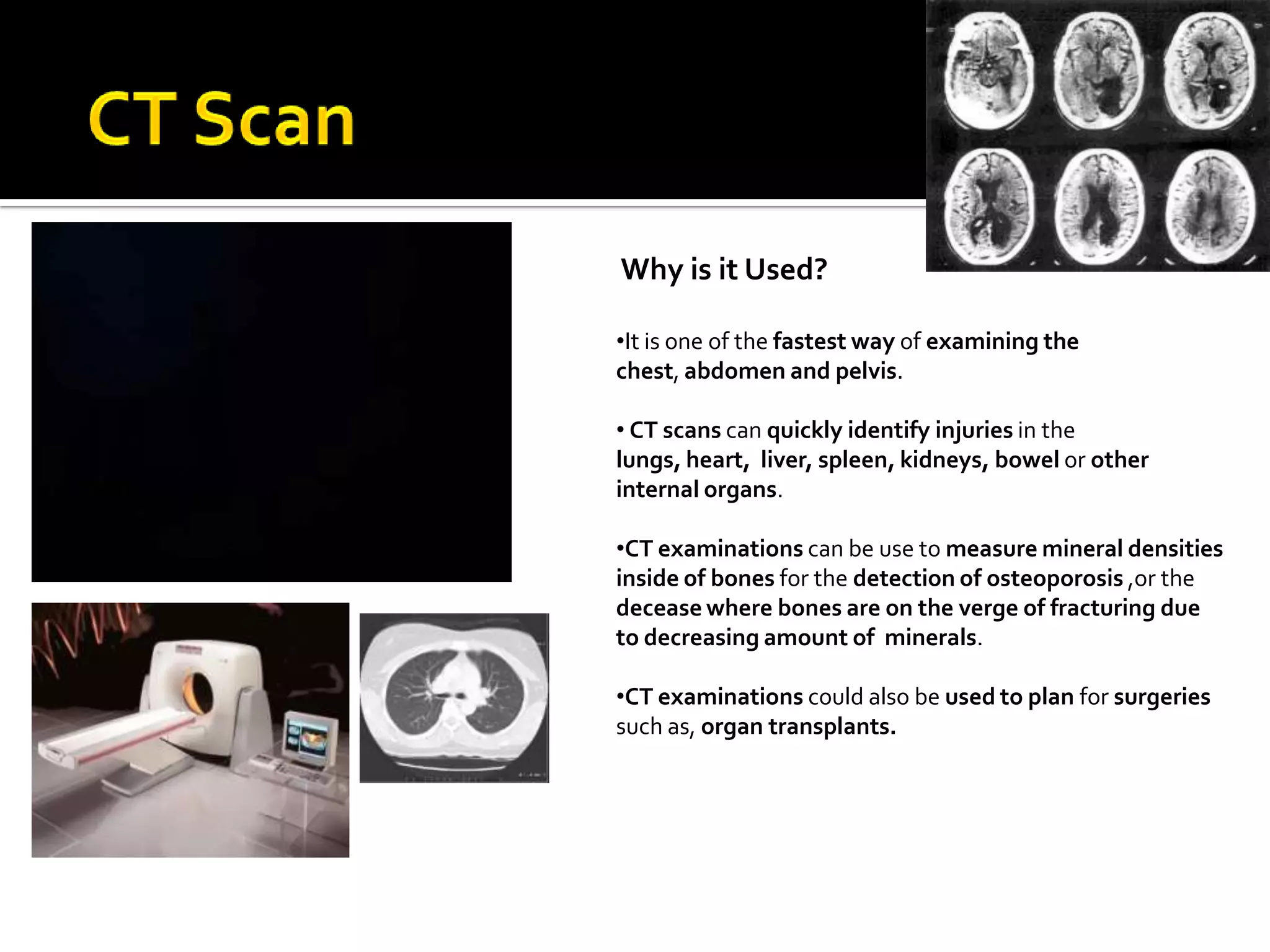
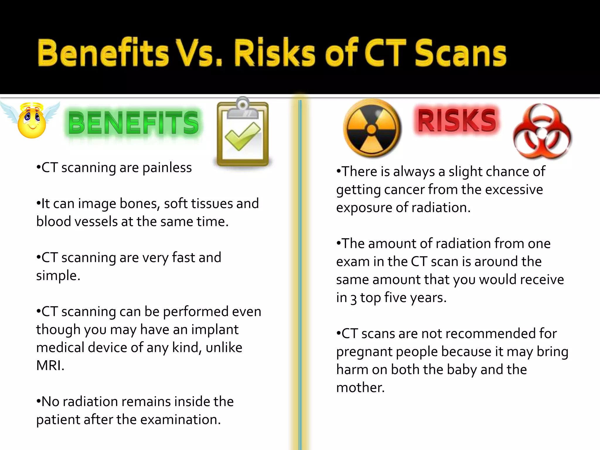
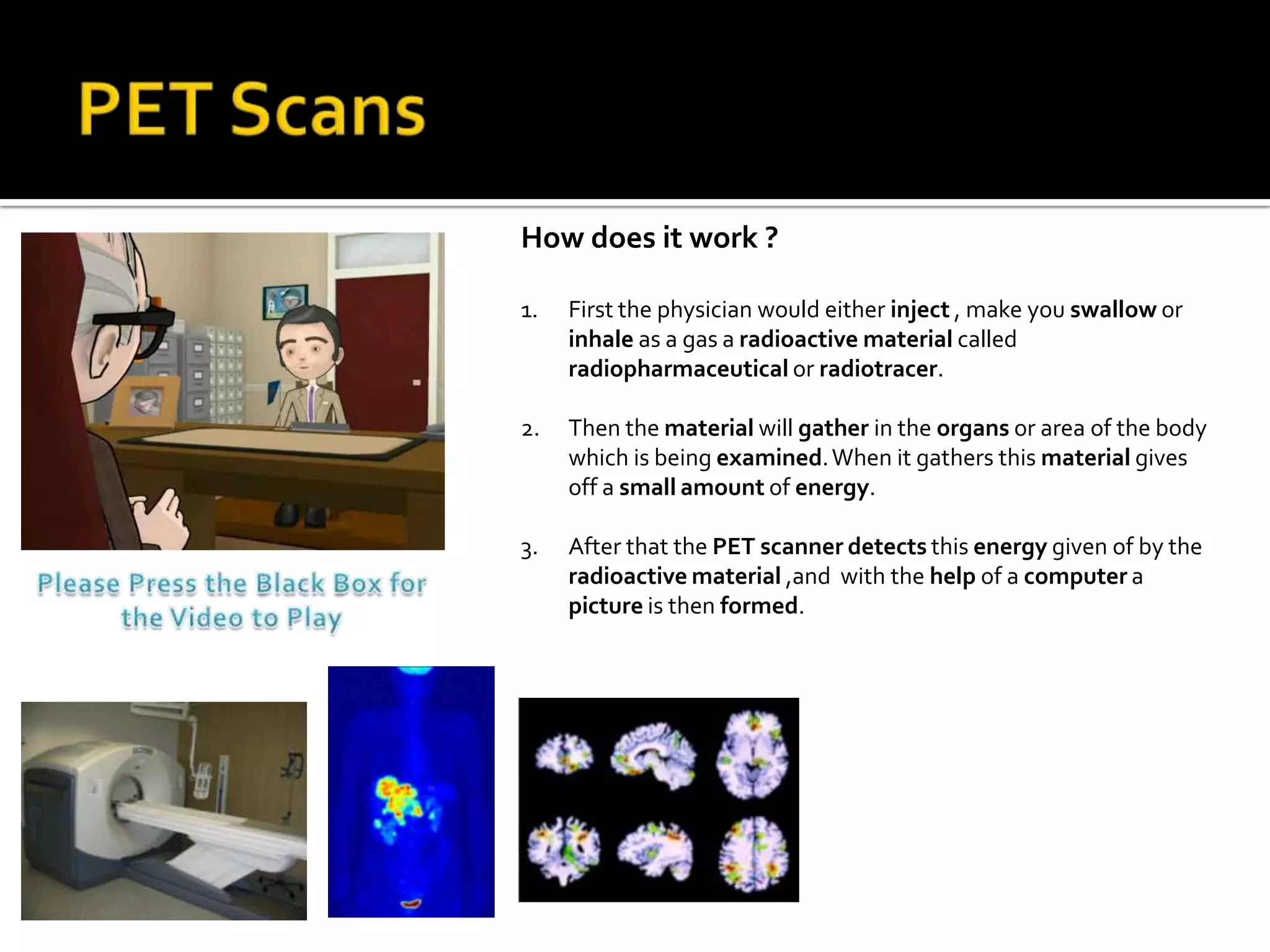
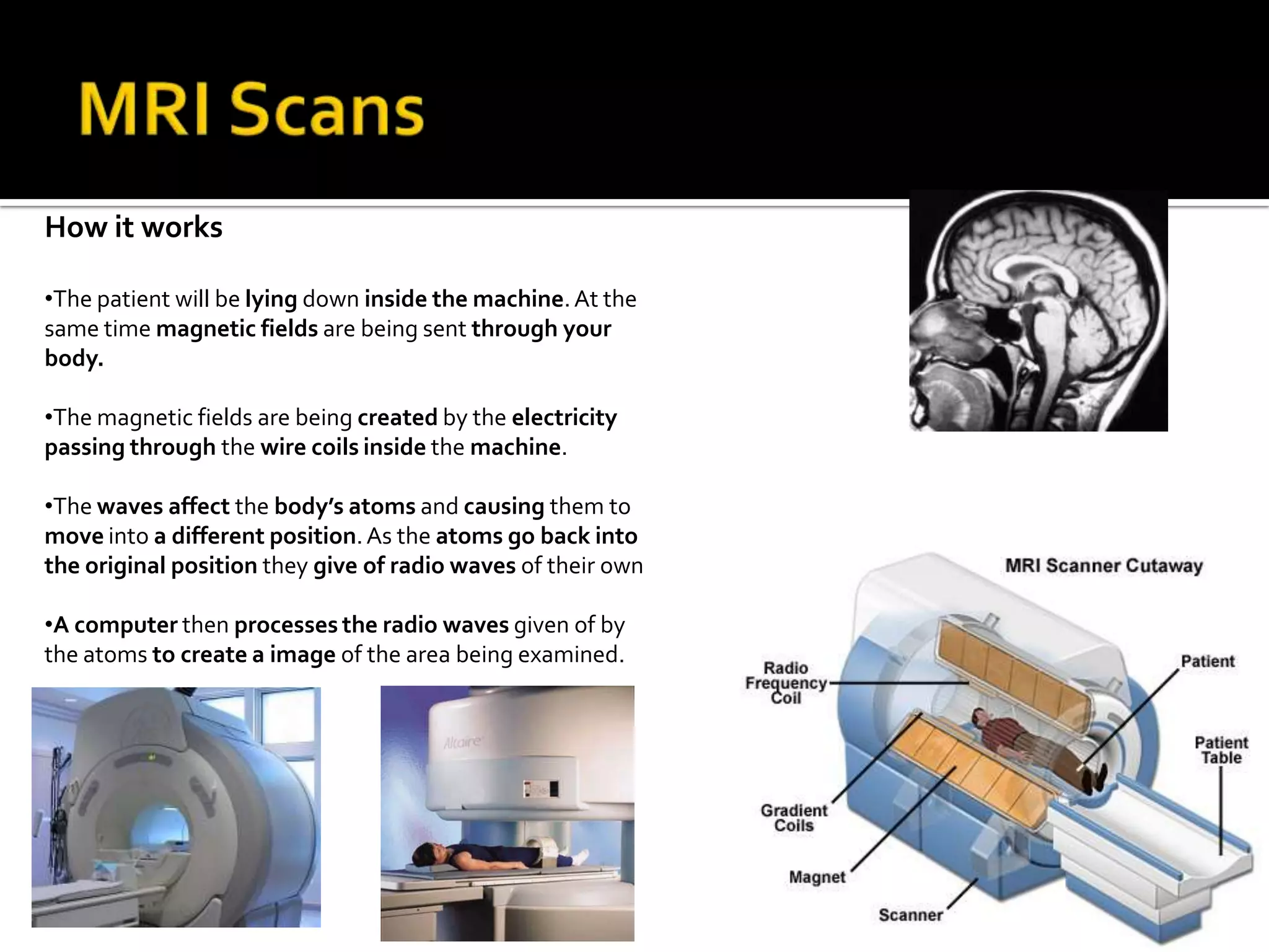
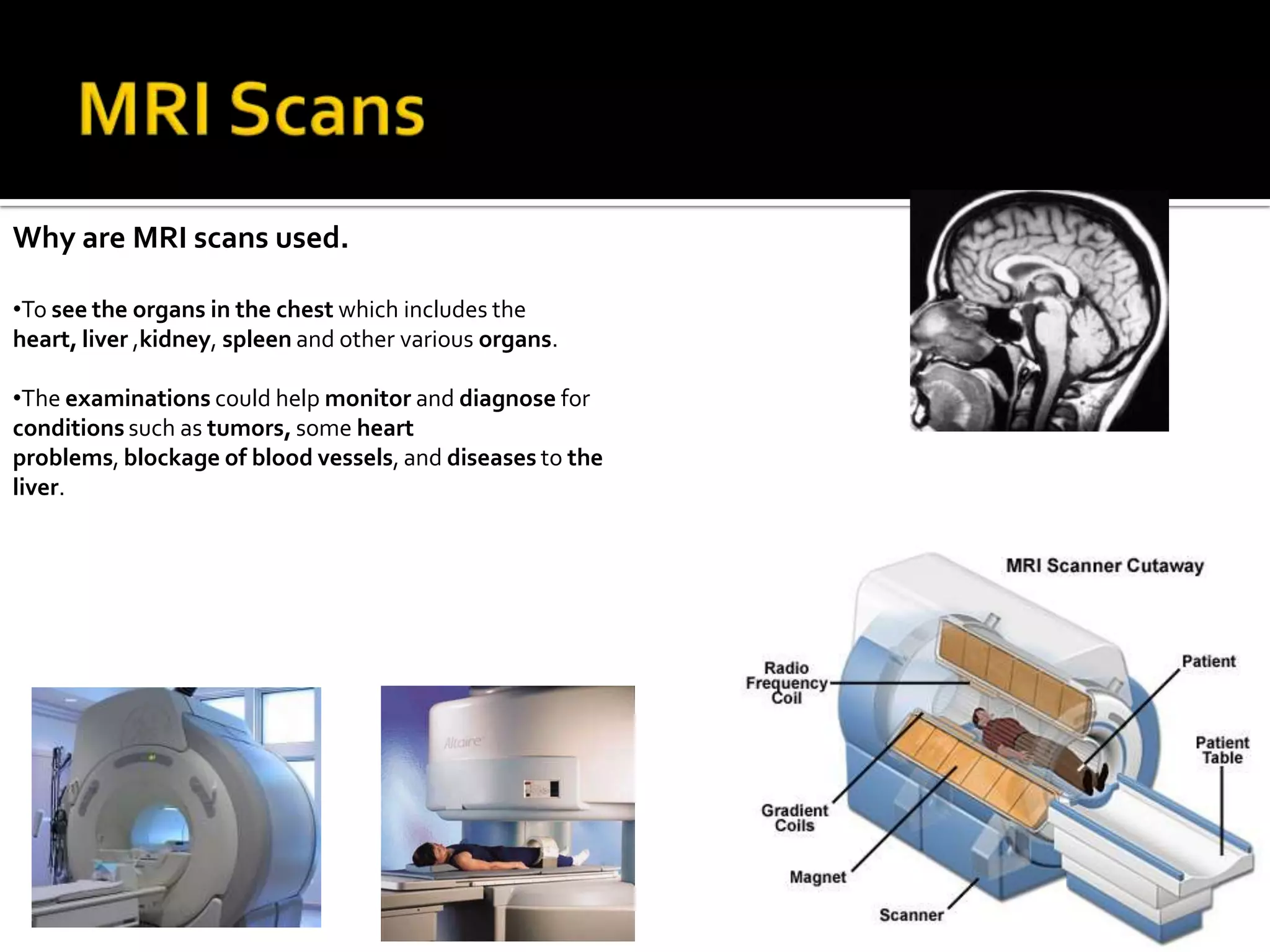
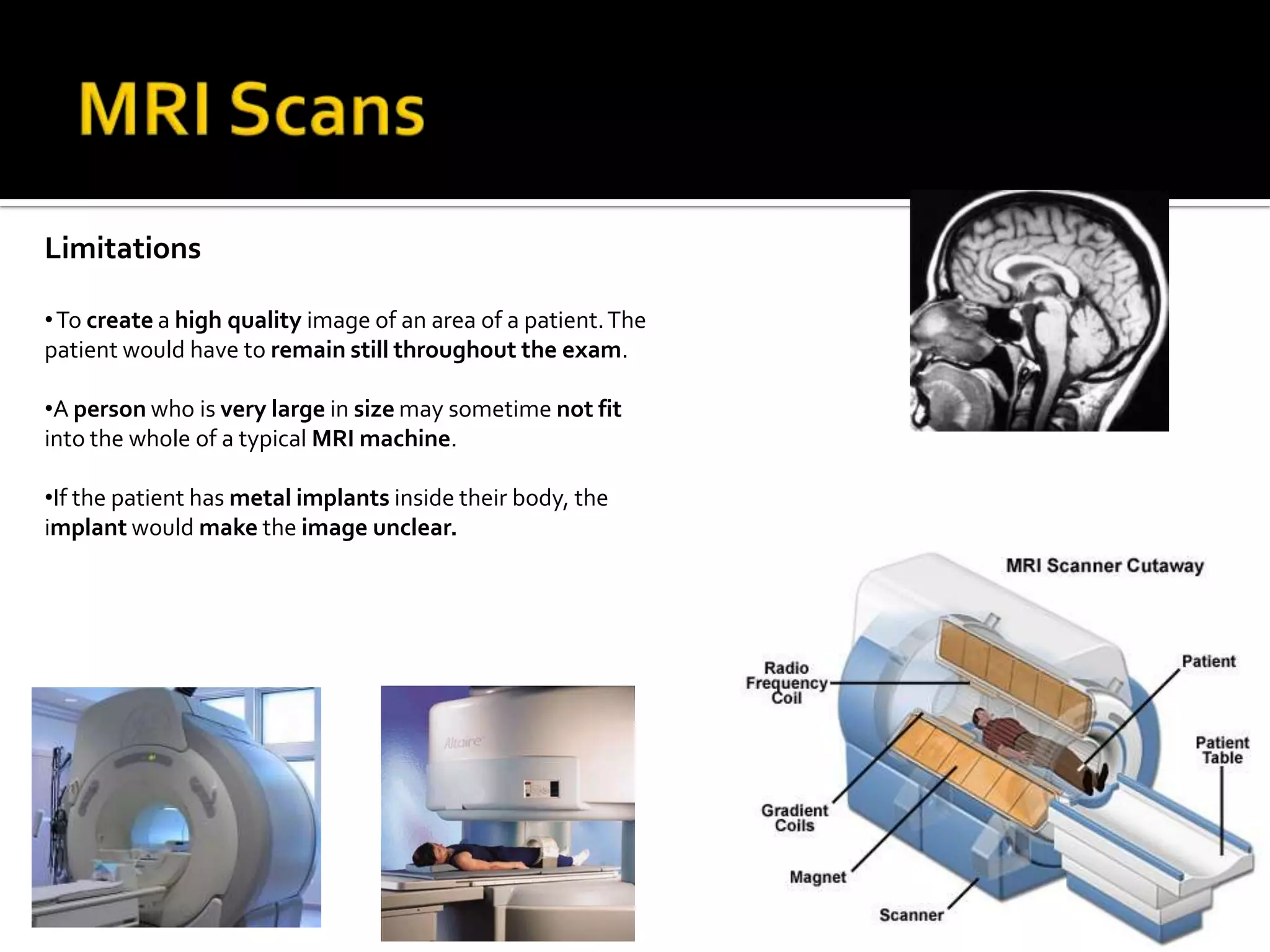
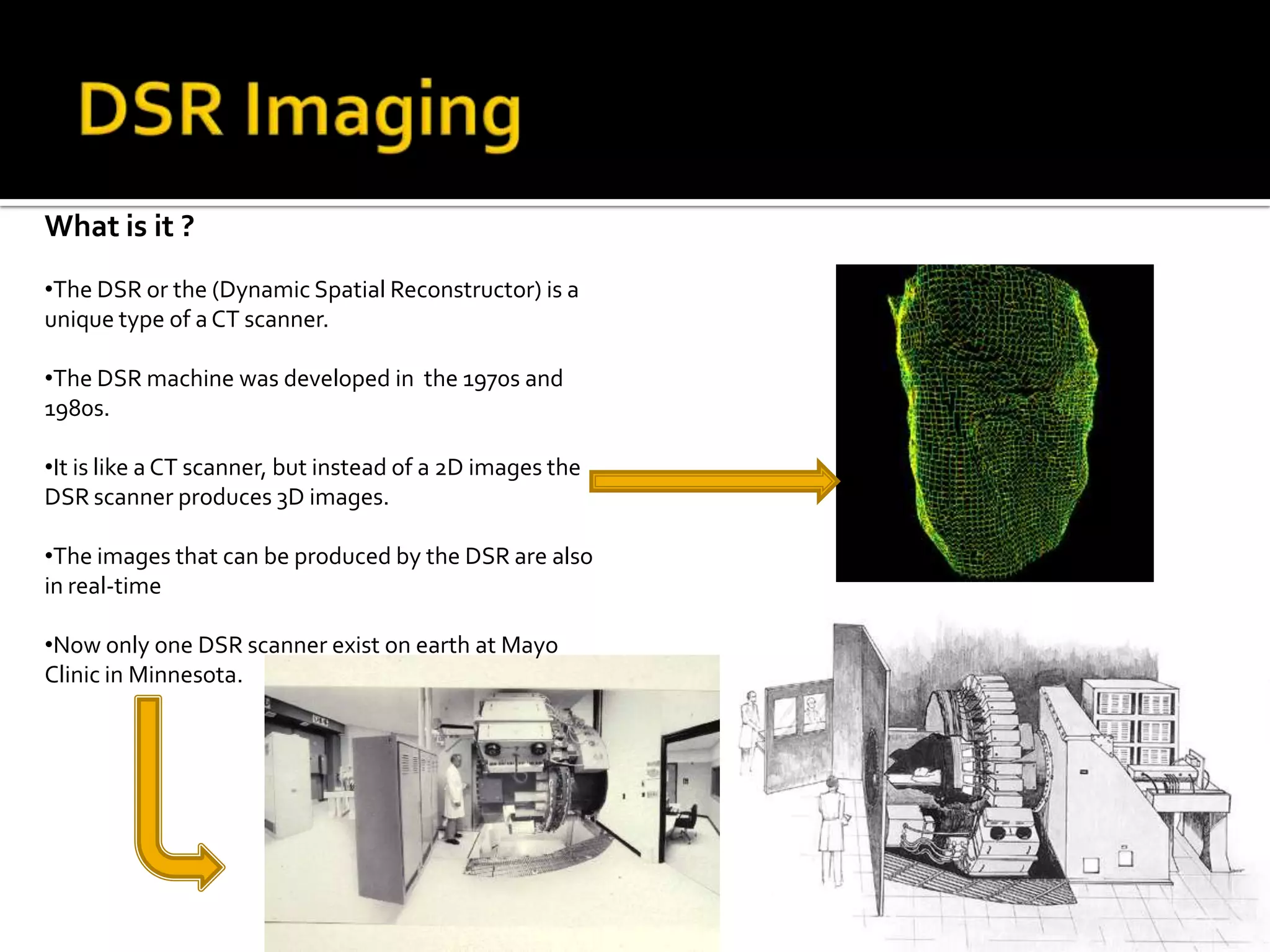
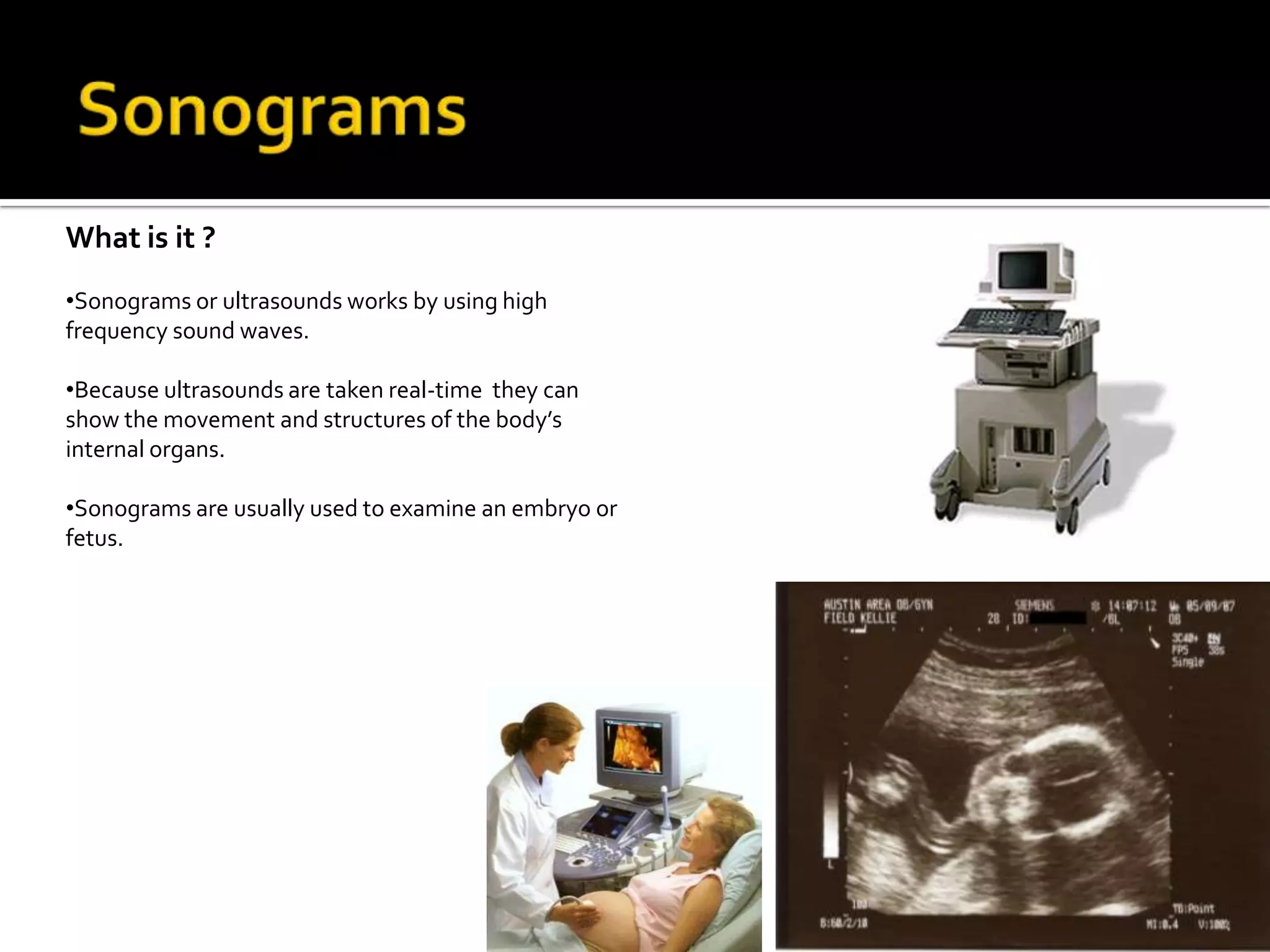
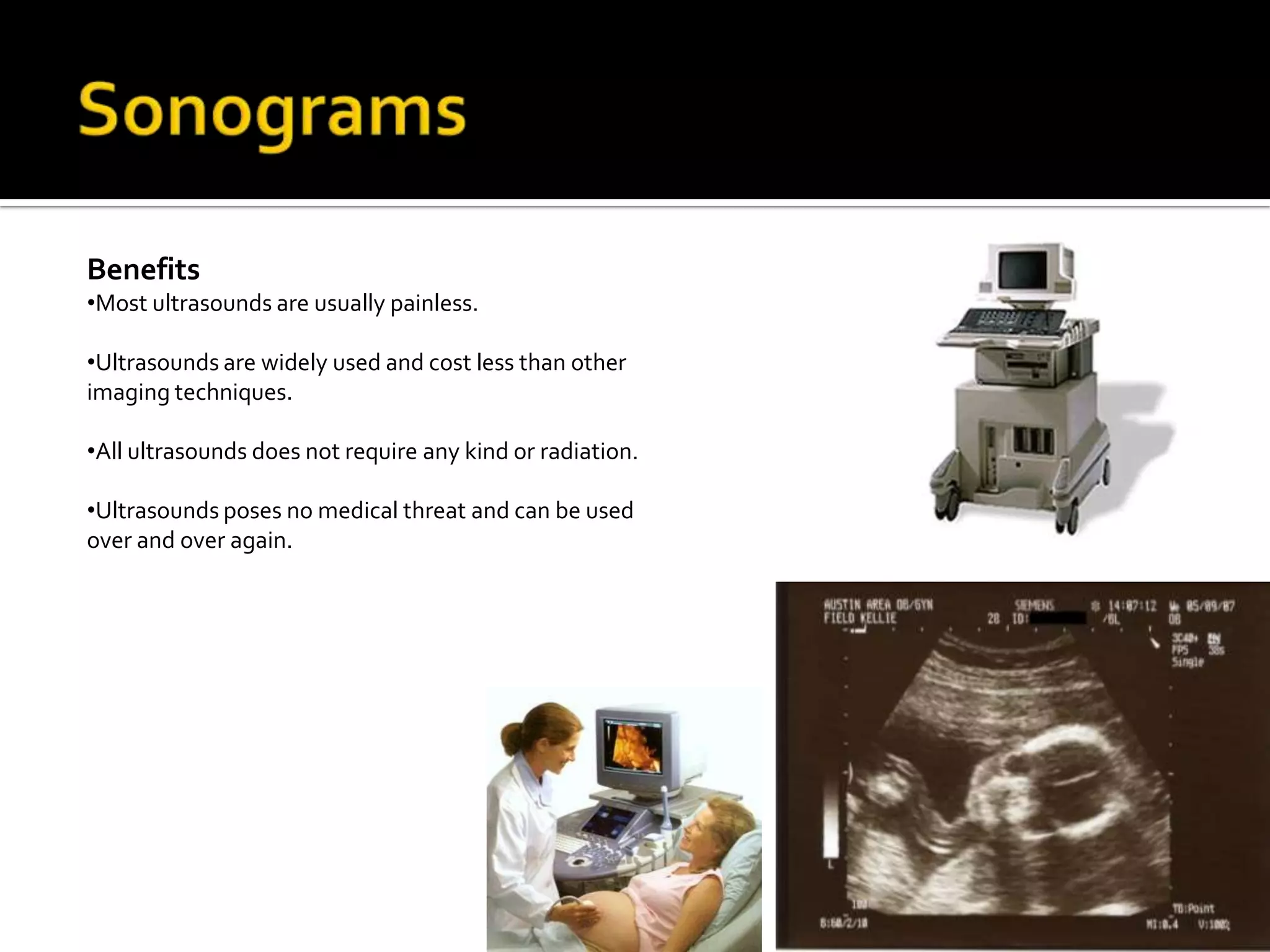
Medical Imaging MachinesCT ScanPET ScanMRI ScanDSR ScanSonogramsBy: Web Khewsubtrakool
Medical Imaging IntroductionPlease Press the Black Box for the Video to Play
CT Scan OverviewCT Scan or computed tomography is a type of medical  imaging that allows the doctors to identify and diagnose the conditions of different parts of your body.
CT scanning is a combination of special x-ray equipment with the help of sophisticated computers to produce different pictures of the body’s insides
CT scans are used to scan parts of your body such as:  organs, bones, soft tissue and blood vessels.
 Doctors sometimes choose the CT scan mostly because it gives clearer details than conventional X-ray machines.
CT scanners have numerous x-ray beams and a set of X-ray detectors which rotates around you inside the doughnut’s interior . Measuring the amount of radiation being absorbed into your body. With these data the computer would take it and process it into a 2D cross-sectional images of your body.CT Scan  Why is it Used?It is one of the fastest way of examining the chest, abdomen and pelvis.
CT scans can quickly identify injuries in the lungs, heart,  liver, spleen, kidneys, bowel or other internal organs.
CT examinations can be use to measure mineral densities inside of bones for the detection of osteoporosis ,or the decease where bones are on the verge of fracturing due to decreasing amount of  minerals.
CT examinations could also be used to plan for surgeries such as, organ transplants.Benefits Vs. Risks of CT ScansBenefits Vs. Risks of CT ScansRISKSBenefitsRISKSBenefitsCT scanning are painless
It can image bones, soft tissues and blood vessels at the same time.
CT scanning are very fast and simple.
CT scanning can be performed even though you may have an implant medical device of any kind, unlike MRI.
No radiation remains inside the patient after the examination.
There is always a slight chance of getting cancer from the excessive exposure of radiation.
The amount of radiation from one exam in the CT scan is around the same amount that you would receive in 3 top five years.
CT scans are not recommended for pregnant people because it may bring harm on both the baby and the mother.PET ScansWhat is it?PET or (Positron Emission Tomography) is a type of nuclear medicine imaging.
Nuclear medicine is a branch of medical imaging that requires small amount of radio active materials to find or treat a variety of deceases.
The machine looks similar to a CT or a MRI scanner. So it looks like a round doughnut with a desk that moves through it. Please Press the Black Box for the Video to Play
PET ScansWhat and Why is it Used for?It is used to findcancerinside your body. When found it could also benefit the doctor, so that the doctors could find a way to cureit.Used to see and identify the spreading of cancercells. So the doctors can tell if it is dangerous or not.It can detect brain disorders such as  Alzheimer, Parkinson’s decease and more.It can determine the effects of a heartattack Please Press the Black Box for the Video to Play

Project 1 (skeletal system multi media) mc sheet

  • 1.
    Medical Imaging MachinesCTScanPET ScanMRI ScanDSR ScanSonogramsBy: Web Khewsubtrakool
  • 2.
    Medical Imaging IntroductionPleasePress the Black Box for the Video to Play
  • 3.
    CT Scan OverviewCTScan or computed tomography is a type of medical imaging that allows the doctors to identify and diagnose the conditions of different parts of your body.
  • 4.
    CT scanning isa combination of special x-ray equipment with the help of sophisticated computers to produce different pictures of the body’s insides
  • 5.
    CT scans areused to scan parts of your body such as: organs, bones, soft tissue and blood vessels.
  • 6.
    Doctors sometimeschoose the CT scan mostly because it gives clearer details than conventional X-ray machines.
  • 7.
    CT scanners havenumerous x-ray beams and a set of X-ray detectors which rotates around you inside the doughnut’s interior . Measuring the amount of radiation being absorbed into your body. With these data the computer would take it and process it into a 2D cross-sectional images of your body.CT Scan Why is it Used?It is one of the fastest way of examining the chest, abdomen and pelvis.
  • 8.
    CT scans canquickly identify injuries in the lungs, heart, liver, spleen, kidneys, bowel or other internal organs.
  • 9.
    CT examinations canbe use to measure mineral densities inside of bones for the detection of osteoporosis ,or the decease where bones are on the verge of fracturing due to decreasing amount of minerals.
  • 10.
    CT examinations couldalso be used to plan for surgeries such as, organ transplants.Benefits Vs. Risks of CT ScansBenefits Vs. Risks of CT ScansRISKSBenefitsRISKSBenefitsCT scanning are painless
  • 11.
    It can imagebones, soft tissues and blood vessels at the same time.
  • 12.
    CT scanning arevery fast and simple.
  • 13.
    CT scanning canbe performed even though you may have an implant medical device of any kind, unlike MRI.
  • 14.
    No radiation remainsinside the patient after the examination.
  • 15.
    There is alwaysa slight chance of getting cancer from the excessive exposure of radiation.
  • 16.
    The amount ofradiation from one exam in the CT scan is around the same amount that you would receive in 3 top five years.
  • 17.
    CT scans arenot recommended for pregnant people because it may bring harm on both the baby and the mother.PET ScansWhat is it?PET or (Positron Emission Tomography) is a type of nuclear medicine imaging.
  • 18.
    Nuclear medicine isa branch of medical imaging that requires small amount of radio active materials to find or treat a variety of deceases.
  • 19.
    The machine lookssimilar to a CT or a MRI scanner. So it looks like a round doughnut with a desk that moves through it. Please Press the Black Box for the Video to Play
  • 20.
    PET ScansWhat andWhy is it Used for?It is used to findcancerinside your body. When found it could also benefit the doctor, so that the doctors could find a way to cureit.Used to see and identify the spreading of cancercells. So the doctors can tell if it is dangerous or not.It can detect brain disorders such as Alzheimer, Parkinson’s decease and more.It can determine the effects of a heartattack Please Press the Black Box for the Video to Play
  • 21.
    PET ScansHow doesit work ?First the physician would either inject , make you swallow or inhale as a gas a radioactive material called radiopharmaceutical or radiotracer.Then the material will gather in the organs or area of the body which is being examined. When it gathers this material gives off a smallamount of energy.After that the PETscannerdetects this energy given of by the radioactivematerial ,and with the help of a computer a picture is then formed.Please Press the Black Box for the Video to Play
  • 22.
    Are PET Scanner orGoodBadBones RadioPRESS ITPress It
  • 23.
    MRI Scans Howit worksThe patient will be lying down inside the machine. At the same time magnetic fields are being sent through your body.
  • 24.
    The magnetic fieldsare being created by the electricity passing through the wire coils inside the machine.
  • 25.
    The waves affectthe body’s atoms and causing them to move into a different position. As the atoms go back into the original position they give of radio waves of their own
  • 26.
    A computer thenprocessesthe radio waves given of by the atoms to create a image of the area being examined.MRI Scans Why are MRI scans used.To see the organs in the chest which includes the heart,liver,kidney, spleen and other various organs.
  • 27.
    The examinations couldhelp monitor and diagnose for conditions such as tumors, some heart problems, blockage of blood vessels, and diseases to the liver.MRI Scans Limitations To create a high quality image of an area of a patient. The patient would have to remainstill throughout the exam.
  • 28.
    A person whois very large in size may sometime not fit into the whole of a typical MRI machine.
  • 29.
    If the patienthasmetal implants inside their body, the implant would make the image unclear.DSR ImagingWhat is it ?The DSR or the (Dynamic Spatial Reconstructor) is a unique type of a CT scanner.
  • 30.
    The DSR machinewas developed in the 1970s and 1980s.
  • 31.
    It is likea CT scanner, but instead of a 2D images the DSR scanner produces 3D images.
  • 32.
    The images thatcan be produced by the DSR are also in real-time
  • 33.
    Now only oneDSR scanner exist on earth at Mayo Clinic in Minnesota. Sonograms What is it ?Sonograms or ultrasounds works by using high frequency sound waves.
  • 34.
    Because ultrasounds aretaken real-time they can show the movement and structures of the body’s internal organs.
  • 35.
    Sonograms are usuallyused to examine an embryo or fetus.Sonograms How does it work?The way that sonograms work is similar to the sonar used by bats, ships, and fishermen.
  • 36.
    The ultrasound machineworks by sending high-density sound waves into the area of the patient being examined.
  • 37.
    When a soundwave hit something it bounces back or echoes.
  • 38.
    The machine thenmeasure the amount of the echo waves and determine the shape of the object as well as it’s size.Sonograms BenefitsMost ultrasounds are usually painless.
  • 39.
    Ultrasounds are widelyused and cost less than other imaging techniques.
  • 40.
    All ultrasounds doesnot require any kind or radiation.
  • 41.
    Ultrasounds poses nomedical threat and can be used over and over again.Credits and BibliographyInfo Sites"CAT Scan." RadiologyInfo - The Radiology Information Resource for Patients. Web. 07 Mar. 2010. <http://www.radiologyinfo.org/en/info.cfm?PG=bodyct>. "Dynamic Spatial Reconstructor | Encyclopedia of Nursing and Allied Health | Find Articles at BNET." Find Articles at BNET | News Articles, Magazine Back Issues & Reference Articles on All Topics. Web. 07 Mar. 2010. <http://findarticles.com/p/articles/mi_gGENH/is_20050229/ai_2699003247/>. "Dynamic Spatial Reconstructor Information on Healthline." Health Search Engine and Free Medical Information - Healthline. Web. 07 Mar. 2010. <http://www.healthline.com/galecontent/dynamic-spatial-reconstructor>. "Magnetic Resonance Imaging (MRI)." RadiologyInfo - The Radiology Information Resource for Patients. Web. 07 Mar. 2010. <http://www.radiologyinfo.org/en/info.cfm?PG=bodymr>."Magnetic Resonance Imaging." Wikipedia, the Free Encyclopedia. Web. 07 Mar. 2010. <http://en.wikipedia.org/wiki/Magnetic_resonance_imaging>. "MRI Scan." NetDoctor.co.uk - The UK's Leading Independent Health Website. Web. 07 Mar. 2010. <http://www.netdoctor.co.uk/health_advice/examinations/mriscan.htm>."Nuclear Medicine, PET." RadiologyInfo - The Radiology Information Resource for Patients. Web. 07 Mar. 2010. <http://www.radiologyinfo.org/en/info.cfm?PG=pet>. "PET Scan: Positron Emission Tomography (PET)." PET Scan: PET Scan Info Reveals ... Web. 07 Mar. 2010. <http://www.petscaninfo.com/zportal/portals/pat/basic>. "Positron Emission Tomography." Wikipedia, the Free Encyclopedia. Web. 06 Mar. 2010. <http://en.wikipedia.org/wiki/PET_imaging>.
  • 42.
    Credits and BibliographyVideos Presentation & Video for Information"Medical Milestones - X-Rays." YouTube-Broadcast Yourself. Web. 06 Mar. 2010. <http://www.youtube.com/watch?v=0oy0fYeV764&feature=related>. "Radiology in Motion: CT." RadiologyInfo - The Radiology Information Resource for Patients. Web. 07 Mar. 2010. <http://www.radiologyinfo.org/en/video/index.cfm?filename='CT'>. "Radiology in Motion: MRI." RadiologyInfo - The Radiology Information Resource for Patients. Web. 06 Mar. 2010. <http://www.radiologyinfo.org/en/video/index.cfm?filename='MR'>. "Radiology in Motion: Nuclear Medicine." RadiologyInfo - The Radiology Information Resource for Patients. Web. 07 Mar. 2010. <http://www.radiologyinfo.org/en/video/index.cfm?filename=nm>. "Radiology in Motion: Ultrasound." RadiologyInfo - The Radiology Information Resource for Patients. Web. 07 Mar. 2010. <http://www.radiologyinfo.org/en/video/index.cfm?filename='US'>.
  • 43.
    Credits and BibliographyPicturesUsed For Presentation (Part 1)"CT SCAN MODEL." Radiology Equipment. Web. 07 Mar. 2010. <http://www.radiology-equipment.com/detail.CFM?LineItemID=763>. "DSR Scan." Yale Image Processing and Analysis Group (IPAG). Web. 07 Mar. 2010. <http://noodle.med.yale.edu/alums/amini/prindirs.html>. "Imaging Techniques." Answers.com: Wiki Q&A Combined with Free Online Dictionary, Thesaurus, and Encyclopedias. Web. 07 Mar. 2010. <http://www.answers.com/topic/imaging-techniques>. "Introduction to Magnetic Resonance Imaging (Full Article)." National High Magnetic Field Laboratory. Web. 06 Mar. 2010. <http://www.magnet.fsu.edu/education/tutorials/magnetacademy/mri/fullarticle.html>. "Mayo Dynamic Spatial Reconstructor (DSR)." Mayo Clinic: Medical Education and Research. Web. 07 Mar. 2010. <http://www.mayo.edu/sppdg/AutoGen/6128.html>. "Medical Equipment: CT Scanner." Medical Imaging Equipment. Web. 06 Mar. 2010. <http://www.amberusa.com/remanufactured/catDetails.asp?catId=11&catname=CT%20Scanner>. "Medical Imaging." Healthcare Technology Online: Healthcare Technology System News, Implementation Trends, Best Practices, & Product Information. Web. 07 Mar. 2010. <http://www.healthcaretechnologyonline.com/solution.mvc/medical-imaging-pacs>. "Open MRI Machine." Klamath: Direct Action! - March 4, 2010 | North Coast Journal | Humboldt County, Calif. Web. 06 Mar. 2010. <http://www.northcoastjournal.com/111804/cover1118.html>.
  • 44.
    Credits and BibliographyPicturesUsed For Presentation (Part 2)"PET Scan." Brookhaven National Laboratory. Web. 07 Mar. 2010. <http://www.bnl.gov/bnlweb/pubaf/pr/PR_display.asp?prID=05-84>. "Positron Emission Tomography." Wikipedia, the Free Encyclopedia. Web. 07 Mar. 2010. <http://en.wikipedia.org/wiki/Positron_emission_tomography>. "Scans." California Personal Injury Law Case Notes : California Personal Injury Lawyer & Attorney : Kristine Meredith : The Danko Law Firm : Car Accidents & Product Liability. Web. 07 Mar. 2010. <http://www.capersonalinjurycaselawnotes.com/2009/07/articles/brain-injuries/mris-pet-scans-and-traumatic-brain-injury/>. "Sonograms." Web. 07 Mar. 2010. <http://www.kayarain.com/pictures/Sonograms/index.htm>. "Ultrasound." Diagnostic Radiology and Interventional Radiology Greenwich Radiological Group. Web. 07 Mar. 2010. <http://www.greenwichradiology.com/ultrasound.php>. "Ultrasound Scans." Web. 07 Mar. 2010. <http://www.acrin.org/PATIENTS/ABOUTXRAYSANDSCANS/ABOUTULTRASOUNDSCANS/tabid/243/Default.aspx>. "X-ray Imaging." Nature Publishing Group. Web. 06 Mar. 2010. <http://www.nature.com/ki/journal/v55/n2/fig_tab/4490600f7.html#figure-title>. "X-ray." Web. 07 Mar. 2010. <http://faceache.wordpress.com/2007/08/29/x-ray-looks-normal/>. "X-ray." Wikipedia, the Free Encyclopedia. Web. 07 Mar. 2010. <http://en.wikipedia.org/wiki/X-ray>.