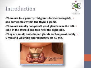
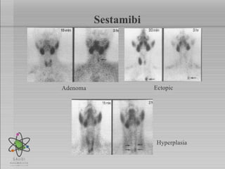
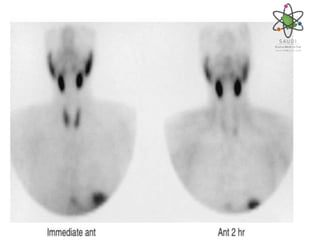
parathyroid scan
by:Zahera Alhajji
Introduction
•-There are four parathyroid glands located alongside
and sometimes within the thyroid gland.
•-There are usually two parathyroid glands near the left
lobe of the thyroid and two near the right lobe.
•-They are small, oval-shaped glands each approximately
6 mm and weighing approximately 30–50 mg.
Indication
•Localization of primary and secondary parathyroid
cancer
•Localization of hyperfunctioning parathyroid tissue
(adenoma/ hyperplasia).
•Localization of hyperfunctioning parathyroid tissue in
patients with persistent or recurrent disease
Contraindication
•-Pregnant women
•-patient on calcium medication
•- patient on thyroid medication (off for 5 days)
•Precaution
•Precaution: Discontinuation of breast feeding for
nursing mothers for 24 hours after imaging.
Radiopharmaceutical
•99mTechnetium(sestamibi-tetrofosmin) adult: 15 mCi
•99mTechnetium-pertechnetate adult: 5-12 mCi
•Child: As per body weight
•Technique of administration
•Intravenous Injection
Examination Time
• Initial injection and images at 15-20 min
• Delayed images at 2 hours .
EQUIPMENT
CAMERA
Small or Large field of view
COLLIMATOR
(pinhole or low energy all purpose or low energy high
Resolution )
.
Patient position& Imaging field
• Patient position: Supine.
• Imaging field: Neck and mediastinum
PROCEDURE
• Prepare the camera and Place patient in supine
position, pillow under shoulders, head back, neck
extended.
• • Position camera anterior over extended neck and
mediastinum.
• Remove any attenuating necklaces or pendants.
• • Instruct patient to remain motionless during
acquisitions.
• Acquire ANT dynamic images.
• Oblique views may be requested by the physician
• Repeat all images at approximately 2hours after
injection. Use the same camera, and as close as
possible to the original patient positions and
collimator positions.
parathyroid scan
parathyroid scan
parathyroid scan

parathyroid scan

  • 1.
  • 2.
    Introduction •-There are fourparathyroid glands located alongside and sometimes within the thyroid gland. •-There are usually two parathyroid glands near the left lobe of the thyroid and two near the right lobe. •-They are small, oval-shaped glands each approximately 6 mm and weighing approximately 30–50 mg.
  • 3.
    Indication •Localization of primaryand secondary parathyroid cancer •Localization of hyperfunctioning parathyroid tissue (adenoma/ hyperplasia). •Localization of hyperfunctioning parathyroid tissue in patients with persistent or recurrent disease
  • 4.
    Contraindication •-Pregnant women •-patient oncalcium medication •- patient on thyroid medication (off for 5 days) •Precaution •Precaution: Discontinuation of breast feeding for nursing mothers for 24 hours after imaging.
  • 5.
    Radiopharmaceutical •99mTechnetium(sestamibi-tetrofosmin) adult: 15mCi •99mTechnetium-pertechnetate adult: 5-12 mCi •Child: As per body weight •Technique of administration •Intravenous Injection
  • 6.
    Examination Time • Initialinjection and images at 15-20 min • Delayed images at 2 hours .
  • 7.
    EQUIPMENT CAMERA Small or Largefield of view COLLIMATOR (pinhole or low energy all purpose or low energy high Resolution ) .
  • 8.
    Patient position& Imagingfield • Patient position: Supine. • Imaging field: Neck and mediastinum
  • 9.
    PROCEDURE • Prepare thecamera and Place patient in supine position, pillow under shoulders, head back, neck extended. • • Position camera anterior over extended neck and mediastinum. • Remove any attenuating necklaces or pendants. • • Instruct patient to remain motionless during acquisitions.
  • 10.
    • Acquire ANTdynamic images. • Oblique views may be requested by the physician • Repeat all images at approximately 2hours after injection. Use the same camera, and as close as possible to the original patient positions and collimator positions.