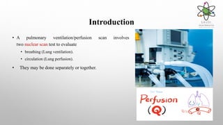
Pulmonary Ventilation /
Perfusion Scan SPECTCT
This Presentation Presented By: Dina Almasoudi
Supervised By: Khalid Alnofaie
The outlines
1. Introduction. 3. Preparation.2. Case History.
4. Technique. 5. Findings 6. Conclusion.
7. Referneces.
Introduction
• A pulmonary ventilation/perfusion scan involves
two nuclear scan test to evaluate
• breathing (Lung ventilation).
• circulation (Lung perfusion).
• They may be done separately or together.
Case History
• 80 years old female patient known case of Hypertenstion (HTN.)
• Newly diagnose case of Burketts lymphoma having high pulmonary artery pressure on echocardiogram
• (ECHO.)
• Indication for perfusion:
• Evaluation of
• pulmonary embolism (PE), the main cause being deep vein thromboses (DVTs),clots from recent surgery, or trauma.
• pulmonary perfusion.
• chest pain or shortness of breath (SOB).
• Indication for ventilation in conjunction with lung perfusion;
• Evaluation of
• pulmonary embolism (PE).
• chronic obstructive pulmonary disease (COPD)
Preparation
• A recent (24 hour) chest radiograph.
• Rehearse the breathing procedure to assure optimal patient cooperation; instruct the patient to breathe
by mouth.
• To assist in distribution to lungs, patient should be asked to cough and take in several deep breaths
before injection
Technique
• View: ANT, POST, LATERAL, 4 OBLIQUES.
• Radiopharmaceutical:
• Ventilation: Tc99m Aerosol (400 MBq).
• Perfusion: Tc99m MAA (100 MBq).
Case History
Finding
A significantly heterogeneous pattern of tracer distribution is noted in left lung both in perfusion and
ventilation with evidence of multiple non-segmental matched defects and no evidence of segmental
or mismatched.
A matched defects noted in Lt upper lobe.
Consultant Radiologist Hatem NasrApproved By:
Conclusion
• VQ scan have many advantages;
• This scan is readily available, with minimal risks involved.
• It’s a relatively good test for blood clots in his-her lungs and current symptoms suggest that you might have a
pulmonary embolism
References
• PSMMC PACS
• HTTPS://MEDLINEPLUS.GOV/ENCY/ARTICLE/003828.HTM
• HTTPS://WWW.UCSFBENIOFFCHILDRENS.ORG/TESTS/003828.HTML
• HTTPS://WWW.INSIDERADIOLOGY.COM.AU/VQ-
SCAN/#:~:TEXT=THE%20BENEFITS%20OF%20A%20VQ%20SCAN%20ARE%3A,MIGHT%20H
AVE%20A%20PULMONARY%20EMBOLISM
Pulmonary Ventilation and Perfusion Scan

Pulmonary Ventilation and Perfusion Scan

  • 1.
    Pulmonary Ventilation / PerfusionScan SPECTCT This Presentation Presented By: Dina Almasoudi Supervised By: Khalid Alnofaie
  • 2.
    The outlines 1. Introduction.3. Preparation.2. Case History. 4. Technique. 5. Findings 6. Conclusion. 7. Referneces.
  • 3.
    Introduction • A pulmonaryventilation/perfusion scan involves two nuclear scan test to evaluate • breathing (Lung ventilation). • circulation (Lung perfusion). • They may be done separately or together.
  • 5.
    Case History • 80years old female patient known case of Hypertenstion (HTN.) • Newly diagnose case of Burketts lymphoma having high pulmonary artery pressure on echocardiogram • (ECHO.) • Indication for perfusion: • Evaluation of • pulmonary embolism (PE), the main cause being deep vein thromboses (DVTs),clots from recent surgery, or trauma. • pulmonary perfusion. • chest pain or shortness of breath (SOB). • Indication for ventilation in conjunction with lung perfusion; • Evaluation of • pulmonary embolism (PE). • chronic obstructive pulmonary disease (COPD)
  • 6.
    Preparation • A recent(24 hour) chest radiograph. • Rehearse the breathing procedure to assure optimal patient cooperation; instruct the patient to breathe by mouth. • To assist in distribution to lungs, patient should be asked to cough and take in several deep breaths before injection
  • 7.
    Technique • View: ANT,POST, LATERAL, 4 OBLIQUES. • Radiopharmaceutical: • Ventilation: Tc99m Aerosol (400 MBq). • Perfusion: Tc99m MAA (100 MBq).
  • 8.
  • 9.
    Finding A significantly heterogeneouspattern of tracer distribution is noted in left lung both in perfusion and ventilation with evidence of multiple non-segmental matched defects and no evidence of segmental or mismatched. A matched defects noted in Lt upper lobe. Consultant Radiologist Hatem NasrApproved By:
  • 10.
    Conclusion • VQ scanhave many advantages; • This scan is readily available, with minimal risks involved. • It’s a relatively good test for blood clots in his-her lungs and current symptoms suggest that you might have a pulmonary embolism
  • 11.
    References • PSMMC PACS •HTTPS://MEDLINEPLUS.GOV/ENCY/ARTICLE/003828.HTM • HTTPS://WWW.UCSFBENIOFFCHILDRENS.ORG/TESTS/003828.HTML • HTTPS://WWW.INSIDERADIOLOGY.COM.AU/VQ- SCAN/#:~:TEXT=THE%20BENEFITS%20OF%20A%20VQ%20SCAN%20ARE%3A,MIGHT%20H AVE%20A%20PULMONARY%20EMBOLISM