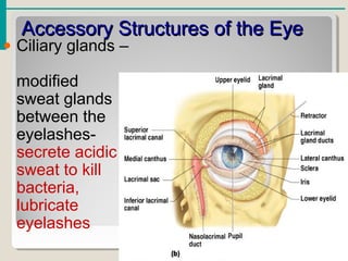
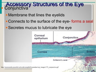
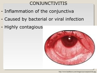
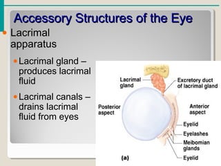
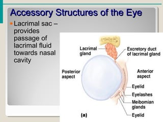
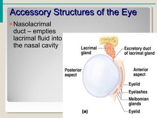
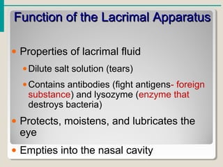
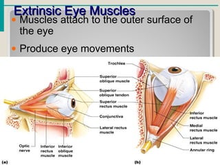
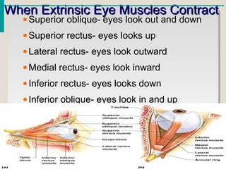
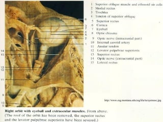
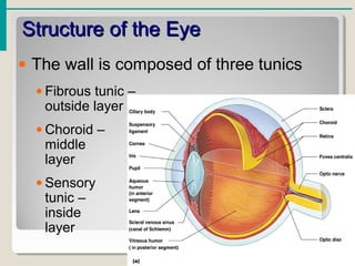
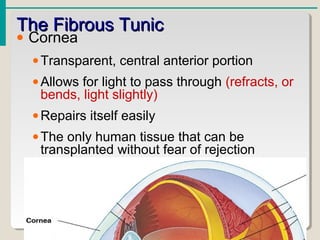
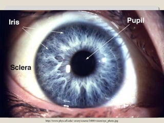
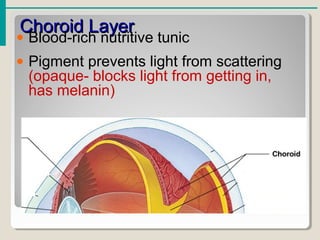
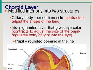
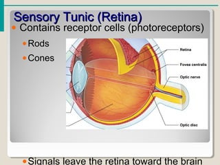
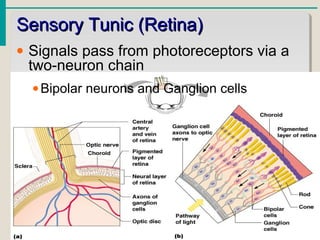
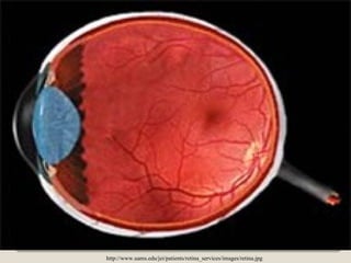
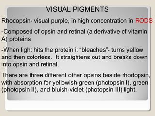
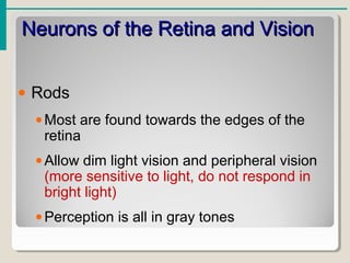
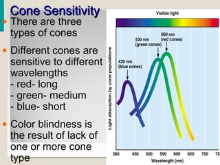
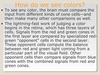
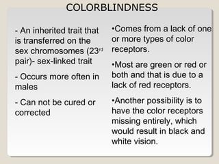
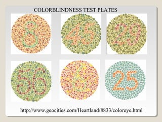
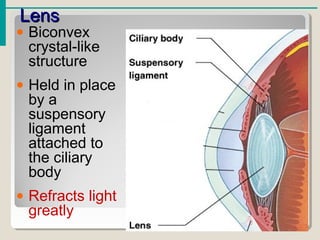
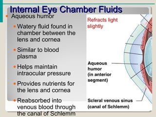
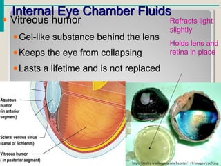
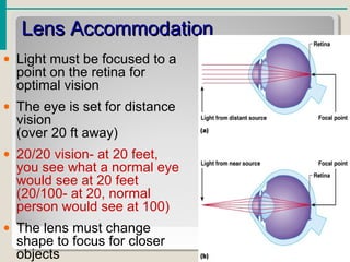
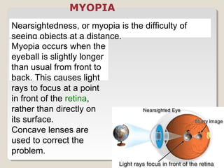
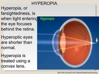
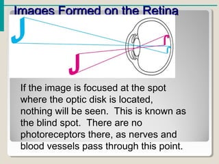
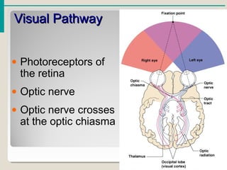
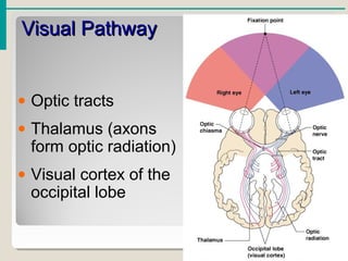
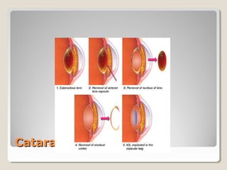
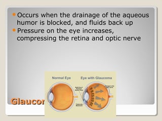
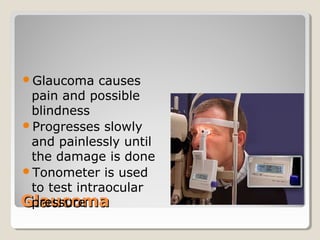
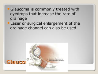
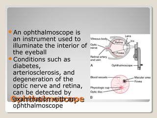
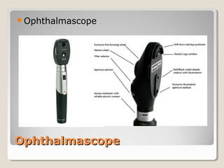
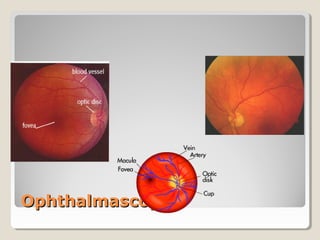
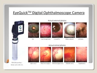
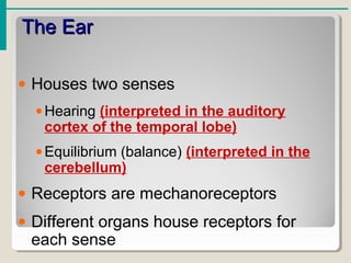
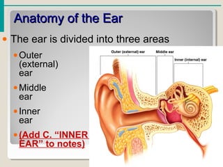
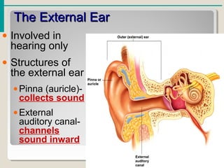
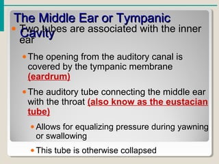
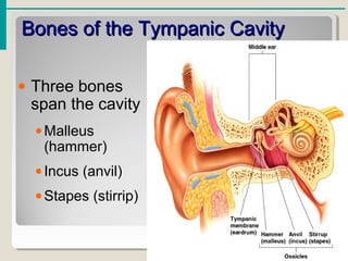
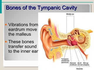
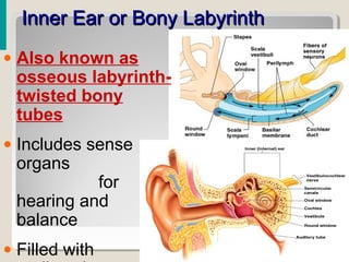
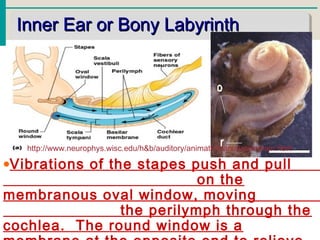
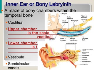
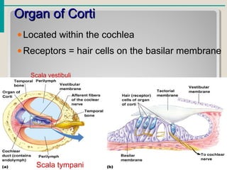
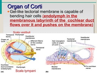
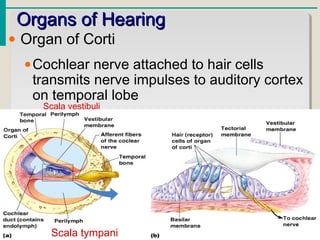
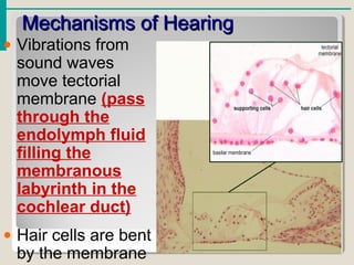
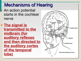
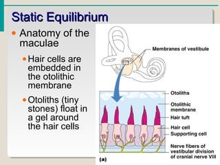
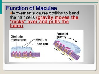
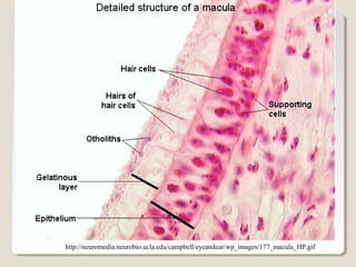
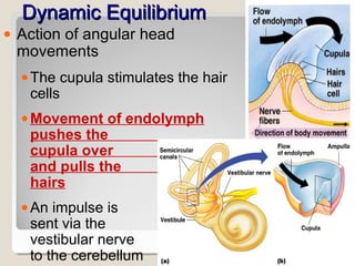
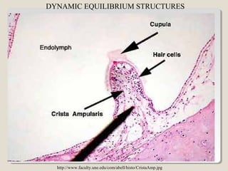
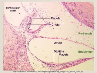
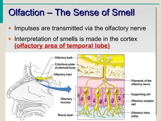
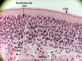
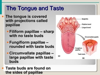
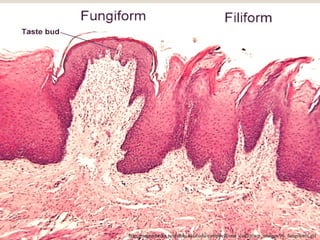
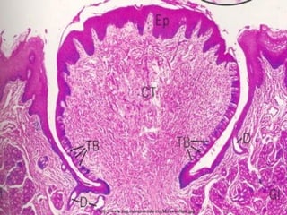
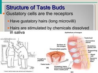
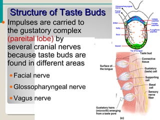
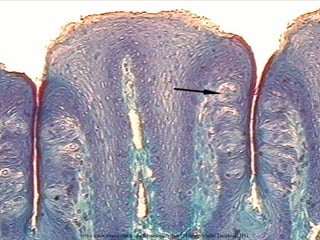
Essentials of Human Anatomy & Physiology
Copyright © 2003 Pearson Education, Inc. publishing as Benjamin Cummings
Slides 8.1 – 8.19
Seventh Edition
Elaine N. Marieb
Chapter 8Chapter 8
Special SensesSpecial Senses
Lecture Slides in PowerPoint by Jerry L. Cook and Melissa Scott
This presentation contains copyright protected materials
The SensesThe Senses
• General senses of touch (tactile)
•Temperature- thermoreceptors (heat)
•Pressure- mechanoreceptors (movement)
•Pain- mechanoreceptors
• Special senses
•Smell- chemoreceptors (chemicals)
•Taste- chemoreceptors
•Sight- photoreceptors (light)
•Hearing- mechanoreceptors
•Equilibrium- (balance) mechanoreceptors
The Eye and VisionThe Eye and Vision
• 70 percent of all sensory receptors are
in the eyes
• Each eye has over a million nerve fibers
• Protection for the eye
•Most of the eye is enclosed in a bony orbit
made up of the lacrimal (medial), ethmoid
(posterior), sphenoid (lateral), frontal
(superior), and zygomatic and maxilla
(inferior)
•A cushion of fat surrounds most of the eye
Accessory Structures of the EyeAccessory Structures of the Eye
• Eyelids-
brush
particles
out of eye
or cover
eye
• Eyelashes-
trap
particles
and keep
them out of
the eye
Accessory Structures of the EyeAccessory Structures of the Eye
• Ciliary glands –
modified
sweat glands
between the
eyelashes-
secrete acidic
sweat to kill
bacteria,
lubricate
eyelashes
Accessory Structures of the EyeAccessory Structures of the Eye
• Conjunctiva
•Membrane that lines the eyelids
•Connects to the surface of the eye- forms a seal
•Secretes mucus to lubricate the eye
http://neuromedia.neurobio.ucla.edu/campbell/eyeandear/wp_images/175_conjunctiva.gif
CONJUNCTIVITIS
- Inflammation of the conjunctiva
- Caused by bacterial or viral infection
- Highly contagious
http://www.healthseva.com/images/eye/conjunctivitis.jpg
Accessory Structures of the EyeAccessory Structures of the Eye
• Lacrimal
apparatus
•Lacrimal gland –
produces lacrimal
fluid
•Lacrimal canals –
drains lacrimal
fluid from eyes
Accessory Structures of the EyeAccessory Structures of the Eye
•Lacrimal sac –
provides
passage of
lacrimal fluid
towards nasal
cavity
Accessory Structures of the EyeAccessory Structures of the Eye
•Nasolacrimal
duct – empties
lacrimal fluid into
the nasal cavity
Function of the Lacrimal ApparatusFunction of the Lacrimal Apparatus
• Properties of lacrimal fluid
•Dilute salt solution (tears)
•Contains antibodies (fight antigens- foreign
substance) and lysozyme (enzyme that
destroys bacteria)
• Protects, moistens, and lubricates the
eye
• Empties into the nasal cavity
Extrinsic Eye MusclesExtrinsic Eye Muscles
• Muscles attach to the outer surface of
the eye
• Produce eye movements
When Extrinsic Eye Muscles ContractWhen Extrinsic Eye Muscles Contract
•Superior oblique- eyes look out and down
•Superior rectus- eyes looks up
•Lateral rectus- eyes look outward
•Medial rectus- eyes look inward
•Inferior rectus- eyes looks down
•Inferior oblique- eyes look in and up
http://www.esg.montana.edu/esg/kla/ta/eyemusc.jpg
Structure of the EyeStructure of the Eye
• The wall is composed of three tunics
• Fibrous tunic –
outside layer
• Choroid –
middle
layer
• Sensory
tunic –
inside
layer
The Fibrous TunicThe Fibrous Tunic
• Sclera
•White connective tissue layer
•Seen anteriorly as the “white of the eye”
•Semi-transparent
The Fibrous TunicThe Fibrous Tunic
• Cornea
•Transparent, central anterior portion
•Allows for light to pass through (refracts, or
bends, light slightly)
•Repairs itself easily
•The only human tissue that can be
transplanted without fear of rejection
http://www.phys.ufl.edu/~avery/course/3400/vision/eye_photo.jpg
Choroid LayerChoroid Layer
• Blood-rich nutritive tunic
• Pigment prevents light from scattering
(opaque- blocks light from getting in,
has melanin)
Choroid LayerChoroid Layer
• Modified interiorly into two structures
•Cilliary body – smooth muscle (contracts to
adjust the shape of the lens)
•Iris- pigmented layer that gives eye color
(contracts to adjust the size of the pupil-
regulates entry of light into the eye)
•Pupil – rounded opening in the iris
Sensory Tunic (Retina)Sensory Tunic (Retina)
• Contains receptor cells (photoreceptors)
•Rods
•Cones
•Signals leave the retina toward the brain
Sensory Tunic (Retina)Sensory Tunic (Retina)
• Signals pass from photoreceptors via a
two-neuron chain
•Bipolar neurons and Ganglion cells
http://www.uams.edu/jei/patients/retina_services/images/retina.jpg
VISUAL PIGMENTS
Rhodopsin- visual purple, in high concentration in RODS
-Composed of opsin and retinal (a derivative of vitamin
A) proteins
-When light hits the protein it “bleaches”- turns yellow
and then colorless. It straightens out and breaks down
into opsin and retinal.
There are three different other opsins beside rhodopsin,
with absorption for yellowish-green (photopsin I), green
(photopsin II), and bluish-violet (photopsin III) light.
Neurons of the Retina and VisionNeurons of the Retina and Vision
• Rods
•Most are found towards the edges of the
retina
•Allow dim light vision and peripheral vision
(more sensitive to light, do not respond in
bright light)
•Perception is all in gray tones
http://www.webvision.med.utah.edu/imageswv/PKCrodb.jpeghttp://webvision.med.utah.edu/imageswv/rod-GC.jpeg
ROD CELLS
Neurons of the Retina and VisionNeurons of the Retina and Vision
• Cones
•Allow for detailed color vision
•Densest in the center of the retina
•Fovea centralis – area of the retina with
only cones
•Respond best in bright light
• No photoreceptor cells are at the
optic disk, or blind spot
http://blc1.kilgore.cc.tx.us/kcap2/images/retina%20100x%20b%20fireworks.jpg
http://www.yorku.ca/eye/rod-cone.gif http://www.secretbeyondmatter.com/ourbrains/theworldinourbrains_files/11-1.jpg
Cone SensitivityCone Sensitivity
• There are three
types of cones
• Different cones are
sensitive to different
wavelengths
- red- long
- green- medium
- blue- short
• Color blindness is
the result of lack of
one or more cone
type
How do we see colors?
• To see any color, the brain must compare the
input from different kinds of cone cells—and
then make many other comparisons as well.
• The lightning-fast work of judging a color
begins in the retina, which has three layers of
cells. Signals from the red and green cones in
the first layer are compared by specialized red-
green "opponent" cells in the second layer.
These opponent cells compute the balance
between red and green light coming from a
particular part of the visual field. Other
opponent cells then compare signals from blue
cones with the combined signals from red and
green cones.
COLORBLINDNESS
- An inherited trait that
is transferred on the
sex chromosomes (23rd
pair)- sex-linked trait
- Occurs more often in
males
- Can not be cured or
corrected
•Comes from a lack of one
or more types of color
receptors.
•Most are green or red or
both and that is due to a
lack of red receptors.
•Another possibility is to
have the color receptors
missing entirely, which
would result in black and
white vision.
http://www.geocities.com/Heartland/8833/coloreye.html
COLORBLINDNESS TEST PLATES
LensLens
• Biconvex
crystal-like
structure
• Held in place
by a
suspensory
ligament
attached to
the ciliary
body
• Refracts light
greatly
Internal Eye Chamber FluidsInternal Eye Chamber Fluids
• Aqueous humor
• Watery fluid found in
chamber between the
lens and cornea
• Similar to blood
plasma
• Helps maintain
intraocular pressure
• Provides nutrients for
the lens and cornea
• Reabsorbed into
venous blood through
the canal of Schlemm
Refracts light
slightly
Internal Eye Chamber FluidsInternal Eye Chamber Fluids
• Vitreous humor
•Gel-like substance behind the lens
•Keeps the eye from collapsing
•Lasts a lifetime and is not replaced
http://faculty.washington.edu/kepeter/119/images/eye3.jpg
Refracts light
slightly
Holds lens and
retina in place
Lens AccommodationLens Accommodation
• Light must be focused to a
point on the retina for
optimal vision
• The eye is set for distance
vision
(over 20 ft away)
• 20/20 vision- at 20 feet,
you see what a normal eye
would see at 20 feet
(20/100- at 20, normal
person would see at 100)
• The lens must change
shape to focus for closer
objects
Nearsightedness, or myopia is the difficulty of
seeing objects at a distance.
Myopia occurs when the
eyeball is slightly longer
than usual from front to
back. This causes light
rays to focus at a point
in front of the retina,
rather than directly on
its surface.
Concave lenses are
used to correct the
problem.
MYOPIA
Hyperopia, or
farsightedness, is
when light entering
the eye focuses
behind the retina.
Hyperoptic eyes
are shorter than
normal.
Hyperopia is
treated using a
convex lens.
http://web.mountain.net/~topeye/images/hyperopia.jpg
HYPEROPIA
Images Formed on the RetinaImages Formed on the Retina
If the image is focused at the spot
where the optic disk is located,
nothing will be seen. This is known as
the blind spot. There are no
photoreceptors there, as nerves and
blood vessels pass through this point.
Visual PathwayVisual Pathway
• Photoreceptors of
the retina
• Optic nerve
• Optic nerve crosses
at the optic chiasma
Visual PathwayVisual Pathway
• Optic tracts
• Thalamus (axons
form optic radiation)
• Visual cortex of the
occipital lobe
Eye ReflexesEye Reflexes
• Internal muscles are controlled by the
autonomic nervous system
•Bright light causes pupils to constrict
through action of radial (iris) and ciliary
muscles
•Viewing close objects causes
accommodation
• External muscles control eye movement
to follow objects- voluntary, controlled at
the frontal eye field
• Viewing close objects causes
CataractsCataracts
In youth, the lens is transparent and a
hardened jelly-like texture
As we age, it becomes increasingly hard
and opaque
CataractsCataracts
Cataracts cause vision to become hazy
and distorted, and eventually cause
blindness
Risk factors: Type II diabetes, exposure
to intense sunlight, heavy smoking
Current treatments: surgical removal,
replacement lens implants or specialized
glasses
Cataract SurgeryCataract Surgery
GlaucomaGlaucoma
Occurs when the drainage of the aqueous
humor is blocked, and fluids back up
Pressure on the eye increases,
compressing the retina and optic nerve
GlaucomaGlaucoma
Glaucoma causes
pain and possible
blindness
Progresses slowly
and painlessly until
the damage is done
Tonometer is used
to test intraocular
pressure
GlaucomaGlaucoma
Glaucoma is commonly treated with
eyedrops that increase the rate of
drainage
Laser or surgical enlargement of the
drainage channel can also be used
OphthalmoscopeOphthalmoscope
An ophthalmoscope is
an instrument used to
illuminate the interior of
the eyeball
Conditions such as
diabetes,
arteriosclerosis, and
degeneration of the
optic nerve and retina,
can be detected by
examination with an
ophthalmoscope
OphthalmascopeOphthalmascope
Ophthalmascope
OphthalmascopeOphthalmascope
Anatomy of the EyeAnatomy of the Eye
The EarThe Ear
• Houses two senses
•Hearing (interpreted in the auditory
cortex of the temporal lobe)
•Equilibrium (balance) (interpreted in the
cerebellum)
• Receptors are mechanoreceptors
• Different organs house receptors for
each sense
Anatomy of the EarAnatomy of the Ear
• The ear is divided into three areas
•Outer
(external)
ear
•Middle
ear
•Inner
ear
•(Add C. “INNER
EAR” to notes)
The External EarThe External Ear
• Involved in
hearing only
• Structures of
the external ear
•Pinna (auricle)-
collects sound
•External
auditory canal-
channels
sound inward
The External Auditory CanalThe External Auditory Canal
• Narrow chamber in the temporal bone-
through the external auditory meatus
• Lined with skin
• Ceruminous (wax) glands are present
• Ends at the tympanic membrane
(eardrum)
The Middle Ear or TympanicThe Middle Ear or Tympanic
CavityCavity
• Air-filled cavity within the temporal bone
• Only involved in the sense of hearing
The Middle Ear or TympanicThe Middle Ear or Tympanic
CavityCavity• Two tubes are associated with the inner
ear
•The opening from the auditory canal is
covered by the tympanic membrane
(eardrum)
•The auditory tube connecting the middle ear
with the throat (also know as the eustacian
tube)
• Allows for equalizing pressure during yawning
or swallowing
• This tube is otherwise collapsed
Bones of the Tympanic CavityBones of the Tympanic Cavity
• Three bones
span the cavity
•Malleus
(hammer)
•Incus (anvil)
•Stapes (stirrip)
http://www.ghorayeb.com/files/STAPES_on_a_Penny_375_SQ.jpg
http://medicine.wustl.edu/~oto/bbears/images/ossic.jpg
Bones of the Tympanic CavityBones of the Tympanic Cavity
• Vibrations from
eardrum move
the malleus
• These bones
transfer sound
to the inner ear
Inner Ear or Bony LabyrinthInner Ear or Bony Labyrinth
• Also known as
osseous labyrinth-
twisted bony
tubes
• Includes sense
organs
for
hearing and
balance
• Filled with
Inner Ear or Bony LabyrinthInner Ear or Bony Labyrinth
•Vibrations of the stapes push and pull
on the
membranous oval window, moving
the perilymph through the
cochlea. The round window is a
http://www.neurophys.wisc.edu/h&b/auditory/animation/animationmain.html
Inner Ear or Bony LabryinthInner Ear or Bony Labryinth
• A maze of bony chambers within the
temporal bone
•Cochlea
• Upper chamber
is the scala
vestibuli
• Lower chamber
is the scala
tympani
•Vestibule
•Semicircular
canals
Organ of CortiOrgan of Corti
•Located within the cochlea
•Receptors = hair cells on the basilar membrane
Scala tympani
Scala vestibuli
•Gel-like tectorial membrane is capable of
bending hair cells (endolymph in the
membranous labyrinth of the cochlear duct
flows over it and pushes on the membrane)
Organ of CortiOrgan of Corti
Scala tympani
Scala vestibuli
Organs of HearingOrgans of Hearing
• Organ of Corti
•Cochlear nerve attached to hair cells
transmits nerve impulses to auditory cortex
on temporal lobe
Scala tympani
Scala vestibuli
Mechanisms of HearingMechanisms of Hearing
• Vibrations from
sound waves
move tectorial
membrane (pass
through the
endolymph fluid
filling the
membranous
labyrinth in the
cochlear duct)
• Hair cells are bent
by the membrane
Mechanisms of HearingMechanisms of Hearing
• An action potential
starts in the cochlear
nerve
• The signal is
transmitted to the
midbrain (for
auditory reflexes
and then directed to
the auditory cortex
of the temporal
lobe)
•Continued stimulation can lead
to adaptation (over
stimulation to the brain
makes it stop interpreting
the sounds)
Mechanisms of HearingMechanisms of Hearing
Organs of EquilibriumOrgans of Equilibrium
• Receptor cells are in two structures
•Vestibule
•Semicircular canals
Organs of EquilibriumOrgans of Equilibrium
• Equilibrium has two functional parts
•Static equilibrium- in the vestibule
•Dynamic equilibrium- in the semicircular
canals
Static EquilibriumStatic Equilibrium
• Maculae –
receptors in
the vestibule
•Report on
the position
of the head
•Send
information
via the
vestibular
nerve
Static EquilibriumStatic Equilibrium
• Anatomy of the
maculae
•Hair cells are
embedded in
the otolithic
membrane
•Otoliths (tiny
stones) float in
a gel around
the hair cells
Function of MaculaeFunction of Maculae
•Movements cause otoliths to bend
the hair cells (gravity moves the
“rocks” over and pulls the
hairs)
http://neuromedia.neurobio.ucla.edu/campbell/eyeandear/wp_images/177_macula_HP.gif
Dynamic EquilibriumDynamic Equilibrium
• Whole structure is the
ampulla
• Crista ampullaris –
receptors in the
semicircular canals
•Tuft of hair cells
•Cupula (gelatinous cap)
covers the hair cells
Dynamic EquilibriumDynamic Equilibrium
• Action of angular head
movements
•The cupula stimulates the hair
cells
•Movement of endolymph
pushes the
cupula over
and pulls the
hairs
•An impulse is
sent via the
vestibular nerve
to the cerebellum
DYNAMIC EQUILIBRIUM STRUCTURES
http://www.faculty.une.edu/com/abell/histo/CristaAmp.jpg
http://neuromedia.neurobio.ucla.edu/campbell/eyeandear/wp_images/177_macula_crista.gif
Chemical Senses – Taste andChemical Senses – Taste and
SmellSmell
• Both senses use chemoreceptors
•Stimulated by chemicals in solution
•Taste has four types of receptors
•Smell can differentiate a large range of
chemicals
• Both senses complement each other
and respond to many of the same
stimuli
Olfaction – The Sense of SmellOlfaction – The Sense of Smell
• Olfactory receptors are in the roof of the nasal
cavity
• Neurons with long cilia
• Chemicals must be dissolved in mucus for
detection
Olfaction – The Sense of SmellOlfaction – The Sense of Smell
• Impulses are transmitted via the olfactory nerve
• Interpretation of smells is made in the cortex
(olfactory area of temporal lobe)
http://asb.aecom.yu.edu/histology/labs/images/slides/A74_OlfactoryEpith_40X.jpg
The Sense of TasteThe Sense of Taste
• Taste buds
house the
receptor
organs
• Location of
taste buds
•Most are on
the tongue
•Soft palate
•Cheeks
The Tongue and TasteThe Tongue and Taste
• The tongue is coveredThe tongue is covered
with projections calledwith projections called
papillaepapillae
• Filiform papillae – sharpFiliform papillae – sharp
with no taste budswith no taste buds
• Fungifiorm papillae –Fungifiorm papillae –
rounded with taste budsrounded with taste buds
• Circumvallate papillae –Circumvallate papillae –
large papillae with tastelarge papillae with taste
budsbuds
• Taste buds are found onTaste buds are found on
the sides of papillaethe sides of papillae
http://neuromedia.neurobio.ucla.edu/campbell/oral_cavity/wp_images/96_fungiform.gif
http://www.esg.montana.edu/esg/kla/ta/vallate.jpg
Structure of Taste BudsStructure of Taste Buds
• Gustatory cells are the receptors
•Have gustatory hairs (long microvilli)
•Hairs are stimulated by chemicals dissolved
in saliva
Structure of Taste BudsStructure of Taste Buds
• Impulses are carried to
the gustatory complex
(pareital lobe) by
several cranial nerves
because taste buds are
found in different areas
•Facial nerve
•Glossopharyngeal nerve
•Vagus nerve
http://www.biosci.ohiou.edu/introbioslab/Bios171/images/lab6/Tastebuds.JPG
Taste SensationsTaste Sensations
• Sweet receptors
• Sugars
• Saccharine
• Some amino acids
• Sour receptors
• Acids
• Bitter receptors
• Alkaloids
• Salty receptors
• Metal ions
• Umami
• Glutamate, aspartate
(MSG, meats) http://instruct1.cit.cornell.edu/courses/psych431/student2000/mle6/tonguebig.gif
Developmental Aspects of theDevelopmental Aspects of the
Special SensesSpecial Senses
• Formed early in embryonic development
• Eyes are outgrowths of the brain
• All special senses are functional at birth

Special Senses

  • 1.
    Essentials of HumanAnatomy & Physiology Copyright © 2003 Pearson Education, Inc. publishing as Benjamin Cummings Slides 8.1 – 8.19 Seventh Edition Elaine N. Marieb Chapter 8Chapter 8 Special SensesSpecial Senses Lecture Slides in PowerPoint by Jerry L. Cook and Melissa Scott This presentation contains copyright protected materials
  • 2.
    The SensesThe Senses •General senses of touch (tactile) •Temperature- thermoreceptors (heat) •Pressure- mechanoreceptors (movement) •Pain- mechanoreceptors • Special senses •Smell- chemoreceptors (chemicals) •Taste- chemoreceptors •Sight- photoreceptors (light) •Hearing- mechanoreceptors •Equilibrium- (balance) mechanoreceptors
  • 3.
    The Eye andVisionThe Eye and Vision • 70 percent of all sensory receptors are in the eyes • Each eye has over a million nerve fibers • Protection for the eye •Most of the eye is enclosed in a bony orbit made up of the lacrimal (medial), ethmoid (posterior), sphenoid (lateral), frontal (superior), and zygomatic and maxilla (inferior) •A cushion of fat surrounds most of the eye
  • 4.
    Accessory Structures ofthe EyeAccessory Structures of the Eye • Eyelids- brush particles out of eye or cover eye • Eyelashes- trap particles and keep them out of the eye
  • 5.
    Accessory Structures ofthe EyeAccessory Structures of the Eye • Ciliary glands – modified sweat glands between the eyelashes- secrete acidic sweat to kill bacteria, lubricate eyelashes
  • 6.
    Accessory Structures ofthe EyeAccessory Structures of the Eye • Conjunctiva •Membrane that lines the eyelids •Connects to the surface of the eye- forms a seal •Secretes mucus to lubricate the eye http://neuromedia.neurobio.ucla.edu/campbell/eyeandear/wp_images/175_conjunctiva.gif
  • 7.
    CONJUNCTIVITIS - Inflammation ofthe conjunctiva - Caused by bacterial or viral infection - Highly contagious http://www.healthseva.com/images/eye/conjunctivitis.jpg
  • 8.
    Accessory Structures ofthe EyeAccessory Structures of the Eye • Lacrimal apparatus •Lacrimal gland – produces lacrimal fluid •Lacrimal canals – drains lacrimal fluid from eyes
  • 9.
    Accessory Structures ofthe EyeAccessory Structures of the Eye •Lacrimal sac – provides passage of lacrimal fluid towards nasal cavity
  • 10.
    Accessory Structures ofthe EyeAccessory Structures of the Eye •Nasolacrimal duct – empties lacrimal fluid into the nasal cavity
  • 11.
    Function of theLacrimal ApparatusFunction of the Lacrimal Apparatus • Properties of lacrimal fluid •Dilute salt solution (tears) •Contains antibodies (fight antigens- foreign substance) and lysozyme (enzyme that destroys bacteria) • Protects, moistens, and lubricates the eye • Empties into the nasal cavity
  • 12.
    Extrinsic Eye MusclesExtrinsicEye Muscles • Muscles attach to the outer surface of the eye • Produce eye movements
  • 13.
    When Extrinsic EyeMuscles ContractWhen Extrinsic Eye Muscles Contract •Superior oblique- eyes look out and down •Superior rectus- eyes looks up •Lateral rectus- eyes look outward •Medial rectus- eyes look inward •Inferior rectus- eyes looks down •Inferior oblique- eyes look in and up
  • 14.
  • 15.
    Structure of theEyeStructure of the Eye • The wall is composed of three tunics • Fibrous tunic – outside layer • Choroid – middle layer • Sensory tunic – inside layer
  • 16.
    The Fibrous TunicTheFibrous Tunic • Sclera •White connective tissue layer •Seen anteriorly as the “white of the eye” •Semi-transparent
  • 17.
    The Fibrous TunicTheFibrous Tunic • Cornea •Transparent, central anterior portion •Allows for light to pass through (refracts, or bends, light slightly) •Repairs itself easily •The only human tissue that can be transplanted without fear of rejection
  • 18.
  • 19.
    Choroid LayerChoroid Layer •Blood-rich nutritive tunic • Pigment prevents light from scattering (opaque- blocks light from getting in, has melanin)
  • 20.
    Choroid LayerChoroid Layer •Modified interiorly into two structures •Cilliary body – smooth muscle (contracts to adjust the shape of the lens) •Iris- pigmented layer that gives eye color (contracts to adjust the size of the pupil- regulates entry of light into the eye) •Pupil – rounded opening in the iris
  • 21.
    Sensory Tunic (Retina)SensoryTunic (Retina) • Contains receptor cells (photoreceptors) •Rods •Cones •Signals leave the retina toward the brain
  • 22.
    Sensory Tunic (Retina)SensoryTunic (Retina) • Signals pass from photoreceptors via a two-neuron chain •Bipolar neurons and Ganglion cells
  • 23.
  • 24.
    VISUAL PIGMENTS Rhodopsin- visualpurple, in high concentration in RODS -Composed of opsin and retinal (a derivative of vitamin A) proteins -When light hits the protein it “bleaches”- turns yellow and then colorless. It straightens out and breaks down into opsin and retinal. There are three different other opsins beside rhodopsin, with absorption for yellowish-green (photopsin I), green (photopsin II), and bluish-violet (photopsin III) light.
  • 25.
    Neurons of theRetina and VisionNeurons of the Retina and Vision • Rods •Most are found towards the edges of the retina •Allow dim light vision and peripheral vision (more sensitive to light, do not respond in bright light) •Perception is all in gray tones
  • 26.
  • 27.
    Neurons of theRetina and VisionNeurons of the Retina and Vision • Cones •Allow for detailed color vision •Densest in the center of the retina •Fovea centralis – area of the retina with only cones •Respond best in bright light • No photoreceptor cells are at the optic disk, or blind spot
  • 28.
  • 29.
  • 30.
    Cone SensitivityCone Sensitivity •There are three types of cones • Different cones are sensitive to different wavelengths - red- long - green- medium - blue- short • Color blindness is the result of lack of one or more cone type
  • 31.
    How do wesee colors? • To see any color, the brain must compare the input from different kinds of cone cells—and then make many other comparisons as well. • The lightning-fast work of judging a color begins in the retina, which has three layers of cells. Signals from the red and green cones in the first layer are compared by specialized red- green "opponent" cells in the second layer. These opponent cells compute the balance between red and green light coming from a particular part of the visual field. Other opponent cells then compare signals from blue cones with the combined signals from red and green cones.
  • 32.
    COLORBLINDNESS - An inheritedtrait that is transferred on the sex chromosomes (23rd pair)- sex-linked trait - Occurs more often in males - Can not be cured or corrected •Comes from a lack of one or more types of color receptors. •Most are green or red or both and that is due to a lack of red receptors. •Another possibility is to have the color receptors missing entirely, which would result in black and white vision.
  • 33.
  • 34.
    LensLens • Biconvex crystal-like structure • Heldin place by a suspensory ligament attached to the ciliary body • Refracts light greatly
  • 35.
    Internal Eye ChamberFluidsInternal Eye Chamber Fluids • Aqueous humor • Watery fluid found in chamber between the lens and cornea • Similar to blood plasma • Helps maintain intraocular pressure • Provides nutrients for the lens and cornea • Reabsorbed into venous blood through the canal of Schlemm Refracts light slightly
  • 36.
    Internal Eye ChamberFluidsInternal Eye Chamber Fluids • Vitreous humor •Gel-like substance behind the lens •Keeps the eye from collapsing •Lasts a lifetime and is not replaced http://faculty.washington.edu/kepeter/119/images/eye3.jpg Refracts light slightly Holds lens and retina in place
  • 37.
    Lens AccommodationLens Accommodation •Light must be focused to a point on the retina for optimal vision • The eye is set for distance vision (over 20 ft away) • 20/20 vision- at 20 feet, you see what a normal eye would see at 20 feet (20/100- at 20, normal person would see at 100) • The lens must change shape to focus for closer objects
  • 38.
    Nearsightedness, or myopiais the difficulty of seeing objects at a distance. Myopia occurs when the eyeball is slightly longer than usual from front to back. This causes light rays to focus at a point in front of the retina, rather than directly on its surface. Concave lenses are used to correct the problem. MYOPIA
  • 39.
    Hyperopia, or farsightedness, is whenlight entering the eye focuses behind the retina. Hyperoptic eyes are shorter than normal. Hyperopia is treated using a convex lens. http://web.mountain.net/~topeye/images/hyperopia.jpg HYPEROPIA
  • 40.
    Images Formed onthe RetinaImages Formed on the Retina If the image is focused at the spot where the optic disk is located, nothing will be seen. This is known as the blind spot. There are no photoreceptors there, as nerves and blood vessels pass through this point.
  • 41.
    Visual PathwayVisual Pathway •Photoreceptors of the retina • Optic nerve • Optic nerve crosses at the optic chiasma
  • 42.
    Visual PathwayVisual Pathway •Optic tracts • Thalamus (axons form optic radiation) • Visual cortex of the occipital lobe
  • 43.
    Eye ReflexesEye Reflexes •Internal muscles are controlled by the autonomic nervous system •Bright light causes pupils to constrict through action of radial (iris) and ciliary muscles •Viewing close objects causes accommodation • External muscles control eye movement to follow objects- voluntary, controlled at the frontal eye field • Viewing close objects causes
  • 44.
    CataractsCataracts In youth, thelens is transparent and a hardened jelly-like texture As we age, it becomes increasingly hard and opaque
  • 45.
    CataractsCataracts Cataracts cause visionto become hazy and distorted, and eventually cause blindness Risk factors: Type II diabetes, exposure to intense sunlight, heavy smoking Current treatments: surgical removal, replacement lens implants or specialized glasses
  • 46.
  • 47.
    GlaucomaGlaucoma Occurs when thedrainage of the aqueous humor is blocked, and fluids back up Pressure on the eye increases, compressing the retina and optic nerve
  • 48.
    GlaucomaGlaucoma Glaucoma causes pain andpossible blindness Progresses slowly and painlessly until the damage is done Tonometer is used to test intraocular pressure
  • 49.
    GlaucomaGlaucoma Glaucoma is commonlytreated with eyedrops that increase the rate of drainage Laser or surgical enlargement of the drainage channel can also be used
  • 50.
    OphthalmoscopeOphthalmoscope An ophthalmoscope is aninstrument used to illuminate the interior of the eyeball Conditions such as diabetes, arteriosclerosis, and degeneration of the optic nerve and retina, can be detected by examination with an ophthalmoscope
  • 51.
  • 52.
  • 53.
    Anatomy of theEyeAnatomy of the Eye
  • 54.
    The EarThe Ear •Houses two senses •Hearing (interpreted in the auditory cortex of the temporal lobe) •Equilibrium (balance) (interpreted in the cerebellum) • Receptors are mechanoreceptors • Different organs house receptors for each sense
  • 55.
    Anatomy of theEarAnatomy of the Ear • The ear is divided into three areas •Outer (external) ear •Middle ear •Inner ear •(Add C. “INNER EAR” to notes)
  • 56.
    The External EarTheExternal Ear • Involved in hearing only • Structures of the external ear •Pinna (auricle)- collects sound •External auditory canal- channels sound inward
  • 57.
    The External AuditoryCanalThe External Auditory Canal • Narrow chamber in the temporal bone- through the external auditory meatus • Lined with skin • Ceruminous (wax) glands are present • Ends at the tympanic membrane (eardrum)
  • 58.
    The Middle Earor TympanicThe Middle Ear or Tympanic CavityCavity • Air-filled cavity within the temporal bone • Only involved in the sense of hearing
  • 59.
    The Middle Earor TympanicThe Middle Ear or Tympanic CavityCavity• Two tubes are associated with the inner ear •The opening from the auditory canal is covered by the tympanic membrane (eardrum) •The auditory tube connecting the middle ear with the throat (also know as the eustacian tube) • Allows for equalizing pressure during yawning or swallowing • This tube is otherwise collapsed
  • 60.
    Bones of theTympanic CavityBones of the Tympanic Cavity • Three bones span the cavity •Malleus (hammer) •Incus (anvil) •Stapes (stirrip)
  • 61.
  • 62.
    Bones of theTympanic CavityBones of the Tympanic Cavity • Vibrations from eardrum move the malleus • These bones transfer sound to the inner ear
  • 63.
    Inner Ear orBony LabyrinthInner Ear or Bony Labyrinth • Also known as osseous labyrinth- twisted bony tubes • Includes sense organs for hearing and balance • Filled with
  • 64.
    Inner Ear orBony LabyrinthInner Ear or Bony Labyrinth •Vibrations of the stapes push and pull on the membranous oval window, moving the perilymph through the cochlea. The round window is a http://www.neurophys.wisc.edu/h&b/auditory/animation/animationmain.html
  • 65.
    Inner Ear orBony LabryinthInner Ear or Bony Labryinth • A maze of bony chambers within the temporal bone •Cochlea • Upper chamber is the scala vestibuli • Lower chamber is the scala tympani •Vestibule •Semicircular canals
  • 66.
    Organ of CortiOrganof Corti •Located within the cochlea •Receptors = hair cells on the basilar membrane Scala tympani Scala vestibuli
  • 67.
    •Gel-like tectorial membraneis capable of bending hair cells (endolymph in the membranous labyrinth of the cochlear duct flows over it and pushes on the membrane) Organ of CortiOrgan of Corti Scala tympani Scala vestibuli
  • 68.
    Organs of HearingOrgansof Hearing • Organ of Corti •Cochlear nerve attached to hair cells transmits nerve impulses to auditory cortex on temporal lobe Scala tympani Scala vestibuli
  • 69.
    Mechanisms of HearingMechanismsof Hearing • Vibrations from sound waves move tectorial membrane (pass through the endolymph fluid filling the membranous labyrinth in the cochlear duct) • Hair cells are bent by the membrane
  • 70.
    Mechanisms of HearingMechanismsof Hearing • An action potential starts in the cochlear nerve • The signal is transmitted to the midbrain (for auditory reflexes and then directed to the auditory cortex of the temporal lobe)
  • 71.
    •Continued stimulation canlead to adaptation (over stimulation to the brain makes it stop interpreting the sounds) Mechanisms of HearingMechanisms of Hearing
  • 72.
    Organs of EquilibriumOrgansof Equilibrium • Receptor cells are in two structures •Vestibule •Semicircular canals
  • 73.
    Organs of EquilibriumOrgansof Equilibrium • Equilibrium has two functional parts •Static equilibrium- in the vestibule •Dynamic equilibrium- in the semicircular canals
  • 74.
    Static EquilibriumStatic Equilibrium •Maculae – receptors in the vestibule •Report on the position of the head •Send information via the vestibular nerve
  • 75.
    Static EquilibriumStatic Equilibrium •Anatomy of the maculae •Hair cells are embedded in the otolithic membrane •Otoliths (tiny stones) float in a gel around the hair cells
  • 76.
    Function of MaculaeFunctionof Maculae •Movements cause otoliths to bend the hair cells (gravity moves the “rocks” over and pulls the hairs)
  • 77.
  • 78.
    Dynamic EquilibriumDynamic Equilibrium •Whole structure is the ampulla • Crista ampullaris – receptors in the semicircular canals •Tuft of hair cells •Cupula (gelatinous cap) covers the hair cells
  • 79.
    Dynamic EquilibriumDynamic Equilibrium •Action of angular head movements •The cupula stimulates the hair cells •Movement of endolymph pushes the cupula over and pulls the hairs •An impulse is sent via the vestibular nerve to the cerebellum
  • 80.
  • 81.
  • 82.
    Chemical Senses –Taste andChemical Senses – Taste and SmellSmell • Both senses use chemoreceptors •Stimulated by chemicals in solution •Taste has four types of receptors •Smell can differentiate a large range of chemicals • Both senses complement each other and respond to many of the same stimuli
  • 83.
    Olfaction – TheSense of SmellOlfaction – The Sense of Smell • Olfactory receptors are in the roof of the nasal cavity • Neurons with long cilia • Chemicals must be dissolved in mucus for detection
  • 84.
    Olfaction – TheSense of SmellOlfaction – The Sense of Smell • Impulses are transmitted via the olfactory nerve • Interpretation of smells is made in the cortex (olfactory area of temporal lobe)
  • 85.
  • 86.
    The Sense ofTasteThe Sense of Taste • Taste buds house the receptor organs • Location of taste buds •Most are on the tongue •Soft palate •Cheeks
  • 87.
    The Tongue andTasteThe Tongue and Taste • The tongue is coveredThe tongue is covered with projections calledwith projections called papillaepapillae • Filiform papillae – sharpFiliform papillae – sharp with no taste budswith no taste buds • Fungifiorm papillae –Fungifiorm papillae – rounded with taste budsrounded with taste buds • Circumvallate papillae –Circumvallate papillae – large papillae with tastelarge papillae with taste budsbuds • Taste buds are found onTaste buds are found on the sides of papillaethe sides of papillae
  • 88.
  • 89.
  • 90.
    Structure of TasteBudsStructure of Taste Buds • Gustatory cells are the receptors •Have gustatory hairs (long microvilli) •Hairs are stimulated by chemicals dissolved in saliva
  • 91.
    Structure of TasteBudsStructure of Taste Buds • Impulses are carried to the gustatory complex (pareital lobe) by several cranial nerves because taste buds are found in different areas •Facial nerve •Glossopharyngeal nerve •Vagus nerve
  • 92.
  • 93.
    Taste SensationsTaste Sensations •Sweet receptors • Sugars • Saccharine • Some amino acids • Sour receptors • Acids • Bitter receptors • Alkaloids • Salty receptors • Metal ions • Umami • Glutamate, aspartate (MSG, meats) http://instruct1.cit.cornell.edu/courses/psych431/student2000/mle6/tonguebig.gif
  • 94.
    Developmental Aspects oftheDevelopmental Aspects of the Special SensesSpecial Senses • Formed early in embryonic development • Eyes are outgrowths of the brain • All special senses are functional at birth