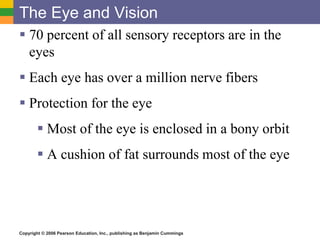
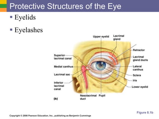
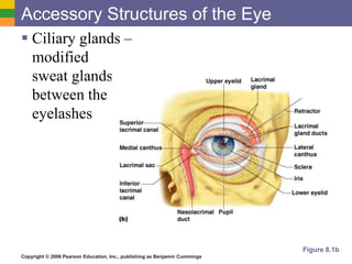
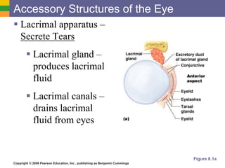
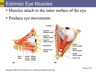
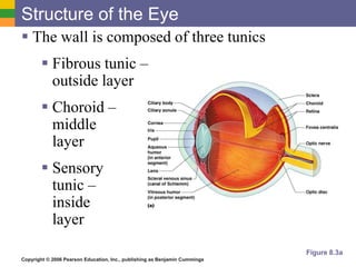
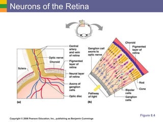
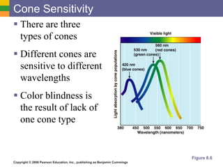
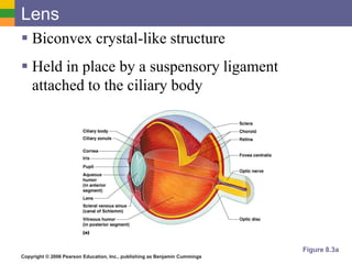
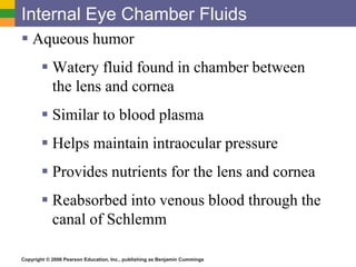
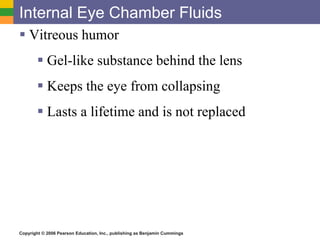
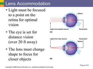
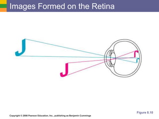
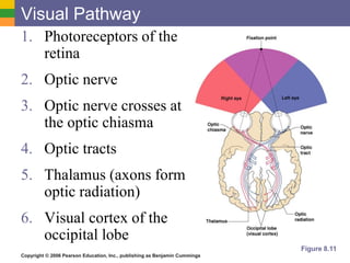
Copyright © 2006 Pearson Education, Inc., publishing as Benjamin Cummings
Special Senses
 Objective 2
 Identify the structures of the
eye, and describe their
functions.
Copyright © 2006 Pearson Education, Inc., publishing as Benjamin Cummings
The Eye and Vision
 70 percent of all sensory receptors are in the
eyes
 Each eye has over a million nerve fibers
 Protection for the eye
 Most of the eye is enclosed in a bony orbit
 A cushion of fat surrounds most of the eye
Copyright © 2006 Pearson Education, Inc., publishing as Benjamin Cummings
Protective Structures of the Eye
 Eyelids
 Eyelashes
Figure 8.1b
Copyright © 2006 Pearson Education, Inc., publishing as Benjamin Cummings
Accessory Structures of the Eye
 Ciliary glands –
modified
sweat glands
between the
eyelashes
Figure 8.1b
Copyright © 2006 Pearson Education, Inc., publishing as Benjamin Cummings
Accessory Structures of the Eye
 Conjunctiva
 Membrane that lines the eyelids
 Connects to the surface of the eye
 Secretes mucus to lubricate the eye
Copyright © 2006 Pearson Education, Inc., publishing as Benjamin Cummings
Accessory Structures of the Eye
 Lacrimal apparatus –
Secrete Tears
 Lacrimal gland –
produces lacrimal
fluid
 Lacrimal canals –
drains lacrimal
fluid from eyes
Figure 8.1a
Copyright © 2006 Pearson Education, Inc., publishing as Benjamin Cummings
Function of the Lacrimal Apparatus
 Properties of lacrimal fluid
 Dilute salt solution (tears)
 Contains antibodies and lysozyme
 Protects, moistens, and lubricates the eye
 Empties into the nasal cavity
Copyright © 2006 Pearson Education, Inc., publishing as Benjamin Cummings
Extrinsic Eye Muscles
 Muscles attach to the outer surface of the eye
 Produce eye movements
Figure 8.2
Copyright © 2006 Pearson Education, Inc., publishing as Benjamin Cummings
Structure of the Eye
 The wall is composed of three tunics
 Fibrous tunic –
outside layer
 Choroid –
middle
layer
 Sensory
tunic –
inside
layer
Figure 8.3a
Copyright © 2006 Pearson Education, Inc., publishing as Benjamin Cummings
The Fibrous Tunic
 Sclera
 White connective tissue layer
 Seen anteriorly as the “white of the eye”
 Cornea
 Transparent, central anterior portion
 Allows for light to pass through
 Repairs itself easily
 The only human tissue that can be transplanted
without fear of rejection
Copyright © 2006 Pearson Education, Inc., publishing as Benjamin Cummings
Choroid Layer
 Blood-rich nutritive tunic
 Pigment prevents light from scattering
 Modified interiorly into two structures
 Cilliary body – smooth muscle that
changes the shape of the lens
 Iris – contracts/expands to let light in
 Pigmented layer that gives eye color
 Pupil – rounded opening in the iris
Copyright © 2006 Pearson Education, Inc., publishing as Benjamin Cummings
Sensory Tunic (Retina)
 Contains receptor cells (photoreceptors)
 Rods
 Cones
 Signals leave the retina toward the brain
through the optic nerve in the back of the eye
Copyright © 2006 Pearson Education, Inc., publishing as Benjamin Cummings
Special Senses
 Objective 3
 Compare and contrast the
structure and functions of
rods and cones.
Copyright © 2006 Pearson Education, Inc., publishing as Benjamin Cummings
Neurons of the Retina and Vision
 Rods
 Most are found towards the edges of the
retina
 Allow dim light vision and peripheral
vision
 Perception is all in gray tones
Copyright © 2006 Pearson Education, Inc., publishing as Benjamin Cummings
Neurons of the Retina and Vision
 Cones
 Allow for detailed color vision
 Densest in the center of the retina
 Fovea centralis – area of the retina with
only cones
 No photoreceptor cells are at the optic disk,
or blind spot
Copyright © 2006 Pearson Education, Inc., publishing as Benjamin Cummings
Neurons of the Retina
Figure 8.4
Copyright © 2006 Pearson Education, Inc., publishing as Benjamin Cummings
Cone Sensitivity
 There are three
types of cones
 Different cones are
sensitive to different
wavelengths
 Color blindness is
the result of lack of
one cone type
Figure 8.6
Copyright © 2006 Pearson Education, Inc., publishing as Benjamin Cummings
Special Senses
 Objective 4
 Trace the pathway of light
through the retina.
Copyright © 2006 Pearson Education, Inc., publishing as Benjamin Cummings
Lens
 Biconvex crystal-like structure
 Held in place by a suspensory ligament
attached to the ciliary body
Figure 8.3a
Copyright © 2006 Pearson Education, Inc., publishing as Benjamin Cummings
Internal Eye Chamber Fluids
 Aqueous humor
 Watery fluid found in chamber between
the lens and cornea
 Similar to blood plasma
 Helps maintain intraocular pressure
 Provides nutrients for the lens and cornea
 Reabsorbed into venous blood through the
canal of Schlemm
Copyright © 2006 Pearson Education, Inc., publishing as Benjamin Cummings
Internal Eye Chamber Fluids
 Vitreous humor
 Gel-like substance behind the lens
 Keeps the eye from collapsing
 Lasts a lifetime and is not replaced
Copyright © 2006 Pearson Education, Inc., publishing as Benjamin Cummings
Lens Accommodation
 Light must be focused
to a point on the
retina for optimal
vision
 The eye is set for
distance vision
(over 20 ft away)
 The lens must change
shape to focus for
closer objects
Figure 8.9
Copyright © 2006 Pearson Education, Inc., publishing as Benjamin Cummings
Images Formed on the Retina
Figure 8.10
Copyright © 2006 Pearson Education, Inc., publishing as Benjamin Cummings
Visual Pathway
1. Photoreceptors of the
retina
2. Optic nerve
3. Optic nerve crosses at
the optic chiasma
4. Optic tracts
5. Thalamus (axons form
optic radiation)
6. Visual cortex of the
occipital lobe
Figure 8.11
Copyright © 2006 Pearson Education, Inc., publishing as Benjamin Cummings
Eye Reflexes
 Internal muscles are controlled by the autonomic
nervous system
 Bright light causes pupils to constrict through
action of radial and ciliary muscles
 Viewing close objects causes accommodation
 External muscles control eye movement to follow
objects
 Viewing close objects causes convergence (eyes
moving medially)

Eye

  • 1.
    Copyright © 2006Pearson Education, Inc., publishing as Benjamin Cummings Special Senses  Objective 2  Identify the structures of the eye, and describe their functions.
  • 2.
    Copyright © 2006Pearson Education, Inc., publishing as Benjamin Cummings The Eye and Vision  70 percent of all sensory receptors are in the eyes  Each eye has over a million nerve fibers  Protection for the eye  Most of the eye is enclosed in a bony orbit  A cushion of fat surrounds most of the eye
  • 3.
    Copyright © 2006Pearson Education, Inc., publishing as Benjamin Cummings Protective Structures of the Eye  Eyelids  Eyelashes Figure 8.1b
  • 4.
    Copyright © 2006Pearson Education, Inc., publishing as Benjamin Cummings Accessory Structures of the Eye  Ciliary glands – modified sweat glands between the eyelashes Figure 8.1b
  • 5.
    Copyright © 2006Pearson Education, Inc., publishing as Benjamin Cummings Accessory Structures of the Eye  Conjunctiva  Membrane that lines the eyelids  Connects to the surface of the eye  Secretes mucus to lubricate the eye
  • 6.
    Copyright © 2006Pearson Education, Inc., publishing as Benjamin Cummings Accessory Structures of the Eye  Lacrimal apparatus – Secrete Tears  Lacrimal gland – produces lacrimal fluid  Lacrimal canals – drains lacrimal fluid from eyes Figure 8.1a
  • 7.
    Copyright © 2006Pearson Education, Inc., publishing as Benjamin Cummings Function of the Lacrimal Apparatus  Properties of lacrimal fluid  Dilute salt solution (tears)  Contains antibodies and lysozyme  Protects, moistens, and lubricates the eye  Empties into the nasal cavity
  • 8.
    Copyright © 2006Pearson Education, Inc., publishing as Benjamin Cummings Extrinsic Eye Muscles  Muscles attach to the outer surface of the eye  Produce eye movements Figure 8.2
  • 9.
    Copyright © 2006Pearson Education, Inc., publishing as Benjamin Cummings Structure of the Eye  The wall is composed of three tunics  Fibrous tunic – outside layer  Choroid – middle layer  Sensory tunic – inside layer Figure 8.3a
  • 10.
    Copyright © 2006Pearson Education, Inc., publishing as Benjamin Cummings The Fibrous Tunic  Sclera  White connective tissue layer  Seen anteriorly as the “white of the eye”  Cornea  Transparent, central anterior portion  Allows for light to pass through  Repairs itself easily  The only human tissue that can be transplanted without fear of rejection
  • 11.
    Copyright © 2006Pearson Education, Inc., publishing as Benjamin Cummings Choroid Layer  Blood-rich nutritive tunic  Pigment prevents light from scattering  Modified interiorly into two structures  Cilliary body – smooth muscle that changes the shape of the lens  Iris – contracts/expands to let light in  Pigmented layer that gives eye color  Pupil – rounded opening in the iris
  • 12.
    Copyright © 2006Pearson Education, Inc., publishing as Benjamin Cummings Sensory Tunic (Retina)  Contains receptor cells (photoreceptors)  Rods  Cones  Signals leave the retina toward the brain through the optic nerve in the back of the eye
  • 13.
    Copyright © 2006Pearson Education, Inc., publishing as Benjamin Cummings Special Senses  Objective 3  Compare and contrast the structure and functions of rods and cones.
  • 14.
    Copyright © 2006Pearson Education, Inc., publishing as Benjamin Cummings Neurons of the Retina and Vision  Rods  Most are found towards the edges of the retina  Allow dim light vision and peripheral vision  Perception is all in gray tones
  • 15.
    Copyright © 2006Pearson Education, Inc., publishing as Benjamin Cummings Neurons of the Retina and Vision  Cones  Allow for detailed color vision  Densest in the center of the retina  Fovea centralis – area of the retina with only cones  No photoreceptor cells are at the optic disk, or blind spot
  • 16.
    Copyright © 2006Pearson Education, Inc., publishing as Benjamin Cummings Neurons of the Retina Figure 8.4
  • 17.
    Copyright © 2006Pearson Education, Inc., publishing as Benjamin Cummings Cone Sensitivity  There are three types of cones  Different cones are sensitive to different wavelengths  Color blindness is the result of lack of one cone type Figure 8.6
  • 18.
    Copyright © 2006Pearson Education, Inc., publishing as Benjamin Cummings Special Senses  Objective 4  Trace the pathway of light through the retina.
  • 19.
    Copyright © 2006Pearson Education, Inc., publishing as Benjamin Cummings Lens  Biconvex crystal-like structure  Held in place by a suspensory ligament attached to the ciliary body Figure 8.3a
  • 20.
    Copyright © 2006Pearson Education, Inc., publishing as Benjamin Cummings Internal Eye Chamber Fluids  Aqueous humor  Watery fluid found in chamber between the lens and cornea  Similar to blood plasma  Helps maintain intraocular pressure  Provides nutrients for the lens and cornea  Reabsorbed into venous blood through the canal of Schlemm
  • 21.
    Copyright © 2006Pearson Education, Inc., publishing as Benjamin Cummings Internal Eye Chamber Fluids  Vitreous humor  Gel-like substance behind the lens  Keeps the eye from collapsing  Lasts a lifetime and is not replaced
  • 22.
    Copyright © 2006Pearson Education, Inc., publishing as Benjamin Cummings Lens Accommodation  Light must be focused to a point on the retina for optimal vision  The eye is set for distance vision (over 20 ft away)  The lens must change shape to focus for closer objects Figure 8.9
  • 23.
    Copyright © 2006Pearson Education, Inc., publishing as Benjamin Cummings Images Formed on the Retina Figure 8.10
  • 24.
    Copyright © 2006Pearson Education, Inc., publishing as Benjamin Cummings Visual Pathway 1. Photoreceptors of the retina 2. Optic nerve 3. Optic nerve crosses at the optic chiasma 4. Optic tracts 5. Thalamus (axons form optic radiation) 6. Visual cortex of the occipital lobe Figure 8.11
  • 25.
    Copyright © 2006Pearson Education, Inc., publishing as Benjamin Cummings Eye Reflexes  Internal muscles are controlled by the autonomic nervous system  Bright light causes pupils to constrict through action of radial and ciliary muscles  Viewing close objects causes accommodation  External muscles control eye movement to follow objects  Viewing close objects causes convergence (eyes moving medially)