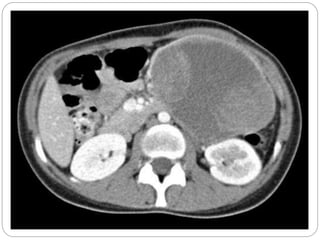
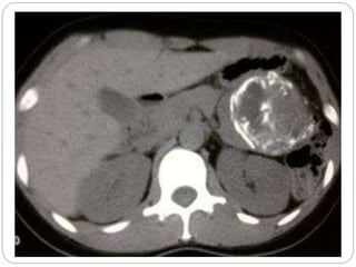
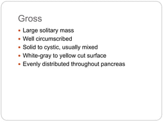
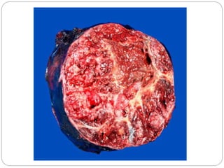
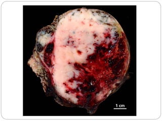
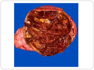
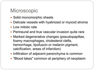
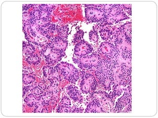
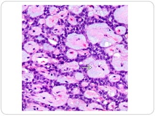
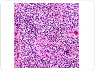
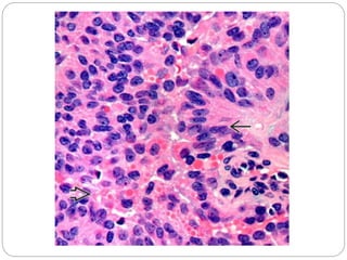
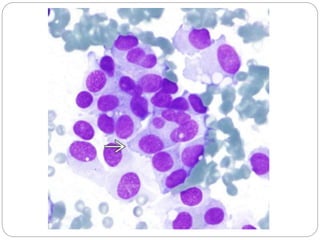
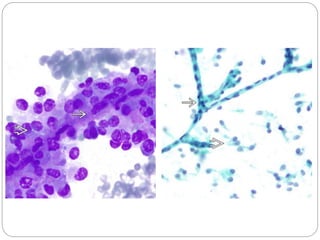
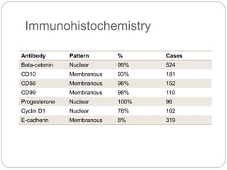
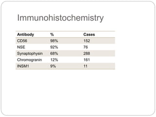
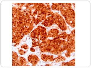
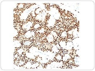
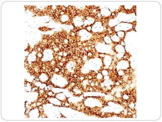
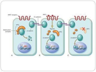
Rawa Muhsin
Solid Pseudopapillary Neoplasm of
Pancreas
Etiology
 Low-grade malignant neoplasm of uncertain
cellular differentiation
 Electron microscopy shows evidence of epithelial
differentiation
 90-100% harbor mutations in CTNNB1 gene
Clinical
 Most patients in 20s and 30s
 Female predominance (10-20:1)
 Evenly distributed throughout pancreas
 Vague abdominal symptoms
 Indolent
 10-15% metastasize (liver, peritoneum, lymph
nodes)
 Prognosis excellent
Gross
 Large solitary mass
 Well circumscribed
 Solid to cystic, usually mixed
 White-gray to yellow cut surface
 Evenly distributed throughout pancreas
Microscopic
 Solid monomorphic sheets
 Delicate vessels with hyalinized or myxoid stroma
 Low mitotic rate
 Perineural and true vascular invasion quite rare
 Marked degenerative changes (pseudopapillae,
foamy macrophages, cholesterol clefts,
hemorrhage, lipofuscin or melanin pigment,
calcification, areas of infarction)
 Infiltration of adjacent parenchyma is common
 "Blood lakes" common at periphery of neoplasm
Cytologic
 Nuclei oriented away from vessels with zone of
cytoplasm separating nuclei from capillaries
 Uniform and round to oval with finely dispersed
nuclear chromatin
 Often with longitudinal nuclear grooves
 Moderate amount of eosinophilic cytoplasm but
can be clear with vacuoles
 Intracytoplasmic eosinophilic hyaline globules
Immunohistochemistry
Antibody Pattern % Cases
Beta-catenin Nuclear 99% 524
CD10 Membranous 93% 181
CD56 Membranous 98% 152
CD99 Membranous 98% 110
Progesterone Nuclear 100% 96
Cyclin D1 Nuclear 78% 162
E-cadherin Membranous 8% 319
Immunohistochemistry
Antibody % Cases
CD56 98% 152
NSE 92% 76
Synaptophysin 68% 288
Chromogranin 12% 161
INSM1 9% 11
Molecular
 Missense mutations in exon 3 of CTNNB1 in
nearly all cases
 Prevents destruction of beta-catenin
Differential diagnoses
 Pancreatic neuroendocrine tumor
 Nuclear features
 IHC panel
 Acinic cell carcinoma
 Pattern
 IHC panel
 Pseudocyst
 Serous neoplasms
Solid Pseudopapillary Neoplasm

Solid Pseudopapillary Neoplasm

  • 1.
  • 2.
    Etiology  Low-grade malignantneoplasm of uncertain cellular differentiation  Electron microscopy shows evidence of epithelial differentiation  90-100% harbor mutations in CTNNB1 gene
  • 3.
    Clinical  Most patientsin 20s and 30s  Female predominance (10-20:1)  Evenly distributed throughout pancreas  Vague abdominal symptoms  Indolent  10-15% metastasize (liver, peritoneum, lymph nodes)  Prognosis excellent
  • 6.
    Gross  Large solitarymass  Well circumscribed  Solid to cystic, usually mixed  White-gray to yellow cut surface  Evenly distributed throughout pancreas
  • 11.
    Microscopic  Solid monomorphicsheets  Delicate vessels with hyalinized or myxoid stroma  Low mitotic rate  Perineural and true vascular invasion quite rare  Marked degenerative changes (pseudopapillae, foamy macrophages, cholesterol clefts, hemorrhage, lipofuscin or melanin pigment, calcification, areas of infarction)  Infiltration of adjacent parenchyma is common  "Blood lakes" common at periphery of neoplasm
  • 14.
    Cytologic  Nuclei orientedaway from vessels with zone of cytoplasm separating nuclei from capillaries  Uniform and round to oval with finely dispersed nuclear chromatin  Often with longitudinal nuclear grooves  Moderate amount of eosinophilic cytoplasm but can be clear with vacuoles  Intracytoplasmic eosinophilic hyaline globules
  • 20.
    Immunohistochemistry Antibody Pattern %Cases Beta-catenin Nuclear 99% 524 CD10 Membranous 93% 181 CD56 Membranous 98% 152 CD99 Membranous 98% 110 Progesterone Nuclear 100% 96 Cyclin D1 Nuclear 78% 162 E-cadherin Membranous 8% 319
  • 21.
    Immunohistochemistry Antibody % Cases CD5698% 152 NSE 92% 76 Synaptophysin 68% 288 Chromogranin 12% 161 INSM1 9% 11
  • 25.
    Molecular  Missense mutationsin exon 3 of CTNNB1 in nearly all cases  Prevents destruction of beta-catenin
  • 27.
    Differential diagnoses  Pancreaticneuroendocrine tumor  Nuclear features  IHC panel  Acinic cell carcinoma  Pattern  IHC panel  Pseudocyst  Serous neoplasms

Editor's Notes

  • #5 Well-circumscribed neoplasm with solid and cystic components Calcifications in ~ 30%
  • #6 Well-circumscribed neoplasm with solid and cystic components Calcifications in ~ 30%
  • #8 This well-demarcated tumor has a soft and friable solid surface with hemorrhagic areas.
  • #9 Hemorrhagic tumor of the head of the pancreas without involvement of the Wirsung.
  • #10 Encapsulated solid hemorrhagic tumor.
  • #11 The tumor can present as a well-demarcated, hemorrhagic cystic mass mimicking a pseudocyst.
  • #13 Solid sheets of tumor cells become dyscohesive and result in a characteristic pseudopapillary appearance with a central fibrovascular-like core surrounded by neoplastic cells.
  • #14 The delicate vessels can have myxoid stroma or may be hyalinized.
  • #16 The sheets of tumor cells have overlapping, round to oval nuclei that are oriented away from the vessels with a rim of cytoplasm toward the capillary.
  • #17 Tumor cells have round to oval nuclei and sometimes exhibit longitudinal nuclear grooves . These intra- and extracytoplasmic eosinophilic hyaline globules stain positive for PASD and α-1-antitrypsin.
  • #18 Typically the neoplastic cells are polygonal with eosinophilic cytoplasm, but these tumors can also have clear cytoplasm or, rarely, be composed of monomorphic spindle cells.
  • #19 Diff-Quik-stained FNA smear of a solid pseudopapillary tumor shows tumor cells with round to oval, eccentrically placed nuclei , condensed nuclear chromatin, and a moderate amount of delicate cytoplasm.
  • #20 Diff-Quik-stained FNA smear of a solid pseudopapillary tumor shows pseudopapillae comprised of a capillary channel bordered by tumor cells . Pap-stained FNA smear of solid pseudopapillary tumor shows "naked" branched capillaries with a background of dispersed tumor cells that have slender cytoplasmic processes .
  • #23 Immunohistochemistry for β-catenin shows nuclear and cytoplasmic staining in > 90% of tumors.
  • #24 Immunohistochemistry for progesterone receptor shows nuclear staining in nearly 80% of solid-pseudopapillary tumors. Staining for estrogen receptor is generally negative.
  • #25 Immunohistochemistry for CD10 shows cytoplasmic staining in solid-pseudopapillary tumors in nearly all the cases. Perinuclear staining may be seen in some instances.
  • #27 Figure 7-31  The  role  of  APC  in  regulating  the  stability  and  function  of  β-catenin.  APC  and  β-catenin  are  components  of  the  WNT  signaling  pathway.  A,  In  resting  colonic  epithelial  cells  (not  exposed  to  WNT),  β-catenin  forms  a  macromolecular  complex  containing  the  APC  protein.  This  complex  leads  to the destruction of β-catenin, and intracellular levels of β-catenin are low. B, When normal colonic epithelial cells are stimulated by WNT molecules, the  destruction complex is  deactivated,  β-catenin degradation does not  occur, and  cytoplasmic levels  increase. β-catenin translocates to the nucleus,  where  it binds to TCF, a transcription factor that activates genes involved in cell cycle progression. C, When APC is mutated or absent, as frequently occurs in colonic  polyps and cancers, the destruction of β-catenin cannot occur. β-catenin translocates to the nucleus and coactivates genes that promote entry into the cell  cycle, and cells behave as if they are under constant stimulation by the WNT pathway.