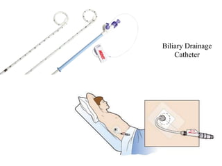
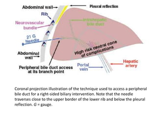
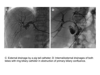
SlideShare a Scribd company logo
Biliary Drainage
OSR
Dr. Yash Kumar Achantani
INTRODUCTION
• Biliary obstruction is caused by many conditions, including
benign and malignant diseases.
• Malignant biliary obstruction (MBO) is usually caused by
cholangiocarcinoma, gall bladder and pancreatic malignancies,
metastatic lymphadenopathy and infrequently by hepatic and
advanced gastric and duodenal malignancies.
• Frequently the tumours are unresectable at diagnosis and only
palliative treatment is possible to improve patients’ quality of
life.
• The key purpose of biliary interventions in these patients is to
decompress the obstructed biliary system and if possible to
develop a communication between the biliary tree and the
bowel allowing physiological bile flow.
• As hepatic dysfunction is a risk factor for major hepatic
resection, biliary drainage helps in improving the liver
function prior to surgery or neoadjuvant chemotherapy.
• Percutaneous transhepatic biliary drainage (PTBD) is in
practice as a part of treatment of biliary obstruction for more
than four decades
INDICATIONS AND
CONTRAINDICATIONS
• Percutaneous drainage is performed as a preoperative
procedure for resectable malignancies, prior to neoadjuvant
chemotherapy or as a palliative technique.
• Indications includes high biliary obstructions, failed
endoscopic drainage, post-operative cases with biliary
obstruction, recurrent malignancies and multiple segment
strictures.
• There are no absolute contraindication, Relative
contraindications are haemorrhagic disorders, allergy to
iodinated contrast agents and ascites.
PRE-PROCEDURE IMAGING AND
PREPARATION
• All patients with surgical obstructive jaundice should undergo
multiphase contrast enhanced CT scan and/or MRI with
MRCP prior to percutaneous management.
• CT scan and MRI are useful in identifying the level and cause
of obstruction and defining the extent of the disease, thus
providing a platform to plan further management.
• MRCP provides information about the extent of biliary
obstruction and dilatation, level of obstruction and presence of
any variations in biliary anatomy
• Ultrasonography is very useful as a screening procedure prior
to percutaneous intervention.
• It helps in assessment of the biliary dilatation, presence of
ascites, volume of the liver lobes/segments and patency of
biliary confluence (primary and secondary) as this decides the
approach used, lobe to be drained.
• Before percutaneous intervention is done, patient’s coagulation
profile should be checked. (PT values should preferably be
less than four seconds above the control level and INR less
than 1.5)
• If coagulation parameters are deranged, vitamin K injection
can be given for three days (in elective cases) and the
procedure should be done after normalization.
• In case of emergencies (patient in cholangitis with risk of
septicaemia), if PT and INR are deranged, fresh frozen plasma
is given before and during the procedure and drainage should
be performed.
• In emergent situations where coagulation profile is not known,
drainage can be done immediately.
• A dose of intravenous antibiotics (preferably cephalosporins)
and fluids are given prior to the procedure.
• In elective cases, the procedure is performed after at least three
hours of fasting.
• Ascites is a relative contraindication and should be drained
before PTBD is performed.
• The procedure is performed under local anaesthesia and light
conscious sedation.
Percutaneous transhepatic biliary
drainage (PTBD)
• The procedure is done with the patient in supine position after
preliminary screening with ultrasonography.
• The skin area, depending on whether the right or left or both
ductal system needs drainage, is cleaned and draped.
• The sectoral duct (usually segment 3 or 6 for left and right
sided PTBD respectively) is punctured by 18G Chiba needle,
using ultrasonography as guidance at approximately 1-3 cm
away from the secondary biliary confluence.
• Once there is backflow of bile, a 0.032/0.035 inch soft “J” tip
guide wire (Terumo) is passed through the needle, which is
then exchanged for a 5F or 6F dilator followed by removal of
the guidewire.
• Cholangiogram is performed slowly to define the biliary
anatomy and type of obstruction (tapered, abrupt, irregular).
• Subsequently, the dilator is exchanged for a biliary
manipulation catheter or an angled tip angiographic catheter
over the wire.
• When the catheter tip is at the site of obstruction, attempt is
made to manipulate the soft hydrophilic guidewire (either
straight or “J” tip) to cross the stricture.
• Once the wire is across the stricture and in the duodenum, the
catheter is pushed over the wire into the duodenum.
• The soft wire is then replaced by Super Stiff Amplatz
guidewire over which the tract and stricture are dilated with 7F
and 8F dilators.
• Then, an 8.3F internal-external drainage catheter (Ring biliary
catheter, Cook) is positioned across the stricture and the
position is confirmed with contrast injection.
• In patients where initial attempt to cross the stricture fails, an
external drainage catheter is left with tip proximal to the
obstruction and internalization is attempted after a gap of two-
four days.
• For strictures distal to the hilum, i.e., with patent primary
biliary confluence, the technique is usually simple and single
drainage is sufficient. Drainage of single or both systems can
be done when the primary biliary confluence is completely
occluded.
• Multiple segment drainage may also be performed when
secondary confluence is involved.
• Chronically obstructed biliary segments with parenchymal
atrophy need not be drained as improvement in liver function
is unlikely.
• Various difficulties are encountered while performing PTBD.
• If biliary dilatation is mild or minimal, a micropuncture access
set with 21G needle and 0.018 wire can be used for initial
puncture.
• In grossly dilated biliary system, it is difficult to identify the
point of obstruction and to obtain stability for passing the
guide wire.
• Here, an external drainage is done initially to decompress the
system which increases stability, reduces edema, straightens
the wire course and also helps in identifying the point for
passing the wire in subsequent sitting
Chiba needle
Biliary Drainage
Catheter
Coronal projection illustration of the technique used to access a peripheral
bile duct for a right-sided biliary intervention. Note that the needle
traverses close to the upper border of the lower rib and below the pleural
reflection. G = gauge.
Ultrasound in preprocedural workup (a) determining the level of obstruction and
degree of intrahepatic biliary radical dilatation: Ultrasonography abdomen showing
an ill-defined heterogeneously hypoechoic mass (arrow) at the porta hepatis
blocking primary and bilateral secondary confluences with biliary radicles dilated till
periphery (curved arrow), suitable for ultrasound-guided puncture, (b) Selection of
target lobe: Excluding lobar atrophy (arrow) and portal venous thrombosis, (c)
ruling out ascites (arrow), which is a relative contraindication
Cholangiograms. A: Initial cholangiogram after puncture showing block below
the primary biliary confluence (arrow); B: Internal/external drainage by
ring biliary catheter;
C: External drainage by a pig tail catheter; D: Internal/external drainages of both
lobes with ring biliary catheter in obstruction of primary biliary confluence.
Brush biopsy needle (arrow) seen inserted through the sheath
to obtain sample from the mass at the level of obstruction.
BILIARY STENTING
• Once the obstruction is traversed, stenting can be done in the
same sitting to reduce the incidence of procedure related
complications.
• Types of stents
Self expandable metallic stents (Covered, Uncovered)
Plastic stents.
• Metallic stents have higher patency rates, lesser overall cost
and shorter hospital stay than plastic stents.
• The stricture is dilated with plastic dilators, if necessary, so
that the stent apparatus could be passed.
• Once the proximal and distal markers of the stent are at
appropriate positions, the stent is released.
• Proximal and distal ends of the stent should be at least 1 cm
and preferably 2 cm from the proximal and distal ends of
obstruction, respectively.
• The metallic stent self expands and results in biliary drainage
and decompression of the biliary system.
• The metallic stents have thermal memory and expand to their
maximum width when they reach the body temperature,
usually occurring in 24-48 h.
• If the expansion is not adequate after 48 h, dilatation of the
stent with balloon catheter may be needed for successful
drainage.
• Uncovered stents are usually preferred due to their lower costs,
very low incidence of migration and lower complications of
cholangitis and pancreatitis. But, there is higher incidence of
blockage due to tumor ingrowth.
• Covered stents are coated with polyurethane material and have
no gaps between the struts which reduce the chances of tumor
in-growth and thus improve stent patency. But they are
associated with increased risks of stent migration and side
branch occlusion.
• Stent blockage may occur due to tumor ingrowth, tumor
overgrowth or sludge. This needs repeat procedure.
• PTBD can be done again and a ring biliary catheter or another
stent can be placed through the blocked stent.
• Another problem is stent fracture which can occur when
balloon dilatation is attempted. This may cause early blockage
of the stent.
Biliary stenting. A: Positioning of the metallic stent with markers (arrows)
for optimal deployment; B: Expanded metallic stent after deployment across
the stricture;
C: Bilobar metallic stent placement (arrows); D: Placement of ring biliary
catheter by the side of blocked plastic stent after failed endoscopic exchange.
Stent block. A: Ring biliary catheter (arrow) placed through a blocked
metallic stent (block arrow) due to disease progression; B: Second
metallic stent (arrow) placed through the initial blocked right sided metallic
stent (block arrow).
COMPLICATIONS
• The complications can be either early (occurring within 30 d)
and late.
• Complications include pain at the site of puncture, bile leak
with risk of biliary peritonitis and biloma formation,
haemorrhage including haemobilia, biliovenous fistula, arterial
injury, cholangitis and septicaemia, acute pancreatitis and
catheter related problems like kinking or dislocation.
• Right sided punctures are also associated with the risk of
pneumothorax and haemothorax.
• Late complications include cholangitis, liver abscess,
septicaemia, drainage catheter or stent blockage.
Hemobilia. A: Cholangiogram in a case of hemobilia shows multiple filling defects
(arrow) within the biliary ducts; B: Cholangiogram showing opacification
of branches of portal vein (arrow) suggesting bilio-venous fistula; C:
Cholangiogram showing opacification of branches of right hepatic artery (arrow).
Catheter related complications. A: Cholangiogram showing pericatheter leak
(black arrow) due to proximally dislodged ring biliary catheter (white arrow); B:
Dislodged external drainage catheter (arrow) with only tip in the left hepatic
duct; C: Fracture (arrow) of ring biliary catheter.
Axial contrast enhanced CT scan showing bulky pancreas with
peripancreatic necrotic collection (a) developing after PTBD in a
patient of carcinoma of gall bladder (arrow)
Axial contrast enhanced CT scan after PTBD showing multiple
abscesses in right lobe of liver (arrows)
Axial CT scan after PTBD showing
perihepatic bilioma (arrow)
Axial CT scans after PTBD showing hyperdense fluid in perihepatic (arrow) region
and pelvis (a) suggesting hemoperitoneum.
Biliary drainage
Biliary drainage

More Related Content

What's hot

Triple phase ct PowerPoint slide PPT pk
Triple phase ct PowerPoint slide PPT pkTriple phase ct PowerPoint slide PPT pk
Triple phase ct PowerPoint slide PPT pk
Dr pradeep Kumar
 
Magnetic resonance cholangiopancreatography ppt
Magnetic resonance cholangiopancreatography pptMagnetic resonance cholangiopancreatography ppt
Magnetic resonance cholangiopancreatography ppt
Anjan Dangal
 
Abdominal CT scan
Abdominal CT scanAbdominal CT scan
Abdominal CT scan
ALAUF JALALUDEEN
 
T-tube Cholangiogram
T-tube CholangiogramT-tube Cholangiogram
T-tube Cholangiogram
ricksw78
 
TPCT - Triphasic CT Scan - Liver
TPCT - Triphasic CT Scan - LiverTPCT - Triphasic CT Scan - Liver
TPCT - Triphasic CT Scan - Liver
drpankajs
 
Sinogram and fistulogram
Sinogram and fistulogramSinogram and fistulogram
Sinogram and fistulogram
InosRagan
 
Excretion Urography / Intravenous Urography (IVU)
Excretion Urography / Intravenous Urography (IVU)Excretion Urography / Intravenous Urography (IVU)
Excretion Urography / Intravenous Urography (IVU)
Sudil Paudyal
 
INTRAVENOUS UROGRAM (IVU)
INTRAVENOUS UROGRAM (IVU)INTRAVENOUS UROGRAM (IVU)
INTRAVENOUS UROGRAM (IVU)
Jai Kumar
 
Enteroclysis
EnteroclysisEnteroclysis
Percutaneous transhepatic choledochography
Percutaneous transhepatic choledochographyPercutaneous transhepatic choledochography
Percutaneous transhepatic choledochography
Maajid Mohi ud din
 
MRCP Magnetic Resonance cholangiopancreatography
 MRCP Magnetic Resonance cholangiopancreatography MRCP Magnetic Resonance cholangiopancreatography
MRCP Magnetic Resonance cholangiopancreatography
Prajjwal Upadhayaya
 
Mrcp radiology
Mrcp radiologyMrcp radiology
Mrcp radiology
Vidya TK
 
PERCITANEOUS NEPHROSTOMY and HYSTEROSALPIONGOGRAPHY
PERCITANEOUS NEPHROSTOMY and HYSTEROSALPIONGOGRAPHYPERCITANEOUS NEPHROSTOMY and HYSTEROSALPIONGOGRAPHY
PERCITANEOUS NEPHROSTOMY and HYSTEROSALPIONGOGRAPHY
SharmaRajan4
 
SELDINGER TECHNIQUE & INTERVENTIONAL RADIOLOGY
SELDINGER TECHNIQUE & INTERVENTIONAL RADIOLOGYSELDINGER TECHNIQUE & INTERVENTIONAL RADIOLOGY
SELDINGER TECHNIQUE & INTERVENTIONAL RADIOLOGY
Riyas M K
 
Operative Cholangiogram
Operative CholangiogramOperative Cholangiogram
Operative CholangiogramStef C
 
Transjugular intrahepatic porto systemic shunt
Transjugular intrahepatic porto systemic shuntTransjugular intrahepatic porto systemic shunt
Transjugular intrahepatic porto systemic shuntairwave12
 
CT Enteroclysis
CT EnteroclysisCT Enteroclysis
CT Enteroclysis
Dr. Yash Kumar Achantani
 
Triphasic CT scan
Triphasic CT scanTriphasic CT scan
Triphasic CT scan
Tahira Aghani
 
MCU- Micturating cysto-urethrogram
MCU- Micturating cysto-urethrogramMCU- Micturating cysto-urethrogram
MCU- Micturating cysto-urethrogram
Dr. Mohit Goel
 

What's hot (20)

Triple phase ct PowerPoint slide PPT pk
Triple phase ct PowerPoint slide PPT pkTriple phase ct PowerPoint slide PPT pk
Triple phase ct PowerPoint slide PPT pk
 
Magnetic resonance cholangiopancreatography ppt
Magnetic resonance cholangiopancreatography pptMagnetic resonance cholangiopancreatography ppt
Magnetic resonance cholangiopancreatography ppt
 
Abdominal CT scan
Abdominal CT scanAbdominal CT scan
Abdominal CT scan
 
T-tube Cholangiogram
T-tube CholangiogramT-tube Cholangiogram
T-tube Cholangiogram
 
TPCT - Triphasic CT Scan - Liver
TPCT - Triphasic CT Scan - LiverTPCT - Triphasic CT Scan - Liver
TPCT - Triphasic CT Scan - Liver
 
Sinogram and fistulogram
Sinogram and fistulogramSinogram and fistulogram
Sinogram and fistulogram
 
Excretion Urography / Intravenous Urography (IVU)
Excretion Urography / Intravenous Urography (IVU)Excretion Urography / Intravenous Urography (IVU)
Excretion Urography / Intravenous Urography (IVU)
 
INTRAVENOUS UROGRAM (IVU)
INTRAVENOUS UROGRAM (IVU)INTRAVENOUS UROGRAM (IVU)
INTRAVENOUS UROGRAM (IVU)
 
Enteroclysis
EnteroclysisEnteroclysis
Enteroclysis
 
Percutaneous transhepatic choledochography
Percutaneous transhepatic choledochographyPercutaneous transhepatic choledochography
Percutaneous transhepatic choledochography
 
T Tube
T TubeT Tube
T Tube
 
MRCP Magnetic Resonance cholangiopancreatography
 MRCP Magnetic Resonance cholangiopancreatography MRCP Magnetic Resonance cholangiopancreatography
MRCP Magnetic Resonance cholangiopancreatography
 
Mrcp radiology
Mrcp radiologyMrcp radiology
Mrcp radiology
 
PERCITANEOUS NEPHROSTOMY and HYSTEROSALPIONGOGRAPHY
PERCITANEOUS NEPHROSTOMY and HYSTEROSALPIONGOGRAPHYPERCITANEOUS NEPHROSTOMY and HYSTEROSALPIONGOGRAPHY
PERCITANEOUS NEPHROSTOMY and HYSTEROSALPIONGOGRAPHY
 
SELDINGER TECHNIQUE & INTERVENTIONAL RADIOLOGY
SELDINGER TECHNIQUE & INTERVENTIONAL RADIOLOGYSELDINGER TECHNIQUE & INTERVENTIONAL RADIOLOGY
SELDINGER TECHNIQUE & INTERVENTIONAL RADIOLOGY
 
Operative Cholangiogram
Operative CholangiogramOperative Cholangiogram
Operative Cholangiogram
 
Transjugular intrahepatic porto systemic shunt
Transjugular intrahepatic porto systemic shuntTransjugular intrahepatic porto systemic shunt
Transjugular intrahepatic porto systemic shunt
 
CT Enteroclysis
CT EnteroclysisCT Enteroclysis
CT Enteroclysis
 
Triphasic CT scan
Triphasic CT scanTriphasic CT scan
Triphasic CT scan
 
MCU- Micturating cysto-urethrogram
MCU- Micturating cysto-urethrogramMCU- Micturating cysto-urethrogram
MCU- Micturating cysto-urethrogram
 

Similar to Biliary drainage

Percutaneous nephrostomy
Percutaneous nephrostomyPercutaneous nephrostomy
Percutaneous nephrostomy
sarfraj Ahmad
 
Bile duct injury.pptx
Bile duct injury.pptxBile duct injury.pptx
Bile duct injury.pptx
Sujan Shrestha
 
Radiographic procedure PTC & PTB..D.pptx
Radiographic procedure PTC & PTB..D.pptxRadiographic procedure PTC & PTB..D.pptx
Radiographic procedure PTC & PTB..D.pptx
justinfan550
 
Biliary injury.pdf
Biliary injury.pdfBiliary injury.pdf
Biliary injury.pdf
AshrafAdam7
 
Bile duct injuries BDI
Bile duct injuries BDIBile duct injuries BDI
Bile duct injuries BDI
Anupshrestha27
 
Vivek vij caudate lobe
Vivek vij caudate lobeVivek vij caudate lobe
Vivek vij caudate lobe
Sujan Shrestha
 
Liver trauma
Liver traumaLiver trauma
Liver trauma
mostafa hegazy
 
Radiographic technique of biliary system
Radiographic technique of biliary systemRadiographic technique of biliary system
Radiographic technique of biliary system
InosRagan
 
Bile duct injuriesCBDstricture, biliary fistula.pptx
Bile duct injuriesCBDstricture, biliary fistula.pptxBile duct injuriesCBDstricture, biliary fistula.pptx
Bile duct injuriesCBDstricture, biliary fistula.pptx
Pradeep Pande
 
Orthotopic neobladder
Orthotopic neobladderOrthotopic neobladder
Orthotopic neobladder
Prateek Laddha
 
Urinary bladder catheters aaron
Urinary bladder catheters aaronUrinary bladder catheters aaron
Urinary bladder catheters aaron
kemboiarn
 
Conduits after esophagectomy for esophageal reconstruction
Conduits after esophagectomy for esophageal reconstruction Conduits after esophagectomy for esophageal reconstruction
Conduits after esophagectomy for esophageal reconstruction
Shahbaz Panhwer
 
Investigation of Biliary Tract
Investigation of Biliary Tract Investigation of Biliary Tract
Investigation of Biliary Tract
Scholars of Medical Imaging BPKIHS Dharan
 
Investigation of Biliary Tract
Investigation of Biliary Tract Investigation of Biliary Tract
Investigation of Biliary Tract
Sujan Poudel
 
Urology surgery. Bladder, Urethra and Prostsate
Urology surgery. Bladder, Urethra and ProstsateUrology surgery. Bladder, Urethra and Prostsate
Urology surgery. Bladder, Urethra and Prostsate
Kishore Rajan
 
Post cholecystectomy syndromes
Post cholecystectomy syndromesPost cholecystectomy syndromes
Post cholecystectomy syndromes
Youttam Laudari
 
Cholecystectomy class
Cholecystectomy classCholecystectomy class
Cholecystectomy class
surgerymgmcri
 
Presentation editing.pptx
Presentation editing.pptxPresentation editing.pptx
Presentation editing.pptx
Abdullah764280
 
HEPATOBILIARY STUDIES.pptx
HEPATOBILIARY STUDIES.pptxHEPATOBILIARY STUDIES.pptx
HEPATOBILIARY STUDIES.pptx
Josephmwanika
 
CT urography
CT urography CT urography
CT urography
SabitaMandal1
 

Similar to Biliary drainage (20)

Percutaneous nephrostomy
Percutaneous nephrostomyPercutaneous nephrostomy
Percutaneous nephrostomy
 
Bile duct injury.pptx
Bile duct injury.pptxBile duct injury.pptx
Bile duct injury.pptx
 
Radiographic procedure PTC & PTB..D.pptx
Radiographic procedure PTC & PTB..D.pptxRadiographic procedure PTC & PTB..D.pptx
Radiographic procedure PTC & PTB..D.pptx
 
Biliary injury.pdf
Biliary injury.pdfBiliary injury.pdf
Biliary injury.pdf
 
Bile duct injuries BDI
Bile duct injuries BDIBile duct injuries BDI
Bile duct injuries BDI
 
Vivek vij caudate lobe
Vivek vij caudate lobeVivek vij caudate lobe
Vivek vij caudate lobe
 
Liver trauma
Liver traumaLiver trauma
Liver trauma
 
Radiographic technique of biliary system
Radiographic technique of biliary systemRadiographic technique of biliary system
Radiographic technique of biliary system
 
Bile duct injuriesCBDstricture, biliary fistula.pptx
Bile duct injuriesCBDstricture, biliary fistula.pptxBile duct injuriesCBDstricture, biliary fistula.pptx
Bile duct injuriesCBDstricture, biliary fistula.pptx
 
Orthotopic neobladder
Orthotopic neobladderOrthotopic neobladder
Orthotopic neobladder
 
Urinary bladder catheters aaron
Urinary bladder catheters aaronUrinary bladder catheters aaron
Urinary bladder catheters aaron
 
Conduits after esophagectomy for esophageal reconstruction
Conduits after esophagectomy for esophageal reconstruction Conduits after esophagectomy for esophageal reconstruction
Conduits after esophagectomy for esophageal reconstruction
 
Investigation of Biliary Tract
Investigation of Biliary Tract Investigation of Biliary Tract
Investigation of Biliary Tract
 
Investigation of Biliary Tract
Investigation of Biliary Tract Investigation of Biliary Tract
Investigation of Biliary Tract
 
Urology surgery. Bladder, Urethra and Prostsate
Urology surgery. Bladder, Urethra and ProstsateUrology surgery. Bladder, Urethra and Prostsate
Urology surgery. Bladder, Urethra and Prostsate
 
Post cholecystectomy syndromes
Post cholecystectomy syndromesPost cholecystectomy syndromes
Post cholecystectomy syndromes
 
Cholecystectomy class
Cholecystectomy classCholecystectomy class
Cholecystectomy class
 
Presentation editing.pptx
Presentation editing.pptxPresentation editing.pptx
Presentation editing.pptx
 
HEPATOBILIARY STUDIES.pptx
HEPATOBILIARY STUDIES.pptxHEPATOBILIARY STUDIES.pptx
HEPATOBILIARY STUDIES.pptx
 
CT urography
CT urography CT urography
CT urography
 

More from Dr. Yash Kumar Achantani

USG of Aorta and Coeliac axis
USG of Aorta and Coeliac axisUSG of Aorta and Coeliac axis
USG of Aorta and Coeliac axis
Dr. Yash Kumar Achantani
 
TACE- Transarterial Chemoembolisation
TACE- Transarterial Chemoembolisation TACE- Transarterial Chemoembolisation
TACE- Transarterial Chemoembolisation
Dr. Yash Kumar Achantani
 
Retrograde Pyelography
Retrograde PyelographyRetrograde Pyelography
Retrograde Pyelography
Dr. Yash Kumar Achantani
 
Imaging of Renal Tumors
Imaging of Renal TumorsImaging of Renal Tumors
Imaging of Renal Tumors
Dr. Yash Kumar Achantani
 
Renal isotope scan
Renal isotope scanRenal isotope scan
Renal isotope scan
Dr. Yash Kumar Achantani
 
Post Processing of CT Thorax
Post Processing of CT ThoraxPost Processing of CT Thorax
Post Processing of CT Thorax
Dr. Yash Kumar Achantani
 
Imaging of Obstructive jaundice
Imaging of Obstructive jaundiceImaging of Obstructive jaundice
Imaging of Obstructive jaundice
Dr. Yash Kumar Achantani
 
HIDA Scan
HIDA ScanHIDA Scan
MRI in CVA
MRI in CVAMRI in CVA
CT Urography
CT UrographyCT Urography
CT Imaging of CA Esophagus
CT Imaging of CA EsophagusCT Imaging of CA Esophagus
CT Imaging of CA Esophagus
Dr. Yash Kumar Achantani
 
CT Cervical Spine
CT Cervical SpineCT Cervical Spine
CT Cervical Spine
Dr. Yash Kumar Achantani
 
CT - Lung Carcinoma
CT - Lung CarcinomaCT - Lung Carcinoma
CT - Lung Carcinoma
Dr. Yash Kumar Achantani
 
Liver cirrhosis USG
Liver cirrhosis USGLiver cirrhosis USG
Liver cirrhosis USG
Dr. Yash Kumar Achantani
 
MRI in Tibial Fractures
MRI in Tibial FracturesMRI in Tibial Fractures
MRI in Tibial Fractures
Dr. Yash Kumar Achantani
 
Basics of Renal Doppler
Basics of Renal DopplerBasics of Renal Doppler
Basics of Renal Doppler
Dr. Yash Kumar Achantani
 
Renal Angiography
Renal AngiographyRenal Angiography
Renal Angiography
Dr. Yash Kumar Achantani
 
Imaging of Large Bowel Polyp
Imaging of Large Bowel PolypImaging of Large Bowel Polyp
Imaging of Large Bowel Polyp
Dr. Yash Kumar Achantani
 
Esophageal stent
Esophageal stentEsophageal stent
Esophageal stent
Dr. Yash Kumar Achantani
 
CTV and MRV
CTV and MRVCTV and MRV

More from Dr. Yash Kumar Achantani (20)

USG of Aorta and Coeliac axis
USG of Aorta and Coeliac axisUSG of Aorta and Coeliac axis
USG of Aorta and Coeliac axis
 
TACE- Transarterial Chemoembolisation
TACE- Transarterial Chemoembolisation TACE- Transarterial Chemoembolisation
TACE- Transarterial Chemoembolisation
 
Retrograde Pyelography
Retrograde PyelographyRetrograde Pyelography
Retrograde Pyelography
 
Imaging of Renal Tumors
Imaging of Renal TumorsImaging of Renal Tumors
Imaging of Renal Tumors
 
Renal isotope scan
Renal isotope scanRenal isotope scan
Renal isotope scan
 
Post Processing of CT Thorax
Post Processing of CT ThoraxPost Processing of CT Thorax
Post Processing of CT Thorax
 
Imaging of Obstructive jaundice
Imaging of Obstructive jaundiceImaging of Obstructive jaundice
Imaging of Obstructive jaundice
 
HIDA Scan
HIDA ScanHIDA Scan
HIDA Scan
 
MRI in CVA
MRI in CVAMRI in CVA
MRI in CVA
 
CT Urography
CT UrographyCT Urography
CT Urography
 
CT Imaging of CA Esophagus
CT Imaging of CA EsophagusCT Imaging of CA Esophagus
CT Imaging of CA Esophagus
 
CT Cervical Spine
CT Cervical SpineCT Cervical Spine
CT Cervical Spine
 
CT - Lung Carcinoma
CT - Lung CarcinomaCT - Lung Carcinoma
CT - Lung Carcinoma
 
Liver cirrhosis USG
Liver cirrhosis USGLiver cirrhosis USG
Liver cirrhosis USG
 
MRI in Tibial Fractures
MRI in Tibial FracturesMRI in Tibial Fractures
MRI in Tibial Fractures
 
Basics of Renal Doppler
Basics of Renal DopplerBasics of Renal Doppler
Basics of Renal Doppler
 
Renal Angiography
Renal AngiographyRenal Angiography
Renal Angiography
 
Imaging of Large Bowel Polyp
Imaging of Large Bowel PolypImaging of Large Bowel Polyp
Imaging of Large Bowel Polyp
 
Esophageal stent
Esophageal stentEsophageal stent
Esophageal stent
 
CTV and MRV
CTV and MRVCTV and MRV
CTV and MRV
 

Recently uploaded

CACJapan - GROUP Presentation 1- Wk 4.pdf
CACJapan - GROUP Presentation 1- Wk 4.pdfCACJapan - GROUP Presentation 1- Wk 4.pdf
CACJapan - GROUP Presentation 1- Wk 4.pdf
camakaiclarkmusic
 
A Survey of Techniques for Maximizing LLM Performance.pptx
A Survey of Techniques for Maximizing LLM Performance.pptxA Survey of Techniques for Maximizing LLM Performance.pptx
A Survey of Techniques for Maximizing LLM Performance.pptx
thanhdowork
 
Biological Screening of Herbal Drugs in detailed.
Biological Screening of Herbal Drugs in detailed.Biological Screening of Herbal Drugs in detailed.
Biological Screening of Herbal Drugs in detailed.
Ashokrao Mane college of Pharmacy Peth-Vadgaon
 
1.4 modern child centered education - mahatma gandhi-2.pptx
1.4 modern child centered education - mahatma gandhi-2.pptx1.4 modern child centered education - mahatma gandhi-2.pptx
1.4 modern child centered education - mahatma gandhi-2.pptx
JosvitaDsouza2
 
Unit 8 - Information and Communication Technology (Paper I).pdf
Unit 8 - Information and Communication Technology (Paper I).pdfUnit 8 - Information and Communication Technology (Paper I).pdf
Unit 8 - Information and Communication Technology (Paper I).pdf
Thiyagu K
 
Digital Artifact 2 - Investigating Pavilion Designs
Digital Artifact 2 - Investigating Pavilion DesignsDigital Artifact 2 - Investigating Pavilion Designs
Digital Artifact 2 - Investigating Pavilion Designs
chanes7
 
Multithreading_in_C++ - std::thread, race condition
Multithreading_in_C++ - std::thread, race conditionMultithreading_in_C++ - std::thread, race condition
Multithreading_in_C++ - std::thread, race condition
Mohammed Sikander
 
Supporting (UKRI) OA monographs at Salford.pptx
Supporting (UKRI) OA monographs at Salford.pptxSupporting (UKRI) OA monographs at Salford.pptx
Supporting (UKRI) OA monographs at Salford.pptx
Jisc
 
"Protectable subject matters, Protection in biotechnology, Protection of othe...
"Protectable subject matters, Protection in biotechnology, Protection of othe..."Protectable subject matters, Protection in biotechnology, Protection of othe...
"Protectable subject matters, Protection in biotechnology, Protection of othe...
SACHIN R KONDAGURI
 
Embracing GenAI - A Strategic Imperative
Embracing GenAI - A Strategic ImperativeEmbracing GenAI - A Strategic Imperative
Embracing GenAI - A Strategic Imperative
Peter Windle
 
Normal Labour/ Stages of Labour/ Mechanism of Labour
Normal Labour/ Stages of Labour/ Mechanism of LabourNormal Labour/ Stages of Labour/ Mechanism of Labour
Normal Labour/ Stages of Labour/ Mechanism of Labour
Wasim Ak
 
Marketing internship report file for MBA
Marketing internship report file for MBAMarketing internship report file for MBA
Marketing internship report file for MBA
gb193092
 
Thesis Statement for students diagnonsed withADHD.ppt
Thesis Statement for students diagnonsed withADHD.pptThesis Statement for students diagnonsed withADHD.ppt
Thesis Statement for students diagnonsed withADHD.ppt
EverAndrsGuerraGuerr
 
Synthetic Fiber Construction in lab .pptx
Synthetic Fiber Construction in lab .pptxSynthetic Fiber Construction in lab .pptx
Synthetic Fiber Construction in lab .pptx
Pavel ( NSTU)
 
special B.ed 2nd year old paper_20240531.pdf
special B.ed 2nd year old paper_20240531.pdfspecial B.ed 2nd year old paper_20240531.pdf
special B.ed 2nd year old paper_20240531.pdf
Special education needs
 
Natural birth techniques - Mrs.Akanksha Trivedi Rama University
Natural birth techniques - Mrs.Akanksha Trivedi Rama UniversityNatural birth techniques - Mrs.Akanksha Trivedi Rama University
Natural birth techniques - Mrs.Akanksha Trivedi Rama University
Akanksha trivedi rama nursing college kanpur.
 
Operation Blue Star - Saka Neela Tara
Operation Blue Star   -  Saka Neela TaraOperation Blue Star   -  Saka Neela Tara
Operation Blue Star - Saka Neela Tara
Balvir Singh
 
Mule 4.6 & Java 17 Upgrade | MuleSoft Mysore Meetup #46
Mule 4.6 & Java 17 Upgrade | MuleSoft Mysore Meetup #46Mule 4.6 & Java 17 Upgrade | MuleSoft Mysore Meetup #46
Mule 4.6 & Java 17 Upgrade | MuleSoft Mysore Meetup #46
MysoreMuleSoftMeetup
 
A Strategic Approach: GenAI in Education
A Strategic Approach: GenAI in EducationA Strategic Approach: GenAI in Education
A Strategic Approach: GenAI in Education
Peter Windle
 
The basics of sentences session 5pptx.pptx
The basics of sentences session 5pptx.pptxThe basics of sentences session 5pptx.pptx
The basics of sentences session 5pptx.pptx
heathfieldcps1
 

Recently uploaded (20)

CACJapan - GROUP Presentation 1- Wk 4.pdf
CACJapan - GROUP Presentation 1- Wk 4.pdfCACJapan - GROUP Presentation 1- Wk 4.pdf
CACJapan - GROUP Presentation 1- Wk 4.pdf
 
A Survey of Techniques for Maximizing LLM Performance.pptx
A Survey of Techniques for Maximizing LLM Performance.pptxA Survey of Techniques for Maximizing LLM Performance.pptx
A Survey of Techniques for Maximizing LLM Performance.pptx
 
Biological Screening of Herbal Drugs in detailed.
Biological Screening of Herbal Drugs in detailed.Biological Screening of Herbal Drugs in detailed.
Biological Screening of Herbal Drugs in detailed.
 
1.4 modern child centered education - mahatma gandhi-2.pptx
1.4 modern child centered education - mahatma gandhi-2.pptx1.4 modern child centered education - mahatma gandhi-2.pptx
1.4 modern child centered education - mahatma gandhi-2.pptx
 
Unit 8 - Information and Communication Technology (Paper I).pdf
Unit 8 - Information and Communication Technology (Paper I).pdfUnit 8 - Information and Communication Technology (Paper I).pdf
Unit 8 - Information and Communication Technology (Paper I).pdf
 
Digital Artifact 2 - Investigating Pavilion Designs
Digital Artifact 2 - Investigating Pavilion DesignsDigital Artifact 2 - Investigating Pavilion Designs
Digital Artifact 2 - Investigating Pavilion Designs
 
Multithreading_in_C++ - std::thread, race condition
Multithreading_in_C++ - std::thread, race conditionMultithreading_in_C++ - std::thread, race condition
Multithreading_in_C++ - std::thread, race condition
 
Supporting (UKRI) OA monographs at Salford.pptx
Supporting (UKRI) OA monographs at Salford.pptxSupporting (UKRI) OA monographs at Salford.pptx
Supporting (UKRI) OA monographs at Salford.pptx
 
"Protectable subject matters, Protection in biotechnology, Protection of othe...
"Protectable subject matters, Protection in biotechnology, Protection of othe..."Protectable subject matters, Protection in biotechnology, Protection of othe...
"Protectable subject matters, Protection in biotechnology, Protection of othe...
 
Embracing GenAI - A Strategic Imperative
Embracing GenAI - A Strategic ImperativeEmbracing GenAI - A Strategic Imperative
Embracing GenAI - A Strategic Imperative
 
Normal Labour/ Stages of Labour/ Mechanism of Labour
Normal Labour/ Stages of Labour/ Mechanism of LabourNormal Labour/ Stages of Labour/ Mechanism of Labour
Normal Labour/ Stages of Labour/ Mechanism of Labour
 
Marketing internship report file for MBA
Marketing internship report file for MBAMarketing internship report file for MBA
Marketing internship report file for MBA
 
Thesis Statement for students diagnonsed withADHD.ppt
Thesis Statement for students diagnonsed withADHD.pptThesis Statement for students diagnonsed withADHD.ppt
Thesis Statement for students diagnonsed withADHD.ppt
 
Synthetic Fiber Construction in lab .pptx
Synthetic Fiber Construction in lab .pptxSynthetic Fiber Construction in lab .pptx
Synthetic Fiber Construction in lab .pptx
 
special B.ed 2nd year old paper_20240531.pdf
special B.ed 2nd year old paper_20240531.pdfspecial B.ed 2nd year old paper_20240531.pdf
special B.ed 2nd year old paper_20240531.pdf
 
Natural birth techniques - Mrs.Akanksha Trivedi Rama University
Natural birth techniques - Mrs.Akanksha Trivedi Rama UniversityNatural birth techniques - Mrs.Akanksha Trivedi Rama University
Natural birth techniques - Mrs.Akanksha Trivedi Rama University
 
Operation Blue Star - Saka Neela Tara
Operation Blue Star   -  Saka Neela TaraOperation Blue Star   -  Saka Neela Tara
Operation Blue Star - Saka Neela Tara
 
Mule 4.6 & Java 17 Upgrade | MuleSoft Mysore Meetup #46
Mule 4.6 & Java 17 Upgrade | MuleSoft Mysore Meetup #46Mule 4.6 & Java 17 Upgrade | MuleSoft Mysore Meetup #46
Mule 4.6 & Java 17 Upgrade | MuleSoft Mysore Meetup #46
 
A Strategic Approach: GenAI in Education
A Strategic Approach: GenAI in EducationA Strategic Approach: GenAI in Education
A Strategic Approach: GenAI in Education
 
The basics of sentences session 5pptx.pptx
The basics of sentences session 5pptx.pptxThe basics of sentences session 5pptx.pptx
The basics of sentences session 5pptx.pptx
 

Biliary drainage

  • 2. INTRODUCTION • Biliary obstruction is caused by many conditions, including benign and malignant diseases. • Malignant biliary obstruction (MBO) is usually caused by cholangiocarcinoma, gall bladder and pancreatic malignancies, metastatic lymphadenopathy and infrequently by hepatic and advanced gastric and duodenal malignancies. • Frequently the tumours are unresectable at diagnosis and only palliative treatment is possible to improve patients’ quality of life.
  • 3. • The key purpose of biliary interventions in these patients is to decompress the obstructed biliary system and if possible to develop a communication between the biliary tree and the bowel allowing physiological bile flow. • As hepatic dysfunction is a risk factor for major hepatic resection, biliary drainage helps in improving the liver function prior to surgery or neoadjuvant chemotherapy. • Percutaneous transhepatic biliary drainage (PTBD) is in practice as a part of treatment of biliary obstruction for more than four decades
  • 4. INDICATIONS AND CONTRAINDICATIONS • Percutaneous drainage is performed as a preoperative procedure for resectable malignancies, prior to neoadjuvant chemotherapy or as a palliative technique. • Indications includes high biliary obstructions, failed endoscopic drainage, post-operative cases with biliary obstruction, recurrent malignancies and multiple segment strictures. • There are no absolute contraindication, Relative contraindications are haemorrhagic disorders, allergy to iodinated contrast agents and ascites.
  • 5. PRE-PROCEDURE IMAGING AND PREPARATION • All patients with surgical obstructive jaundice should undergo multiphase contrast enhanced CT scan and/or MRI with MRCP prior to percutaneous management. • CT scan and MRI are useful in identifying the level and cause of obstruction and defining the extent of the disease, thus providing a platform to plan further management. • MRCP provides information about the extent of biliary obstruction and dilatation, level of obstruction and presence of any variations in biliary anatomy
  • 6. • Ultrasonography is very useful as a screening procedure prior to percutaneous intervention. • It helps in assessment of the biliary dilatation, presence of ascites, volume of the liver lobes/segments and patency of biliary confluence (primary and secondary) as this decides the approach used, lobe to be drained. • Before percutaneous intervention is done, patient’s coagulation profile should be checked. (PT values should preferably be less than four seconds above the control level and INR less than 1.5)
  • 7. • If coagulation parameters are deranged, vitamin K injection can be given for three days (in elective cases) and the procedure should be done after normalization. • In case of emergencies (patient in cholangitis with risk of septicaemia), if PT and INR are deranged, fresh frozen plasma is given before and during the procedure and drainage should be performed. • In emergent situations where coagulation profile is not known, drainage can be done immediately.
  • 8. • A dose of intravenous antibiotics (preferably cephalosporins) and fluids are given prior to the procedure. • In elective cases, the procedure is performed after at least three hours of fasting. • Ascites is a relative contraindication and should be drained before PTBD is performed. • The procedure is performed under local anaesthesia and light conscious sedation.
  • 9. Percutaneous transhepatic biliary drainage (PTBD) • The procedure is done with the patient in supine position after preliminary screening with ultrasonography. • The skin area, depending on whether the right or left or both ductal system needs drainage, is cleaned and draped. • The sectoral duct (usually segment 3 or 6 for left and right sided PTBD respectively) is punctured by 18G Chiba needle, using ultrasonography as guidance at approximately 1-3 cm away from the secondary biliary confluence.
  • 10. • Once there is backflow of bile, a 0.032/0.035 inch soft “J” tip guide wire (Terumo) is passed through the needle, which is then exchanged for a 5F or 6F dilator followed by removal of the guidewire. • Cholangiogram is performed slowly to define the biliary anatomy and type of obstruction (tapered, abrupt, irregular). • Subsequently, the dilator is exchanged for a biliary manipulation catheter or an angled tip angiographic catheter over the wire. • When the catheter tip is at the site of obstruction, attempt is made to manipulate the soft hydrophilic guidewire (either straight or “J” tip) to cross the stricture.
  • 11. • Once the wire is across the stricture and in the duodenum, the catheter is pushed over the wire into the duodenum. • The soft wire is then replaced by Super Stiff Amplatz guidewire over which the tract and stricture are dilated with 7F and 8F dilators. • Then, an 8.3F internal-external drainage catheter (Ring biliary catheter, Cook) is positioned across the stricture and the position is confirmed with contrast injection. • In patients where initial attempt to cross the stricture fails, an external drainage catheter is left with tip proximal to the obstruction and internalization is attempted after a gap of two- four days.
  • 12. • For strictures distal to the hilum, i.e., with patent primary biliary confluence, the technique is usually simple and single drainage is sufficient. Drainage of single or both systems can be done when the primary biliary confluence is completely occluded. • Multiple segment drainage may also be performed when secondary confluence is involved. • Chronically obstructed biliary segments with parenchymal atrophy need not be drained as improvement in liver function is unlikely.
  • 13. • Various difficulties are encountered while performing PTBD. • If biliary dilatation is mild or minimal, a micropuncture access set with 21G needle and 0.018 wire can be used for initial puncture. • In grossly dilated biliary system, it is difficult to identify the point of obstruction and to obtain stability for passing the guide wire. • Here, an external drainage is done initially to decompress the system which increases stability, reduces edema, straightens the wire course and also helps in identifying the point for passing the wire in subsequent sitting
  • 16. Coronal projection illustration of the technique used to access a peripheral bile duct for a right-sided biliary intervention. Note that the needle traverses close to the upper border of the lower rib and below the pleural reflection. G = gauge.
  • 17. Ultrasound in preprocedural workup (a) determining the level of obstruction and degree of intrahepatic biliary radical dilatation: Ultrasonography abdomen showing an ill-defined heterogeneously hypoechoic mass (arrow) at the porta hepatis blocking primary and bilateral secondary confluences with biliary radicles dilated till periphery (curved arrow), suitable for ultrasound-guided puncture, (b) Selection of target lobe: Excluding lobar atrophy (arrow) and portal venous thrombosis, (c) ruling out ascites (arrow), which is a relative contraindication
  • 18. Cholangiograms. A: Initial cholangiogram after puncture showing block below the primary biliary confluence (arrow); B: Internal/external drainage by ring biliary catheter;
  • 19. C: External drainage by a pig tail catheter; D: Internal/external drainages of both lobes with ring biliary catheter in obstruction of primary biliary confluence.
  • 20. Brush biopsy needle (arrow) seen inserted through the sheath to obtain sample from the mass at the level of obstruction.
  • 21. BILIARY STENTING • Once the obstruction is traversed, stenting can be done in the same sitting to reduce the incidence of procedure related complications. • Types of stents Self expandable metallic stents (Covered, Uncovered) Plastic stents. • Metallic stents have higher patency rates, lesser overall cost and shorter hospital stay than plastic stents.
  • 22. • The stricture is dilated with plastic dilators, if necessary, so that the stent apparatus could be passed. • Once the proximal and distal markers of the stent are at appropriate positions, the stent is released. • Proximal and distal ends of the stent should be at least 1 cm and preferably 2 cm from the proximal and distal ends of obstruction, respectively. • The metallic stent self expands and results in biliary drainage and decompression of the biliary system.
  • 23. • The metallic stents have thermal memory and expand to their maximum width when they reach the body temperature, usually occurring in 24-48 h. • If the expansion is not adequate after 48 h, dilatation of the stent with balloon catheter may be needed for successful drainage. • Uncovered stents are usually preferred due to their lower costs, very low incidence of migration and lower complications of cholangitis and pancreatitis. But, there is higher incidence of blockage due to tumor ingrowth.
  • 24. • Covered stents are coated with polyurethane material and have no gaps between the struts which reduce the chances of tumor in-growth and thus improve stent patency. But they are associated with increased risks of stent migration and side branch occlusion. • Stent blockage may occur due to tumor ingrowth, tumor overgrowth or sludge. This needs repeat procedure. • PTBD can be done again and a ring biliary catheter or another stent can be placed through the blocked stent. • Another problem is stent fracture which can occur when balloon dilatation is attempted. This may cause early blockage of the stent.
  • 25. Biliary stenting. A: Positioning of the metallic stent with markers (arrows) for optimal deployment; B: Expanded metallic stent after deployment across the stricture;
  • 26. C: Bilobar metallic stent placement (arrows); D: Placement of ring biliary catheter by the side of blocked plastic stent after failed endoscopic exchange.
  • 27. Stent block. A: Ring biliary catheter (arrow) placed through a blocked metallic stent (block arrow) due to disease progression; B: Second metallic stent (arrow) placed through the initial blocked right sided metallic stent (block arrow).
  • 28. COMPLICATIONS • The complications can be either early (occurring within 30 d) and late. • Complications include pain at the site of puncture, bile leak with risk of biliary peritonitis and biloma formation, haemorrhage including haemobilia, biliovenous fistula, arterial injury, cholangitis and septicaemia, acute pancreatitis and catheter related problems like kinking or dislocation. • Right sided punctures are also associated with the risk of pneumothorax and haemothorax. • Late complications include cholangitis, liver abscess, septicaemia, drainage catheter or stent blockage.
  • 29. Hemobilia. A: Cholangiogram in a case of hemobilia shows multiple filling defects (arrow) within the biliary ducts; B: Cholangiogram showing opacification of branches of portal vein (arrow) suggesting bilio-venous fistula; C: Cholangiogram showing opacification of branches of right hepatic artery (arrow).
  • 30. Catheter related complications. A: Cholangiogram showing pericatheter leak (black arrow) due to proximally dislodged ring biliary catheter (white arrow); B: Dislodged external drainage catheter (arrow) with only tip in the left hepatic duct; C: Fracture (arrow) of ring biliary catheter.
  • 31. Axial contrast enhanced CT scan showing bulky pancreas with peripancreatic necrotic collection (a) developing after PTBD in a patient of carcinoma of gall bladder (arrow)
  • 32. Axial contrast enhanced CT scan after PTBD showing multiple abscesses in right lobe of liver (arrows)
  • 33. Axial CT scan after PTBD showing perihepatic bilioma (arrow)
  • 34. Axial CT scans after PTBD showing hyperdense fluid in perihepatic (arrow) region and pelvis (a) suggesting hemoperitoneum.