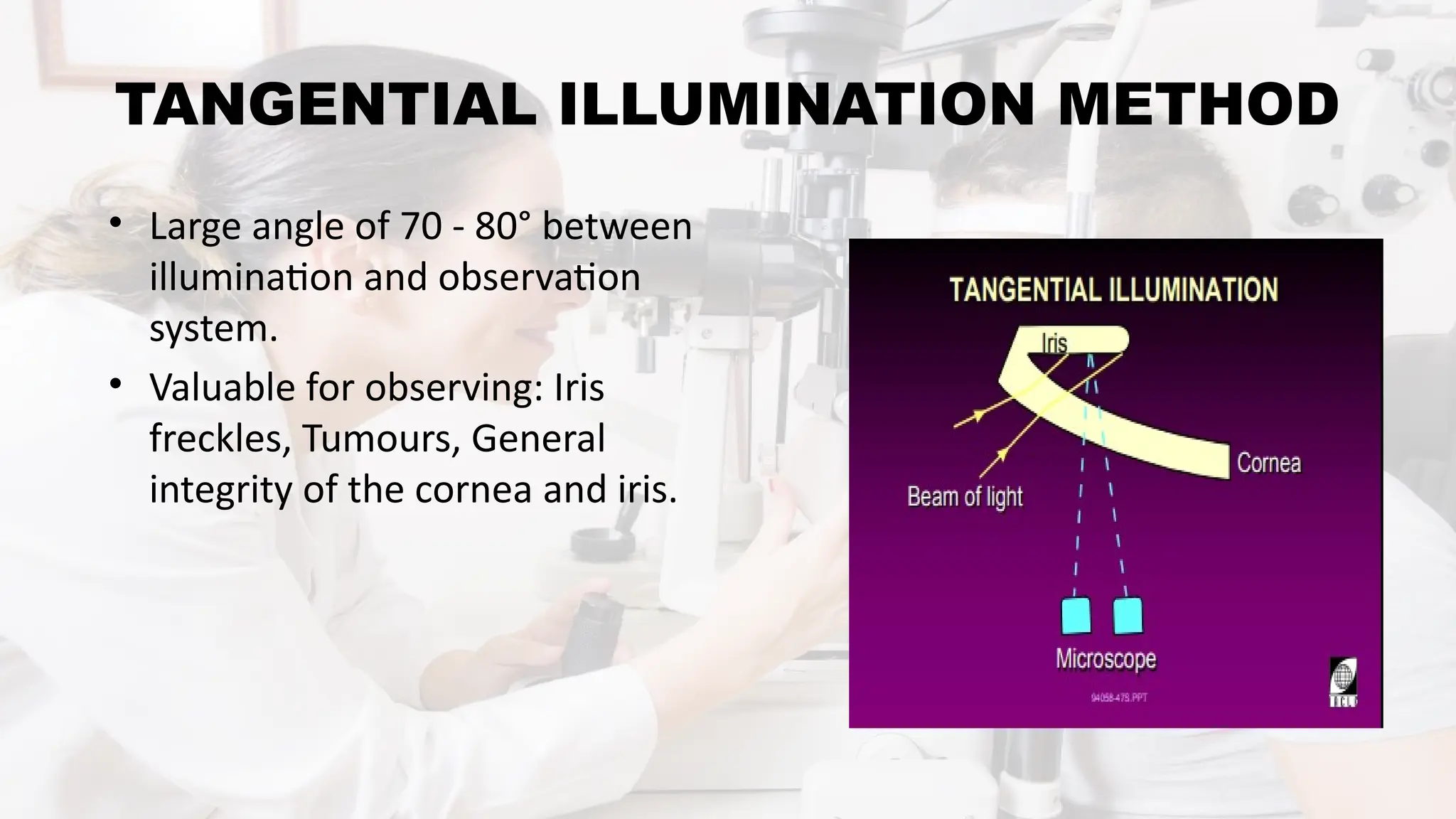
SLIT LAMP
BIOMICROSCOPE
MS. MEGHNA VERMA
Department of Optometry
RAMA University
CONTENTS
• INTRODUCTION
• PARTS OF SLIT LAMP
• PREPARATION
• TECHNIQUE
• METHODS OF ILLUMINATION
INTRODUCTION
• In 1911, GULLSTRAND is credited with the invention of slit
lamp.
• Slit lamp is the most important equipment in the present day.
• Modern slit lamp with its additional device provides qualitative
and quantitative measurements and photographic records.
• Qualitative measurements - magnified view of every part of
the eye from cornea to retina.
• Quantitative measurement - IOP, endothelial cells counts,
pupil size, corneal thickness, anterior chamber depth.
PRINCIPLE
• A “slit” beam of very bright light produced by lamp.
• This beam is focused on to the eye which is then viewed
under magnification with a microscope.
ILLUMIATIO
N SYSTEM
MECHANICAL
SYSTEM
OBSERVATIO
N SYSTEM
PARTS OF SLIT LAMP
OBSERVATION SYSTEM (MICROSCOPE)
• It is a compound microscope.
• It consists of two optical elements i.e. an objective and an eyepiece.
• It provides to the observer an enlarged view of a near object.
• An objective lens consists of two plano-convex lens, with their
convexities put together, providing a composite power of +22 Dioptre.
• An eyepiece lens is of + 10 D.
• In slit lamp, uses a pair of prism is placed between the objective and
eyepiece, to re-invert the image.
• Prism is used to overcome the problem of inverted image, produced by
compound microscope.
ILLUMINATION SYSTEM
• Illumination system is provide a bright and fine focused adjustable slit of
light at the eye.
LIGHT SOURCE
CONDENSER
LENS SYSTEM
SLIT &
DIAPHRAGM
FILTERS
PROJECTION
LENS MIRRORS
& PRISMS
LIGHT SOURCE –
• Nitra lamp, arc lamp, mercury vapour lamp and halogen lamp.
• It provides an illumination of 2X 10 to 4 X 10 LUX.
CONDENSER LENS SYSTEM
• Two plano-convex lenses, their convexities put together.
SLIT AND DIAPHRAGMS
• Height & width of slit can be varied by knobs.
• It provides, examination of fundus and angle of anterior chamber.
FILTERS
• Cobalt blue and red free filters.
PROJECTION LENS
• It forms an image of the slit.
• It keeps lesser the aberration,
better the image quality.
MIRRORS/ PRISMS
• Normally arranged vertical axis with either mirror/ prism, reflecting the
light along horizontal axis.
MECHANICAL SYSTEM
1. JOYSTICK ARRANGEMENT
2. UP AND DOWN MOVEMENT ARRANGEMENT
3. PATIENT SUPPORT ARRANGEMENT
4. FIXATION TARGET
5. MECHANICAL COUPLING
JOYSTICK ARRANGEMENT
• Movement of the microscope and illumination system towards or away
from the eye and from side to side is usually achieved.
UP AND DOWN MOVEMENT ARRANGEMENT
• It moves the whole illumination and viewing system up and down
relative to chin rest.
PATIENT SUPPORT ARRANGEMENT
• A vertically movable chin rest and adjust the height of the table.
FIXATION TARGET
• A movable fixation target greatly facilitates the examination under some
conditions.
MECHANICAL COUPLING
• It not only provides a support but also a coupling of microscope and
illumination system along a common axis of rotation.
BEGINNING OF SLIT LAMP EXAMINATION
Examination
should be in semi
dark room
Start with the
diffuse
illumination
Start with the
Low
magnification
Low to medium
to high
Do not use any
medication
TECHNIQUE OF BIOMICROSCOPY
• The patient should be positioned
comfortably in front of the slit lamp with
his/her chin resting on the chin rest and
forehead against to head rest.
PATIENT
ADJUSTME
NT
•The microscope and illumination system should be
aligned with the patient’s eye to be examined.
•The height can be varied using knobs as per patients
height.
•Fixation target should be placed at the required
position.
INSTRUMENT
ADJUSTMENT
METHODS OF ILLUMINATION
• Diffuse illumination
• Direct illumination
• Indirect illumination
• Retro illumination
• Sclerotic scatter
• Oscillating illumination
DIFFUSE ILLMINATION
• 45 degree angle between light and
microscope
• Fully open slit
• Diffusing filter
• Variable magnification (low to
medium to high)
• Variable illumination (medium to
high)
• Overall view of - lids and lashes,
conjunctiva, cornea, sclera, iris, pupil
DIRECT ILLUMINATION
• Vary angle of illumination
• Low to high magnification
• Vary width and height of light
source
• Variable illumination
Optic Section:
• Narrow, focused slit.
• Corneal curvature, corneal thickness.
Parallelepiped:
• wider beam
• observe corneal stroma,
epithelial breakdown, lens
surface and endothelium.
Conical Beam:
• Small, bright, circular light
source.
• Use with high magnification.
• Used for observation of flare
and cells in the anterior
chamber.
INDIRECT ILLUMINATION
• Observation and illumination systems
are not focused at the same point.
• Vary angle of illumination
• Slit beam is offset
• Vary beam width
• Low to high magnification
• Valuable for observing: Iris pathology,
Epithelial vesicles, Epithelial erosions,
Iris sphincter.
RETRO ILLUMINATION
• Vary angle of illumination
• Moderately wide beam
• Slit beam is offset
• Medium to high magnification
• The cornea is illuminated by light
reflected from the iris, crystalline lens or
fundus.
• Valuable for observing: Epithelial
oedema, Microcysts, Vacuoles,
Dystrophies, lens opacities, Contact lens
deposits.
SPECULAR REFLECTION
• The angle of incident light is equal
to the angle of reflected light.
• Valuable for observing: Endothelial
cells, Tear film debris, Tear film lipid
layer.
SCLEROTIC SCATTER
• Illumination of the cornea by
total internal reflection of a wide
angle light source.
• The light beam is directed at the
limbal region while observing
the cornea.
• Valuable for observing: Localized
epithelial oedema, Corneal scars,
Foreign bodies in the cornea.
TANGENTIAL ILLUMINATION METHOD
• Large angle of 70 - 80° between
illumination and observation
system.
• Valuable for observing: Iris
freckles, Tumours, General
integrity of the cornea and iris.
CLINICAL USES
• Anterior segment evaluation.
• Tear evaluation.
• Measures corneal thickness.
• Evaluate intra ocular pressure.
• Examine angle of anterior chamber.
• Removal of foreign particle.
• Contact lens fitting.
• Evaluation of post fitting contact lens.
• Epilation of lashes.
DIAGNOSTIC SLIT LAMP BIO-MICROSCOPE.pptx

DIAGNOSTIC SLIT LAMP BIO-MICROSCOPE.pptx

  • 1.
    SLIT LAMP BIOMICROSCOPE MS. MEGHNAVERMA Department of Optometry RAMA University
  • 2.
    CONTENTS • INTRODUCTION • PARTSOF SLIT LAMP • PREPARATION • TECHNIQUE • METHODS OF ILLUMINATION
  • 3.
    INTRODUCTION • In 1911,GULLSTRAND is credited with the invention of slit lamp. • Slit lamp is the most important equipment in the present day. • Modern slit lamp with its additional device provides qualitative and quantitative measurements and photographic records. • Qualitative measurements - magnified view of every part of the eye from cornea to retina. • Quantitative measurement - IOP, endothelial cells counts, pupil size, corneal thickness, anterior chamber depth.
  • 5.
    PRINCIPLE • A “slit”beam of very bright light produced by lamp. • This beam is focused on to the eye which is then viewed under magnification with a microscope.
  • 6.
  • 8.
    OBSERVATION SYSTEM (MICROSCOPE) •It is a compound microscope. • It consists of two optical elements i.e. an objective and an eyepiece. • It provides to the observer an enlarged view of a near object. • An objective lens consists of two plano-convex lens, with their convexities put together, providing a composite power of +22 Dioptre. • An eyepiece lens is of + 10 D. • In slit lamp, uses a pair of prism is placed between the objective and eyepiece, to re-invert the image. • Prism is used to overcome the problem of inverted image, produced by compound microscope.
  • 11.
    ILLUMINATION SYSTEM • Illuminationsystem is provide a bright and fine focused adjustable slit of light at the eye. LIGHT SOURCE CONDENSER LENS SYSTEM SLIT & DIAPHRAGM FILTERS PROJECTION LENS MIRRORS & PRISMS
  • 14.
    LIGHT SOURCE – •Nitra lamp, arc lamp, mercury vapour lamp and halogen lamp. • It provides an illumination of 2X 10 to 4 X 10 LUX. CONDENSER LENS SYSTEM • Two plano-convex lenses, their convexities put together. SLIT AND DIAPHRAGMS • Height & width of slit can be varied by knobs. • It provides, examination of fundus and angle of anterior chamber.
  • 15.
    FILTERS • Cobalt blueand red free filters. PROJECTION LENS • It forms an image of the slit. • It keeps lesser the aberration, better the image quality. MIRRORS/ PRISMS • Normally arranged vertical axis with either mirror/ prism, reflecting the light along horizontal axis.
  • 16.
    MECHANICAL SYSTEM 1. JOYSTICKARRANGEMENT 2. UP AND DOWN MOVEMENT ARRANGEMENT 3. PATIENT SUPPORT ARRANGEMENT 4. FIXATION TARGET 5. MECHANICAL COUPLING
  • 18.
    JOYSTICK ARRANGEMENT • Movementof the microscope and illumination system towards or away from the eye and from side to side is usually achieved. UP AND DOWN MOVEMENT ARRANGEMENT • It moves the whole illumination and viewing system up and down relative to chin rest. PATIENT SUPPORT ARRANGEMENT • A vertically movable chin rest and adjust the height of the table.
  • 19.
    FIXATION TARGET • Amovable fixation target greatly facilitates the examination under some conditions. MECHANICAL COUPLING • It not only provides a support but also a coupling of microscope and illumination system along a common axis of rotation.
  • 20.
    BEGINNING OF SLITLAMP EXAMINATION Examination should be in semi dark room Start with the diffuse illumination Start with the Low magnification Low to medium to high Do not use any medication
  • 21.
    TECHNIQUE OF BIOMICROSCOPY •The patient should be positioned comfortably in front of the slit lamp with his/her chin resting on the chin rest and forehead against to head rest. PATIENT ADJUSTME NT •The microscope and illumination system should be aligned with the patient’s eye to be examined. •The height can be varied using knobs as per patients height. •Fixation target should be placed at the required position. INSTRUMENT ADJUSTMENT
  • 22.
    METHODS OF ILLUMINATION •Diffuse illumination • Direct illumination • Indirect illumination • Retro illumination • Sclerotic scatter • Oscillating illumination
  • 23.
    DIFFUSE ILLMINATION • 45degree angle between light and microscope • Fully open slit • Diffusing filter • Variable magnification (low to medium to high) • Variable illumination (medium to high) • Overall view of - lids and lashes, conjunctiva, cornea, sclera, iris, pupil
  • 24.
    DIRECT ILLUMINATION • Varyangle of illumination • Low to high magnification • Vary width and height of light source • Variable illumination
  • 25.
    Optic Section: • Narrow,focused slit. • Corneal curvature, corneal thickness.
  • 26.
    Parallelepiped: • wider beam •observe corneal stroma, epithelial breakdown, lens surface and endothelium.
  • 27.
    Conical Beam: • Small,bright, circular light source. • Use with high magnification. • Used for observation of flare and cells in the anterior chamber.
  • 28.
    INDIRECT ILLUMINATION • Observationand illumination systems are not focused at the same point. • Vary angle of illumination • Slit beam is offset • Vary beam width • Low to high magnification • Valuable for observing: Iris pathology, Epithelial vesicles, Epithelial erosions, Iris sphincter.
  • 29.
    RETRO ILLUMINATION • Varyangle of illumination • Moderately wide beam • Slit beam is offset • Medium to high magnification • The cornea is illuminated by light reflected from the iris, crystalline lens or fundus. • Valuable for observing: Epithelial oedema, Microcysts, Vacuoles, Dystrophies, lens opacities, Contact lens deposits.
  • 30.
    SPECULAR REFLECTION • Theangle of incident light is equal to the angle of reflected light. • Valuable for observing: Endothelial cells, Tear film debris, Tear film lipid layer.
  • 31.
    SCLEROTIC SCATTER • Illuminationof the cornea by total internal reflection of a wide angle light source. • The light beam is directed at the limbal region while observing the cornea. • Valuable for observing: Localized epithelial oedema, Corneal scars, Foreign bodies in the cornea.
  • 32.
    TANGENTIAL ILLUMINATION METHOD •Large angle of 70 - 80° between illumination and observation system. • Valuable for observing: Iris freckles, Tumours, General integrity of the cornea and iris.
  • 33.
    CLINICAL USES • Anteriorsegment evaluation. • Tear evaluation. • Measures corneal thickness. • Evaluate intra ocular pressure. • Examine angle of anterior chamber. • Removal of foreign particle. • Contact lens fitting. • Evaluation of post fitting contact lens. • Epilation of lashes.