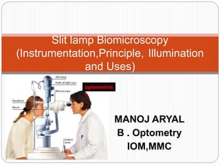
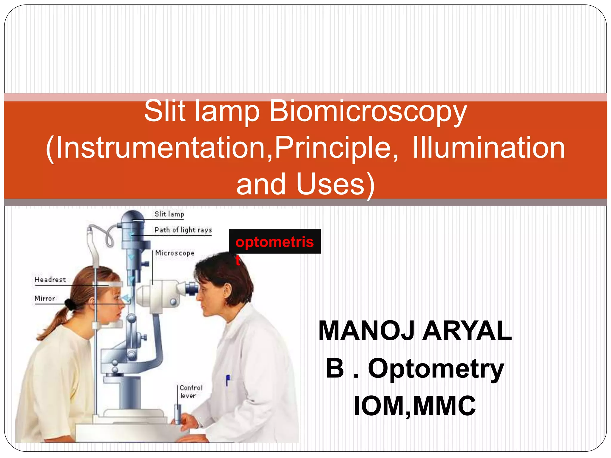
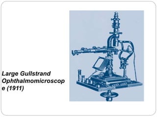
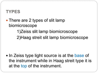
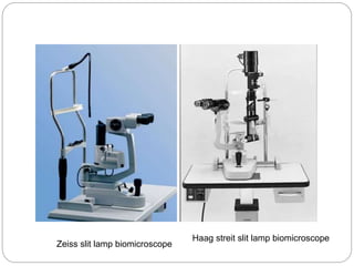
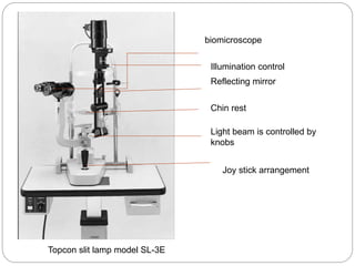
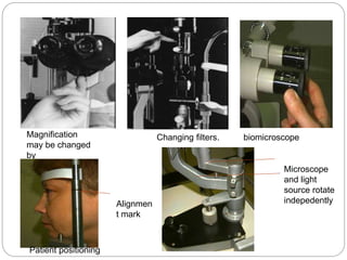
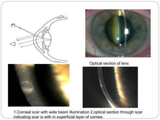
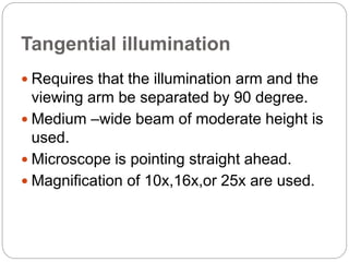
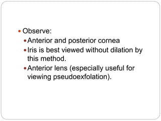
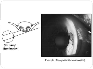
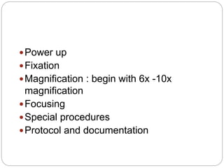
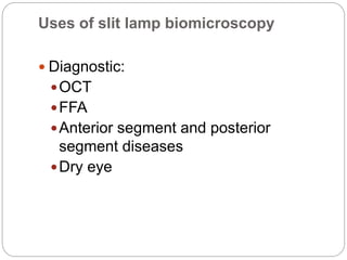
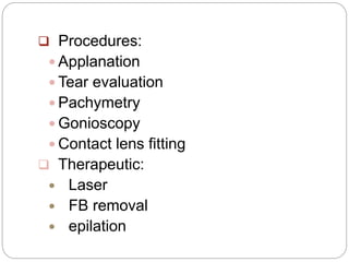
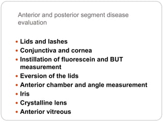
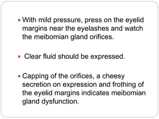
. Introduction Biomicroscope derives its name from the fact that it enables the practitioner to observe the living tissue of eye under magnification. It not only provides magnified view of every part of eye but also allows quantitative measurements and photography of every part for documentation.
3. • The lamp facilitates an examination which looks at anterior segment, or frontal structures, of the human eye, which includes the –Eyelid –Cornea –Sclera –Conjunctiva –Iris –Aqueous –Natural crystalline lens and –Anterior vitreous.
4. Important historical landmarks De Wecker 1863 devised a portable ophthalmomicroscope . Albert and Greenough 1891,developed a binocular microscope which provided stereoscopic view. Gullstrand ,1911 introduced the illumination system which had for the first time a slit diapharm in it Therefore Gullstrand is credited with the invention of slit lamp.
all about slit lamp. special features, basic designs, principal of observation system, principal of illumination, animations are done for the beginners for easy understanding,pitfalls.
SLIT LAMP AND ITS DIFFERENT ILLUMINATION TECHNIQUES.pptxAbhishek Kashyap
This presentation explains in detail about different illumination techniques and filters used in slit lamp examination and the procedure to perform slit lamp examination.
astigmatism
AstigmatismAstigmatism Walter Huang, ODWalter Huang, OD Yuanpei UniversityYuanpei University Department of OptometryDepartment of Optometry
2. DefinitionDefinition When parallel rays of light enter the eyeWhen parallel rays of light enter the eye ((with accommodation relaxedwith accommodation relaxed) and do) and do notnot come to a single point focus on or nearcome to a single point focus on or near the retinathe retina
3. OpticsOptics Power in thePower in the horizontalhorizontal plane projects aplane projects a verticalvertical focal line imagefocal line image Power in thePower in the verticalvertical plane projects aplane projects a horizontalhorizontal focal line imagefocal line image
4. OpticsOptics Refraction of light taking place at a toricRefraction of light taking place at a toric surface: the conoid of Sturmsurface: the conoid of Sturm
TONOMETRY • Tonometry is the procedure performed to determine the intraocular pressure (IOP).
3. CLASSIFICATION TONOMETRY DIRECT INDIRECT Indentation Applanation Manometer
4. APPLANATION Contact Non-contact Goldmann Perkins Air-puff Pulse air
5. INDENTATION TONOMETER • It is based on fundamental fact that plunger will indent a soft eye more than hard eye. • The indentation tonometer in current use is that of Schiotz . • It was devised in 1905 and continued to refine it through 1927.
6. PROCEDURE • Patient should be anaesthetising with 4% lignocaine or 0.5% proparacaine. • with the patient in supine position, looking up at a fixation target while examiners separates the lids and lower the tonometer plate to rest on the cornea so that plunger is free to move. •
1. Introduction Gross anatomy Layers Blood supply, drainage and nerve supply
2. INTRODUCTION • Sclera forms posterior 5/6th of external tunic , connective tissue coat of eyeball. • it continues with duramater and cornea • Its whole surface covered by tenon’s capsule • Anteriorly covered by- bulbar conjunctiva • Inner surface lies in contact with choroid • With a potential suprachoroidal space in between
3. Equa THICKNESS OF SCLERA
4. • Thickness varies with individual, with age • Thinner- children, elder, F> M • Thickest posteriorly • Gradually becomes thinner when traced anteriorly • Thin at insertion of extraocular muscle
The pupil is an opening located in the center of the iris that allows light to enter the retina. • Its function is to control the amount of light entering the eye and it does this via contraction (miosis) and dilation (mydriasis) under the influence of the autonomic nervous system
3. • The iris is a contractile structure, consisting mainly of smooth muscle, surrounding the pupil. Light enters the eye through the pupil, and the iris regulates the amount of light by controlling the size of the pupil.
4. The iris contains two groups of smooth muscles: a circular group called the sphincter pupillae. and a radial group called the dilator pupillae.
5. Parasympathetic pathway • First Order – Retina to Pretectal Nucleus in B/S (at level of Superior colliculus) Second Order – Pretectal nucleus to E/W nucleus (bilateral innervation!) Third Order – E/W nucleus to Ciliary Ganglion Fourth Order – Ciliary Ganglion to Sphincter pupillae (via short ciliary nerves) • • •
The tear film constitutes Three layers :- An outermost lipid (oily) layer An aqueous (watery) layer that makes up 90% of the tear film volume; and A mucin layer that coats the corneal surface.
3. To form smooth optical surface on cornea. To keep the surface of cornea & conjunctiva moist It serve as lubricant It transfer oxygen Provide antibacterial action Wash debris out It provides a pathway for WBC in case of injury
4. Functions of lipid layer Retards evaporation of tear film Prevents the overflow of tears
5. Function of Aqueous Layer Flushes, buffers and lubricates the corneal surface Delivers oxygen and other nutrients to the corneal surface Wash out debris Delivers antibacterial enzymes and antibodies such as lysozyme.
6. Functions of Mucin Layer Spreads tears over corneal surface. Protects the cornea against foreign substances . Makes corneal surface smooth by filling in surface irregularities
Introduction Transparent,avascular,watch-glass like structure. Forms 1/6th part of outer fibrous coat (Sclera) It is the major refracting surface of the eye
3. Dimensions + Avg horizontal dia =11.75 mm (ant surface) + Avg vertical dia = 11 mm (ant surface ) + Avg dia (post surface)= 11.5 mm + Thickness(centre) =0.52mm + Thickness(peripheral) = 0.67mm + Radius of curvature (ant surface) = 7.8mm + Radius of curvature (post surface)= 6.5mm + Refractive power (ant surface) = +48D + Refractive power(post surface)= - 5D + R.I = 1.376
4. Histology + Epithelium + Bowman’s membrane + Stroma + Dua’s layer + Descemet’s membrane + Endothelium
5. Epithelium + Made up of stratified squamous epithelium + Thickness - 50-90 um + 5-6 layers of cells + Regenerative, entire epithelial layer is replaced every 6-8 days + Made up of 3 types of cells - basal,wing, flattened cells + Cells are attached by to each other by means of desmosomes & maculi occludents
6. Bowman’s membrane + Acellular,Non regenerative + Made up of condensed collagen fibrils. + Thickness - 12um + Resistant to infection & injury.
LIMBUS… • The limbus forms the border between the transparent cornea and opaque sclera, contains the pathways of aqueous humour outflow, and is the site of surgical incisions for cataract and glaucoma
2. Anatomical Limbus: Circumcorneal transitional zone of the conjunctivocorneal & corneoscleral junction Conjunctivo-corneal junction: • Bulbar conjunctiva is firmly adherent to underlying structures • Substantia propria of the conjunctiva stops here but its epithelium continues with that of the cornea. Sclero-corneal junction: • Transparent corneal lamellae become continuous • With the oblique, circular and opaque fibres of sclera
3. CONTINUE…. • In the area near limbus, the conjunctiva, tenon’s capsule & the episcleral tissue are fused into a dense tissue which is strongly adherent to corneo scleral junction.It is preferred site for obtaining a firm hold of the eyeball during ocular surgery. • The limbus is a common site for the occurrence of corneal epithelial neoplasm. • The Limbus contains radially oriented fibrovascular ridge known as the palisades of Vogt that may harbour a stem cell population. The palisades of Vogt are more common in the superior and inferior quadrants around the eye
Diagnosis, Management, and Surgery by Adam J. Cohen, Michael Mercandetti & Brian G. Brazzo. The dry eye , a practical approach by Sudi Patel & Kenny J Blades. Jack J Kanski’s clinical ophthalmology Clinical Anatomy of the Eye by Richard S. Snell & Michael A. Lemp.
3. It is concerned with the tear formation & transport. Lacrimal passage includes : Lacrimal gland Conjunctival sac Lacrimal puncta Lacrimal canaliculi Lacrimal sac Nasolacrimal duct
4. The following components of the lacrimal apparatus are discussed : Embryology Osteology Secretory system Excretory system Physiology
5. Ectodermal origin Solid epithelial buds(first 2 months) Supero
Extraocular musles(EOM) They are six in number Four recti: Superior rectus Inferior rectus Medial rectus Lateral rectus Two oblique muscles: Superior oblique Inferior oblique
3. SUPERIOR RECTUS MUSCLE . Origin Superior part of common annular tendon of Zinn Course Passes anterolaterally beneath the levator At 23 degrees with the globe ‘s AP axis Pierces Tenon s capsule Insertion into sclera by flat tendinous 10 mm broad insertion 7.7 mm behind sclero-corneal junction. 42 mm long 9 mm wide
4. Nerve supply Sup division of 3rd N Blood Supply Lateral Muscular br. of Ophthalmic A APPLIED: SR loosely bound to LPS muscle. • During SR resection- eyelid may be pulled forward narr owing palpebral fissure • In hypotropia pseudoptosis may be present Origin of SR and MR are closely attached to the dural sheat h of the optic nerve pain during upward & inward movements of the globe in RETROBULBAR NEURITIS
Diseases of sclera
2. anatomy • Sclera posterior 5/6th opaque part of the external fibrous tunic of the eyeball.
3. • outer surface }covered by Tenon's capsule. • anterior part } covered by bulbar conjunctiva.
4. Its inner surface lies in contact with choroid with a potential suprachoroidal space in between
5. Thickness of sclera. • thinner }children and in females Sclera • thickest} posteriorly (1mm) • gradually becomes thin when traced anteriorly. • thinnest } insertion of extraocular muscles (0.3 mm). • Lamina cribrosa is a sieve-like sclera from which fibres of optic nerve pass.
6. Apertures of sclera • Anterior • Anterior ciliary vessels • Middle • four vortex veins (vena verticosae) • Posterior • Optic nerve • Long & short ciliary nerves
7. Layers of sclera sclera episclera Sclera proper Lamina fusca thin, dense vascularised layer of connective tissue fibroblasts, macrophages and lymphocytes avascular structure dense bundles of collagen fibres. innermost blends with suprachoroidal and supraciliary laminae of the uveal tract. brownish in colour presence of pigmented cells.
Main physiologic function of cornea is to act as a major refracting medium, so that a clear retinal image is formed. • Normal corneal transparency is result of • 1.anatomical factor such as uniform and regular arrangement of corneal epithelium, peculiar arrangement of corneal lamella and corneal vascularity 2.Physiological factor [ie] relative state of corneal dehydration.
3. • Therefore, any process which upsets the anatomy or physiology of cornea will cause LOSS OF TRANSPARENCY to some degree.
4. FACTORS AFFECTING CORNEAL TRANSPARENCY • CORNEAL EPITHELIUM &TEAR FLIM • ARRANGEMENT OF STROMAL LAMELLA • CORNEAL VASCULARIZATION • CORNEAL HYDRATION • CELLULAR FACTORS AFFECTING TRANSPARENCY
CONJUNCTIVA: ANATOMY , PHYSIOLOGY, SYMPTOMATOLOGY AND CLASSIFICATION Pranay Shinde DNB Resident Deen Dayal Upadhyay Hospital,New Delhi
2. ANATOMY It is the mucous membrane covering the under surface of the lids and anterior part of the eyeball upto the cornea.
3. Parts of conjunctiva • Palpebral; covering the lids—firmly adherent. • Forniceal; covering the fornices—loose—thrown into folds. • Bulbar; covering the eyeball—loosely attached except at limbus.
4. Palpebral conjunctiva • Subtarsal sulcus 2mm from posterior edge of the lid margin. • Richly vascular. • Extremely thin. • Strongly bound to the tarsal plate.
5. Palpebral conjunctiva is subdivided into three parts: 1)Marginal 2)Tarsal 3)Orbital
6. Conjunctival fornices • Transitional region between palpebral and bulbar conjunctivae. • Superior fornix 10 mm from limbus. • Inferior fornix 8 mm from limbus. • Lateral fornix 14mm from limbus. • Medially absent. • Ducts of lacrimal glands open into lateral part of superior fornix.
q Colour Vision Deficiency Presented by : Optometrist (intern) Asma Al-Jroudi Saudi Arabia, Riyadh, King Abdulaziz University Hospital 30 Dec 14
2. • What Is Color Vision Deficiency? • Causes Of Color Vision Deficiency • Types Of Color Vision Deficiency • Tretments Of Color Vision Deficiency • Ishihara’s Test • Conclousion
3. What is Colour Blindness? • Color blindness, or color vision deficiency, is the inability or decreased ability to see color, or perceive color differences, under normal lighting conditions. •This condition results from an absence of color- sensitive pigment in the cone cells of the retina, the nerve layer at the back of the eye.
4. What is Colour Blindness? • Cones are the coulored light receptors in back of the eye: Red light receptors, Blue light receptors and Green light receptors. • Colour blindness occurs when one or more of the cone types are defected.
5. Causes of Color Blindness • Genetic: Many more men are affected than women. • Acquired : Chronic illness, Accidents, Medications and Age.
ANATOMY & PHYSIOLOGY Lecturer: Tatyana V. Ryazantseva
2. Outer eye: Eyelids The eyelids fulfill two main functions: protection of the eyeball secretion, distribution and drainage of tears
3. Lid movement The levator extends from an attachment at the orbital apex to attachments at the tarsal plate and skin. ● The lids are securely attached at either end to the bony orbital margin by the medial and lateral palpebral ligaments. Trauma to the medial ligament causes the lid to flop forward and laterally, impairing function and cosmesis.
4. Innervation - Sensory innervation is from the trigeminal (fifth) cranial nerve, via the ophthalmic division (upper lid) and maxillary division (lower lid). - The orbicularis oculi is innervated by the facial (seventh) nerve. - The levator muscle in the upper lid is supplied by the oculomotor (third) nerve.
5. Blood supply and lymphatics The eyelids are supplied by an extensive network of blood vessels which form an anastomosis between branches derived from the external carotid artery via the face and from the internal carotid artery via the orbit.
6. Blood supply and lymphatics Lymphatic fluid drains into the preauricular and submandibular nodes. Preauricular lymphadenopathy is a useful sign of infective eyelid swelling (especially viral).
Anatomy and Physiology of Aqueous Humor Sumit Singh Maharjan
2. Anatomy
3. Angle of anterior chamber
4. Angle of the Anterior chamber
5. Gonioscopic grading of Angle
6. Aqueous Outflow system
7. Trabecular meshwork
8. Functions of Aqueous Humor • Maintenance of Intraocular pressure • Metabolic role cornea lens vitreous and retina • Optical function • Clearing function
9. Physicochemical properties • volume: 0.31ml (0.25ml in Ant. Chamber and 0.06 in post chamber) • Refractive index: 1.336 • Density: slightly greater than water, its viscocity is 1.025-1.040 • Osmotic pressure: slightly hyperosmotic to plasma by 3-5mosm/l • PH: 7.2 • Rate of formation: 2-2.5microliter/min
10. Biochemical composition • Water: 99.9% • Proteins: 5-16mg/100ml • Amino acids: aqueous/plasma concentration varies from 0.08-3.14 • Non colloidal constituents: conc. of ascorbate, pyruvate, lactate in higher am
Vitreous humour
1. Vitreous Humour
2. General features Vitreous humour is an inert ,transparent , colourless, jellylike, hydrophilic gel that serves the optical functions and also acts as important supporting structures for the eyeball. The vitreous cavity is bounded by anteriorly by the lens and ciliary body and posteriorly by the retina Its weighs nearly 4g Vitreous is an extacellular material composed of approximately 99 per cent water
3. Structure The vitreous body is the largest and simplest connective tissue present as a single piece in the human body Divided into three parts- 1. The hyaloid layer or membrane 2. The cortical vitreous and 3. The medullary vitreous
Vitamins all
1. Vitamins. Definition - Organic compound required in small amounts. Vitamin A Vitamin B1, B2, B3, B5, B6, B7, B9, B12 Vitamin D Vitamin E Vitamin K A few wordsabout each.
2. Sourcesin diet - Many plants(photoreceptors), also meat, especially liver. Fat soluble, so you can get too much, or too littleif absorption isaproblem. Vitamin A - Retinol Retinol (vitamin A) Someuses: Vision (11-cis-retinol bound to rhodopsin detectslight in our eyes). Regulating genetranscription (retinoic acid receptorson cell nuclei arepart of a system for regulating transcription of mRNAsfor anumber of genes).
Tear film
1. TEAR FILM
2. The outer most layer of the cornea. It is the exposed part of the eyeball. FUNCTION It provide smooth optical surface It serves to keep the surface of cornea and conjunctiva moist. It serves as a lubricant for the preocular surface and lids It transfer oxygen from the air to the cornea Prevent infection due to the presence of antibacterial substance like lysozymes,and other protein. It wash away debris and irritants Provides pathway to WBC in case of injury.
3. LAYERS OF TEAR FILM It consist of three layers: 1.Lipid layer 2.Aqueous layer 3.Mucoid layer 1.LIPID LAYER
Synthesis
1. Synthesis of Fatty acids
2. Fatty acids are synthesized mainly by de novo synthetic pathway operating in the cytoplasm . It is called as extramitochondrial or cytoplasmic fatty acid synthase system. The major fatty acids synthesised de novo is palmitic acid, the 16C saturated fatty acids. The process occurs in liver ,adipose tissue ,kidney and mammary glands. Fatty acids synthase (FAS) complex : This system exists as multi- enzyme complex .This enzyme form a dimer with identical subunits. Each subunits of the complex is organised into 3 domains with 7 enzyme.
More from C L GUPTA EYE INSTITUTE MORADABAD UTTER PRADESH (20)
Antibiotic Stewardship by Anushri Srivastava.pptxAnushriSrivastav
Stewardship is the act of taking good care of something.
Antimicrobial stewardship is a coordinated program that promotes the appropriate use of antimicrobials (including antibiotics), improves patient outcomes, reduces microbial resistance, and decreases the spread of infections caused by multidrug-resistant organisms.
WHO launched the Global Antimicrobial Resistance and Use Surveillance System (GLASS) in 2015 to fill knowledge gaps and inform strategies at all levels.
ACCORDING TO apic.org,
Antimicrobial stewardship is a coordinated program that promotes the appropriate use of antimicrobials (including antibiotics), improves patient outcomes, reduces microbial resistance, and decreases the spread of infections caused by multidrug-resistant organisms.
ACCORDING TO pewtrusts.org,
Antibiotic stewardship refers to efforts in doctors’ offices, hospitals, long term care facilities, and other health care settings to ensure that antibiotics are used only when necessary and appropriate
According to WHO,
Antimicrobial stewardship is a systematic approach to educate and support health care professionals to follow evidence-based guidelines for prescribing and administering antimicrobials
In 1996, John McGowan and Dale Gerding first applied the term antimicrobial stewardship, where they suggested a causal association between antimicrobial agent use and resistance. They also focused on the urgency of large-scale controlled trials of antimicrobial-use regulation employing sophisticated epidemiologic methods, molecular typing, and precise resistance mechanism analysis.
Antimicrobial Stewardship(AMS) refers to the optimal selection, dosing, and duration of antimicrobial treatment resulting in the best clinical outcome with minimal side effects to the patients and minimal impact on subsequent resistance.
According to the 2019 report, in the US, more than 2.8 million antibiotic-resistant infections occur each year, and more than 35000 people die. In addition to this, it also mentioned that 223,900 cases of Clostridoides difficile occurred in 2017, of which 12800 people died. The report did not include viruses or parasites
VISION
Being proactive
Supporting optimal animal and human health
Exploring ways to reduce overall use of antimicrobials
Using the drugs that prevent and treat disease by killing microscopic organisms in a responsible way
GOAL
to prevent the generation and spread of antimicrobial resistance (AMR). Doing so will preserve the effectiveness of these drugs in animals and humans for years to come.
being to preserve human and animal health and the effectiveness of antimicrobial medications.
to implement a multidisciplinary approach in assembling a stewardship team to include an infectious disease physician, a clinical pharmacist with infectious diseases training, infection preventionist, and a close collaboration with the staff in the clinical microbiology laboratory
to prevent antimicrobial overuse, misuse and abuse.
to minimize the developme
Global launch of the Healthy Ageing and Prevention Index 2nd wave – alongside...ILC- UK
The Healthy Ageing and Prevention Index is an online tool created by ILC that ranks countries on six metrics including, life span, health span, work span, income, environmental performance, and happiness. The Index helps us understand how well countries have adapted to longevity and inform decision makers on what must be done to maximise the economic benefits that comes with living well for longer.
Alongside the 77th World Health Assembly in Geneva on 28 May 2024, we launched the second version of our Index, allowing us to track progress and give new insights into what needs to be done to keep populations healthier for longer.
The speakers included:
Professor Orazio Schillaci, Minister of Health, Italy
Dr Hans Groth, Chairman of the Board, World Demographic & Ageing Forum
Professor Ilona Kickbusch, Founder and Chair, Global Health Centre, Geneva Graduate Institute and co-chair, World Health Summit Council
Dr Natasha Azzopardi Muscat, Director, Country Health Policies and Systems Division, World Health Organisation EURO
Dr Marta Lomazzi, Executive Manager, World Federation of Public Health Associations
Dr Shyam Bishen, Head, Centre for Health and Healthcare and Member of the Executive Committee, World Economic Forum
Dr Karin Tegmark Wisell, Director General, Public Health Agency of Sweden
How many patients does case series should have In comparison to case reports.pdfpubrica101
Pubrica’s team of researchers and writers create scientific and medical research articles, which may be important resources for authors and practitioners. Pubrica medical writers assist you in creating and revising the introduction by alerting the reader to gaps in the chosen study subject. Our professionals understand the order in which the hypothesis topic is followed by the broad subject, the issue, and the backdrop.
https://pubrica.com/academy/case-study-or-series/how-many-patients-does-case-series-should-have-in-comparison-to-case-reports/
Navigating Challenges: Mental Health, Legislation, and the Prison System in B...Guillermo Rivera
This conference will delve into the intricate intersections between mental health, legal frameworks, and the prison system in Bolivia. It aims to provide a comprehensive overview of the current challenges faced by mental health professionals working within the legislative and correctional landscapes. Topics of discussion will include the prevalence and impact of mental health issues among the incarcerated population, the effectiveness of existing mental health policies and legislation, and potential reforms to enhance the mental health support system within prisons.
CHAPTER 1 SEMESTER V PREVENTIVE-PEDIATRICS.pdfSachin Sharma
This content provides an overview of preventive pediatrics. It defines preventive pediatrics as preventing disease and promoting children's physical, mental, and social well-being to achieve positive health. It discusses antenatal, postnatal, and social preventive pediatrics. It also covers various child health programs like immunization, breastfeeding, ICDS, and the roles of organizations like WHO, UNICEF, and nurses in preventive pediatrics.
QA Paediatric dentistry department, Hospital Melaka 2020Azreen Aj
QA study - To improve the 6th monthly recall rate post-comprehensive dental treatment under general anaesthesia in paediatric dentistry department, Hospital Melaka
Dehradun ❤CALL Girls 8901183002 ❤ℂall Girls IN Dehradun ESCORT SERVICE❤
Slit lump biomicroscope
1. MANOJ ARYAL
B . Optometry
IOM,MMC
Slit lamp Biomicroscopy
(Instrumentation,Principle, Illumination
and Uses)
optometris
t
2. Introduction
Biomicroscope derives its name from the
fact that it enables the practitioner to
observe the living tissue of eye under
magnification.
It not only provides magnified view of every
part of eye but also allows quantitative
measurements and photography of every
part for documentation.
3. • The lamp facilitates an examination
which looks at anterior segment, or
frontal structures, of the human eye,
which includes the
–Eyelid
–Cornea
–Sclera
–Conjunctiva
–Iris
–Aqueous
–Natural crystalline lens and
–Anterior vitreous.
4. Important historical landmarks
De Wecker 1863 devised a portable
ophthalmomicroscope .
Albert and Greenough 1891,developed
a binocular microscope which provided
stereoscopic view.
Gullstrand ,1911 introduced the
illumination system which had for the
first time a slit diapharm in it
Therefore Gullstrand is credited with
the invention of slit lamp.
6. TYPES
There are 2 types of slit lamp
biomicroscope
1)Zeiss slit lamp biomicroscope
2)Haag streit slit lamp biomicroscope
In Zeiss type light source is at the base of
the instrument while in Haag streit type it is
at the top of the instrument.
8. PRINCIPLE
A "slit" beam of very bright light
produced by lamp. This beam is focused
on to the eye which is then viewed
under magnification with a microscope
11. Illumination system
It consist of:
A bright ,focal source of light with a slit
mechanism
Provides an illumination of 2*10^5 to 4*10^5
lux.
The beam of light can be changed in
intensity,height,width,direction or angle and
color during the examination with the flick of
lever.
12. Condensing lens system:
Consist of a couple of planoconvex
lenses with their convex surface in
apposition.
Slit and other diapharm:
Height and width of slit can be varied by
using knobs.
13. Projection lens:
Form an image of slit at eye.
Advantages,
1.keeps the aberration of lens down.
2.increase the depth of focus of slit.
14. Reflecting mirrors and prisms
Filters
Yellow barrier filter
Red free filter
Neutral density filter
Cobalt blue filter
diffuser
15. Observation system(microscope)
Observation system is essentially a compound
microscope composed of two optical elements
1.an objective ,2.an eyepiece
It presents to the observer an enlarged image of
a near object.
The objective lens consists of two planoconvex
lenses with their convexities put together
providing a composite power of +22D.
Microscope is binocular i.e. it has two
eyepieces giving binocular observer a
16. The eye piece has a lens of +10D.
To overcome the problem of inverted
image produced by compound
microscope ,slit lamp microscope uses a
pair of prisms b/w the objective and
eyepiece to reinvert the image.
Most slit lamp provide a range of
magnification from 6x to 40x
17. Mechanical system
Joystick arrangement
Movement of microscope and illumination
system towards and away from the eye
and from side and side is achieved via
joystick arrangement.
Up and down movement arrangement
Obtained via some sort or screw
devices.
Patient support arrangement
Vertically movable chin rest and the
18. Fixation target:
A movable fixation target greatly faciliates
the examination under some conditions.
Mechanical coupling :
Provides a coupling of microscope and
the illumination system along a common
axis of rotation that coincides their focal
planes.
This ensures that light falls on the point
where the microscope is focused
Has advantages when using the slit lamp
for routine examination of anterior
19. Magnification control :
Including two or pair of readily
changeable objective lenses and two
sets of eyepieces.
An on and off switch and illumination
control .
20. Topcon slit lamp model SL-3E
Light beam is controlled by
knobs
Joy stick arrangement
Chin rest
Reflecting mirror
biomicroscope
Illumination control
21. Magnification
may be changed
by
flipping a lever...
Changing filters. biomicroscope
Patient positioning
Alignmen
t mark
Microscope
and light
source rotate
indepedently
22. Filters used in slit lamp biomicroscopy
Cobalt blue filter
Used in conjunction with fluorescein stain
Dye pods in area where the corneal epithelium
is broken or absent.
The dye absorbs blue light and emits green.
Uses:
Ocular staining
RGP lenses fitting
Tear layer
23. Red free(green)filter:
Obscure any thing that is red hence
the red free light , thus blood vessels
or haemorrhages appears black.
This increases contrast ,revealing the
path and pattern of inflammed blood
vessels.
Fleischer ring can also be viewed
satisfactorily with the red green filter.
26. Diffuse illumination
Angle between microscope and
illumination system should be 30-45
degree.
Slit width should be widest.
Filter to be used is diffusing filter.
Magnification: low to medium
Illumination: medium to high.
27. Applications:
General view of anterior of eye:
lids,lashes,sclera,cornea ,iris, pupil,
Gross pathology and media opacities
Contact lens fitting.
Assessment of lachrymal reflex.
28. Optics of diffuse illumination Diffuse illumination with slit beam and
background illumination
29. Direct illumination
Involves placing the light source at an
angle of about 40-50 degree from
microscope.
This arrangement permits both light beam
and microscope to be sharply focused on
the ocular tissue being observed.
Wide beam direct illumination is commonly
used as a preliminary technique to
evaluate large area.
30. it is particularly suitable for assessment
of cataracts,scars,nerves,vessels etc.
It is also of great importance for the
determination of stabilization of axis of
toric contact lens.
31. Parallelepiped:
Constructed by narrowing the beam to 1-
2mm in width to illuminate a rectangular
area of cornea.
Microscope is placed directly in front of
patients cornea.
Light source is approximately 45 degree
from straight ahead position.
32. Applications:
Used to detect and examine corneal
structures and defects.
Used to detect corneal striae that
develop when corneal edema occurs
with hydrogel lens wear and in
keratoconus.
Higher magnification than that used with
wide beam illumination is preferred to
evaluate both depth and extent of
corneal ,scarring or foreign bodies.
33.
34. Conical beam(pinpoint)
Produced by narrowing the vertical height of a
parallelepiped to produce a small circular or
square spot of light.
Light source is 45-60 degree temporally and
directed into pupil.
Biomicroscope: directly in front of eye.
Magnification: high(16-25x)
Intensity of light source to heighest setting.
35. Focusing:
Beam is focused between cornea and
anterior lens surface and dark zone
between cornea and anterior lens
observed.
Principle is same as that of beam of sun
light streaming through a room
,illuminating airborne dust particles.
This occurance is called tyndall
phenomenon.
Most useful when examining the
36. Tyndall phenomenon
Cells, pigment or proteins in the
aqueous humour reflect the light like a
faint fog.
To visualise this the slit illuminator is
adjusted to the smallest circular beam
and is projected through the anterior
chamber from a 42° to 90° angle.
The strongest reflection is possible at
90°.
37.
38. Optic section
Optic section is a very thin parallelepiped and
optically cuts a very thin slice of the cornea.
Axes of illuminating and viewing path intersect
in the area of anterior eye media to be
examined e.g. the individual corneal layers.
Angle between illuminating and viewing path
is 45 degree.
Slit length should be kept small to minimize
dazzling the patient.
39. With narrow slit the depth and portion of
different objects(penetration depth of
foreign bodies, shape of lens etc) can be
resolved more easily.
With wider slit their extension and shape
are visible more clearly.
Magnification: maximum.
Examination of AC depth is performed by
wider slit width .1-.3mm .
40. Used to localize:
Nerve fibers
Blood vessels
Infiltrates
Cataracts
AC depth.
41. Optical section of lens
1.Corneal scar with wide beam illumination 2.optical section through scar
indicating scar is with in superficial layer of cornea.
42. Tangential illumination
Requires that the illumination arm and the
viewing arm be separated by 90 degree.
Medium –wide beam of moderate height is
used.
Microscope is pointing straight ahead.
Magnification of 10x,16x,or 25x are used.
43. Observe:
Anterior and posterior cornea
Iris is best viewed without dilation by
this method.
Anterior lens (especially useful for
viewing pseudoexfolation).
45. Specular reflection
Established by separating the microscope and
slit beam by equal angles from normal to cornea.
Position of illuminator about 30 degree to one
side and the microscope 30 degree to otherside.
Angle of illuminator to microscope must be equal
and opposite.
Angle of light should be moved until a very bright
reflex obtained from corneal surface which is
called zone of specular reflection.
46. Irregularities ,deposits ,or excavasation in these
smooth surface will fail to reflect light and these
appears darker than surrounding.
Under specular reflection anterior corneal
surface appears as white uniform surface and
corneal endothelium takes on a mosaic pattern.
Used to observe:
Evaluate general appearance of corneal
endothelium
Lens surfaces
Corneal epithelium
48. Indirect illumination
The beam is focused in an area adjacent to
ocular tissue to be observed.
Main application:
Examination of objects in direct vicinity of
corneal areas of reduced transparency e,g,
infiltrates,corneal scars,deposits,epithelial and
stromal defects
Illumination:
Narrow to medium slit beam
Decentred beam
Magnification: approx. m=12x (depending upon
object size)
49. Retroillumination
Formed by reflecting light of slit beam from
a structure more posterior than the
structure under observation.
A vertical slit beam 1-4mm wide can be
used.
Purpose:
Place object of regard against a bright
background allowing object to appear
dark or black.
50. Used most often in searching for keratic
precipitates and other debris on corneal
endothelium.
The crystalline lens can also be
retroilluminated for viewing of water
clefts and vacuoles of anterior lens and
posterior subcapsular cataract
51. Direct retroillumination from iris:
Used to view corneal pathology.
A moderately wide slit beam is aimed
towards the iris directly behind the
corneal anomaly.
Use magnification of 16x to 25x and
direct the light from 45 degree.
Microscope is directed straight ahead .
53. Indirect retroillumination from iris:
Performed as with direct
retroillumination but the beam is
directed to an area of the iris bordering
the portion of iris behind pathology.
It provides dark background allowing
corneal opacities to be viewed with
more contrast.
Observe:
Cornea, angles.
54.
55. Retroillumination from fundus(red
reflex photography)
The slit illuminator is positioned in an
almost coaxial position with the
biomicroscope.
A wide slit beam is decentered and
adjusted to a half circle by using the slit
width and
The decentred slit beam is projected near
the pupil margin through a dilated pupil.
57. Sclerotic scatter
It is formed by focusing a bright but narrow slit
beam on the limbus and using microscope on
low magnification.
Such an illumination technique causes cornea
to take on total internal reflection.
The slit beam should be placed approximately
40-60 degree from the microscope.
When properly positioned this technique will
produce halo glow of light around the limbus
as the light is transmitted around the cornea.
Corneal changes or abnormalities can be
visualized by reflecting the scattered light.
58. Used to observe:
Central corneal epithelial edema
Corneal abrasions
Corneal nebulae and maculae.
60. Proximal illumination
This illumination technique is used to
observe internal detail, depth, and
density.
Use a short,fairly narrow slit beam.
Place the beam at the border of the
structure or pathology.
The light will be scattered into the
surrounding tissue, creating a light
background that highlights the edges of
61. Depending on the density of the
abnormality, the light from behind may
reflect through, allowing detailed
examination of the internal structure of
the pathology.
Observe: corneal opacities (edema,
infiltrates, vessels, foreign bodies), lens,
iris
62. Transillumination
In transillumination, a structure (in the
eye, the iris) is evaluated by how light
passes through it.
Iris transillumination:
This technique also takes advantage of
the red reflex.
The pupil must be at mid mydriasis (3to
4 mm when light stimulated).
Place the light source coaxial (directly
in line) with the microscope.
63. Use a full circle beam of light equal to
the size of the pupil.
Project the light through the pupil and
into the eye .
Focus the microscope on the iris.
Magnification of 10X to 16X is adequate
Normally the iris pigment absorbs the
light, but pigmentation defects let the red
fundus light pass through..
Observe: iris defects (they will glow with
the orange light reflected from the
fundus)
64.
65. Basic slit lamp examination
Patient positioning:
Head support unit
Adjust height of table or chair
Adjust height of chin rest such that
patients lateral canthus is aligned with
the mark.
Adjust ocular eyepieces.
69. Anterior and posterior segment disease
evaluation
Lids and lashes
Conjunctiva and cornea
Instillation of fluorescein and BUT
measurement
Eversion of the lids
Anterior chamber and angle measurement
Iris
Crystalline lens
Anterior vitreous
71. Evertion of lids
This technique is used to examine the
inferior and superior palpebral
conjunctiva, particularly in contact lens
wear and when looking for allergic
conjunctival changes, papillae, and
foreign bodies.
1. Ask the patient to look down and
grasp the superior eyelashes.
72. 2. Press gently on the superior margin of
the tarsal plate using a cotton swab (or
the index finger of the other hand), and
at the same time pull the eyelashes
upwards.
3. To evert the lower eyelid, pull the
eyelid down and press under the eyelid
margin while moving finger upwards.
The eyelid will evert over finger.
73. Meibomian gland evaluation
With the patient at the biomicroscope, use
white light and medium magnification to
inspect the lower eyelid margins.
Look for capping of the meibomian gland
orifices (yellow mounds), notching of the
eyelid margins (indentations) and frothing of
the tears on the eyelid margins.
Pull the lower eyelid down and look for
concretions in the palpebral conjunctiva.
74. With mild pressure, press on the eyelid
margins near the eyelashes and watch
the meibomian gland orifices.
Clear fluid should be expressed.
Capping of the orifices, a cheesy
secretion on expression and frothing of
the eyelid margins indicates meibomian
gland dysfunction.
75. CENTRAL RETINA PHOTOGRAPHS
WITH A 90-DIOPTER LENS
A moderate slit
beam in the
almost coaxial
position gives
the best results.
76. References
Clinical procedure in optometry
Primary care optometry
Borishs clinical refraction
Theory and practice of optics and refraction:AK
Khurana
internet