Npm : 201243501163
Nama : Hamim Suyuti
Kelas : R7H
Mata Kuliah : Komputer Grafik
Dosen : Nahot Frastian , M.Kom
Program Studi : Teknik Informatika
Universitas : Universitas Indraprasta PGRI
Npm : 2012 4350 1163
Nama : Hamim Suyuti
Kelas : R7H
Mata Kuliah : Komputer Grafik
Dosen : Nahot Frastian, M.Kom
Program Studi : Teknik Informatika
Universitas : Universitas Indraprasta PGRI
. Introduction Biomicroscope derives its name from the fact that it enables the practitioner to observe the living tissue of eye under magnification. It not only provides magnified view of every part of eye but also allows quantitative measurements and photography of every part for documentation.
3. • The lamp facilitates an examination which looks at anterior segment, or frontal structures, of the human eye, which includes the –Eyelid –Cornea –Sclera –Conjunctiva –Iris –Aqueous –Natural crystalline lens and –Anterior vitreous.
4. Important historical landmarks De Wecker 1863 devised a portable ophthalmomicroscope . Albert and Greenough 1891,developed a binocular microscope which provided stereoscopic view. Gullstrand ,1911 introduced the illumination system which had for the first time a slit diapharm in it Therefore Gullstrand is credited with the invention of slit lamp.
SLIT LAMP AND ITS DIFFERENT ILLUMINATION TECHNIQUES.pptxAbhishek Kashyap
This presentation explains in detail about different illumination techniques and filters used in slit lamp examination and the procedure to perform slit lamp examination.
The prostate is an exocrine gland of the male mammalian reproductive system
It is a walnut-sized gland that forms part of the male reproductive system and is located in front of the rectum and just below the urinary bladder
Function is to store and secrete a clear, slightly alkaline fluid that constitutes 10-30% of the volume of the seminal fluid that along with the spermatozoa, constitutes semen
A healthy human prostate measures (4cm-vertical, by 3cm-horizontal, 2cm ant-post ).
It surrounds the urethra just below the urinary bladder. It has anterior, median, posterior and two lateral lobes
It’s work is regulated by androgens which are responsible for male sex characteristics
Generalised disease of the prostate due to hormonal derangement which leads to non malignant enlargement of the gland (increase in the number of epithelial cells and stromal tissue)to cause compression of the urethra leading to symptoms (LUTS
- Video recording of this lecture in English language: https://youtu.be/lK81BzxMqdo
- Video recording of this lecture in Arabic language: https://youtu.be/Ve4P0COk9OI
- Link to download the book free: https://nephrotube.blogspot.com/p/nephrotube-nephrology-books.html
- Link to NephroTube website: www.NephroTube.com
- Link to NephroTube social media accounts: https://nephrotube.blogspot.com/p/join-nephrotube-on-social-media.html
Explore natural remedies for syphilis treatment in Singapore. Discover alternative therapies, herbal remedies, and lifestyle changes that may complement conventional treatments. Learn about holistic approaches to managing syphilis symptoms and supporting overall health.
Pulmonary Thromboembolism - etilogy, types, medical- Surgical and nursing man...VarunMahajani
Disruption of blood supply to lung alveoli due to blockage of one or more pulmonary blood vessels is called as Pulmonary thromboembolism. In this presentation we will discuss its causes, types and its management in depth.
Ozempic: Preoperative Management of Patients on GLP-1 Receptor Agonists Saeid Safari
Preoperative Management of Patients on GLP-1 Receptor Agonists like Ozempic and Semiglutide
ASA GUIDELINE
NYSORA Guideline
2 Case Reports of Gastric Ultrasound
Title: Sense of Taste
Presenter: Dr. Faiza, Assistant Professor of Physiology
Qualifications:
MBBS (Best Graduate, AIMC Lahore)
FCPS Physiology
ICMT, CHPE, DHPE (STMU)
MPH (GC University, Faisalabad)
MBA (Virtual University of Pakistan)
Learning Objectives:
Describe the structure and function of taste buds.
Describe the relationship between the taste threshold and taste index of common substances.
Explain the chemical basis and signal transduction of taste perception for each type of primary taste sensation.
Recognize different abnormalities of taste perception and their causes.
Key Topics:
Significance of Taste Sensation:
Differentiation between pleasant and harmful food
Influence on behavior
Selection of food based on metabolic needs
Receptors of Taste:
Taste buds on the tongue
Influence of sense of smell, texture of food, and pain stimulation (e.g., by pepper)
Primary and Secondary Taste Sensations:
Primary taste sensations: Sweet, Sour, Salty, Bitter, Umami
Chemical basis and signal transduction mechanisms for each taste
Taste Threshold and Index:
Taste threshold values for Sweet (sucrose), Salty (NaCl), Sour (HCl), and Bitter (Quinine)
Taste index relationship: Inversely proportional to taste threshold
Taste Blindness:
Inability to taste certain substances, particularly thiourea compounds
Example: Phenylthiocarbamide
Structure and Function of Taste Buds:
Composition: Epithelial cells, Sustentacular/Supporting cells, Taste cells, Basal cells
Features: Taste pores, Taste hairs/microvilli, and Taste nerve fibers
Location of Taste Buds:
Found in papillae of the tongue (Fungiform, Circumvallate, Foliate)
Also present on the palate, tonsillar pillars, epiglottis, and proximal esophagus
Mechanism of Taste Stimulation:
Interaction of taste substances with receptors on microvilli
Signal transduction pathways for Umami, Sweet, Bitter, Sour, and Salty tastes
Taste Sensitivity and Adaptation:
Decrease in sensitivity with age
Rapid adaptation of taste sensation
Role of Saliva in Taste:
Dissolution of tastants to reach receptors
Washing away the stimulus
Taste Preferences and Aversions:
Mechanisms behind taste preference and aversion
Influence of receptors and neural pathways
Impact of Sensory Nerve Damage:
Degeneration of taste buds if the sensory nerve fiber is cut
Abnormalities of Taste Detection:
Conditions: Ageusia, Hypogeusia, Dysgeusia (parageusia)
Causes: Nerve damage, neurological disorders, infections, poor oral hygiene, adverse drug effects, deficiencies, aging, tobacco use, altered neurotransmitter levels
Neurotransmitters and Taste Threshold:
Effects of serotonin (5-HT) and norepinephrine (NE) on taste sensitivity
Supertasters:
25% of the population with heightened sensitivity to taste, especially bitterness
Increased number of fungiform papillae
MANAGEMENT OF ATRIOVENTRICULAR CONDUCTION BLOCK.pdfJim Jacob Roy
Cardiac conduction defects can occur due to various causes.
Atrioventricular conduction blocks ( AV blocks ) are classified into 3 types.
This document describes the acute management of AV block.
ARTIFICIAL INTELLIGENCE IN HEALTHCARE.pdfAnujkumaranit
Artificial intelligence (AI) refers to the simulation of human intelligence processes by machines, especially computer systems. It encompasses tasks such as learning, reasoning, problem-solving, perception, and language understanding. AI technologies are revolutionizing various fields, from healthcare to finance, by enabling machines to perform tasks that typically require human intelligence.
These simplified slides by Dr. Sidra Arshad present an overview of the non-respiratory functions of the respiratory tract.
Learning objectives:
1. Enlist the non-respiratory functions of the respiratory tract
2. Briefly explain how these functions are carried out
3. Discuss the significance of dead space
4. Differentiate between minute ventilation and alveolar ventilation
5. Describe the cough and sneeze reflexes
Study Resources:
1. Chapter 39, Guyton and Hall Textbook of Medical Physiology, 14th edition
2. Chapter 34, Ganong’s Review of Medical Physiology, 26th edition
3. Chapter 17, Human Physiology by Lauralee Sherwood, 9th edition
4. Non-respiratory functions of the lungs https://academic.oup.com/bjaed/article/13/3/98/278874
2. Slit lamp assessment is considered to be the
gold standard device for the assessment of
the anterior segment of the eye in clinical
practice
This is because they provide…
Excellent image quality
Stereoscopic image
Flexible illumination
Flexible magnification
Therefore there are many different uses
Even more when attachments are added
3. What are the uses of slit lamp
ownOn their With accessories
Routine examination
of anterior segment
Adnexa through to
anterior vitreous
Problem-based
examination of
anterior segment
Gonioscopy
Fundoscopy
Ocular photography
Contact tonometry
(Goldmann)
Direct contact
goniolenses
4. • The lamp facilitates an examination of
the anterior segment, or frontal
structures of the human eye which
includes the :
1. Eyelid &eye lashes
2. Cornea
3. Sclera
4. Conjunctiva
5. Iris
6. Aqueous
7. Natural crystalline lens and
8. Anterior vitreous.
5.
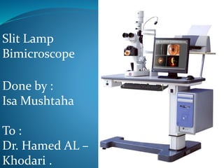
6. Basic Design
1. Viewing arm
Biomicroscope
Adjustable focus eyepieces
Magnification dial
2. Illumination arm
The “slit lamp”
Slit size, shape and filter controls
Variable size, shape, colour and brightness
3. Biomicroscope and illumination are mechanically
coupled around central pivot point (copivotal)
Both focus at the same point (parfocal)
Both arms can swing independently 180º along horizontal
– there is a scale in degrees
Both always central regardless of angle (isocentric)
4. Moveable base plate and joystick control
7. Types of illumination incorporated in slit lamp
1. Slit width
Wide- survey globe/cornea
Narrow- depth, width & position of small
abnormalities
beam as wide as cornea is thick
forms a parallelepiped volume: a box of
illuminated tissue is seen
Thin (slit)- narrowest beam forms an optical
section
so thin it's just discernible التمييز سهل
valuating small changes in clarity & pinpointing
الدقة depth of pathology
8. 2. Light-source intensity
Medium to high: most purposes
High: optical section
3. Filters
neutral, cobalt blue (for fluorescein), red-free
and red free filter (green filter).
4. Magnification
low power (~10x) is used for survey
medium to high (16-40x) for optic section &
parallelepiped
Higher than (40x) for specular reflection
normally, light is focused at same point as
microscope (“parfocal”)
9.
10. 1. Start with neutralizing the eye piece at zero if
the examiner has no refractive error or wearing
his glasses & lower powered objective lens .
2. Use lowest voltage setting on transformer
ensure open aperture
3. Select the longest slit length
4. Adjust chin rest :
Patient's eyes approximately with level of the
marker on head rest .
5. Lamp height of the slit beam centered vertically
on Patient's medial canthus
6. Focus by moving joystick
How the examiner can adjust and use the slit lamp
11. locking nut: loose for free
movementOcular focus to 0
adjust beam height for tall,
narrow vertical beam
adjust width for narrow beam w/
good illumination
12. Methods of examination of slit lamp
without attachments
There are six basic methods of
illumination
1. Diffuse illumination
2. Direct focal illumination
3. Indirect illumination
4. Sclerotic scatter
5. Retro-illumination
6. Specular reflection
13. 1. Diffuse Illumination :
This method is done by using a wide slit
which slightly out of focus and this method is
used in examination of the iris and the adnexa
of the eye .
The slit should be wide and the magnification
should be low as possible to prevent loss of
field and enable large field of view .
14.
15. 2. Direct focal illumination :
•Illumination and observation are focused in the
same plane
•slit width narrow to broad
•Illumination angle 45° to 60°
•Magnification 10x-40x
This method is used to examine the cornea in
details , the anterior chamber , crystalline lens ,
anterior part of the vitreous and in determine if
there is flare , pus or blood cells in the anterior
chamber .
16.
17. 3. Indirect illumination :
•The beam is focused in an area
adjacent to ocular tissue to be observed
•Decentered beam
•Illumination 2 to 4mm slit
•Magnification: Low to medium
(depending upon object size).
This method is used to determine if
there is infiltrates رشح , corneal scars ,
deposits ترسبات and epithelial and
stromal defects .
18.
19. 4. Sclerotic scatter :
•Light incident on the limbus with 2-
4mm slit at an angle of 45° - 60°
•Decentered slit
•The microscope focused centrally
•Total internal reflection of the
incoming light at inner corneal
boundaries (endothelium and
epithelium) .
This method is used in examination of
scars, foreign bodies, corneal defects
and irregularities in the cornea .
20. 5. Retro-Illumination from the Fundus :
This technique is used to observe media claritiesصفاءand
opacities
•The pupil is dilated
•the slit beam and microscope are made co-axial and light
strikes the fundus and creates a glow behind the opacity in
the media
•The media opacity creates a shadow in the glow
Applications
•abnormities in the anterior vitreous, lens, anterior
chamber, cornea
21.
22. 6. Specular Reflection: it is useful slit lamp
technique for scrutinsing the corneal endothelium ,
the tear film surface , crystalline
lens and other surfaces .
A bright reflection will be observed in the anterior
surface and less bright reflection will be observed in
posterior surface of the cornea .
The illumination is narrow parallelepiped and the
microscope is placed directly in front of the eye with
the light source 25degree from the microscope .
The endothelium will appear clear and rough surface (
due to individual endothelial cells ) .
23.
24. Attachments of the Slit lamp
There are many different attachments and
accessories,of these :
1. video attachment :used to facilitate teaching of slit
lamp .
2. Goldman tonometer :used in measuring the intra
ocular pressure of the anterior chamber .
3. Gold 3-mirror lens : used in measuring the angle
of the anterior chamber and center of the retina .
25. 4. Volk double aspheric lens
(+60,+78,+90) :used in examination
the fundus os the patient indirectly .
5. Direct contact goniolenses : for
examination of the periphrey of the
fundus .