Downloaded 744 times

![The littmann Galilean telescope principleThe Galilean magnification changer developed by littmann [1950] It is completely separate optics that sit nearly b\w the objective and eye piece lenses and does not require either of them to change .It provides a larger range of magnifications than the other technique .](https://image.slidesharecdn.com/slitlamp-110828091356-phpapp01/85/Slit-lamp-14-320.jpg)


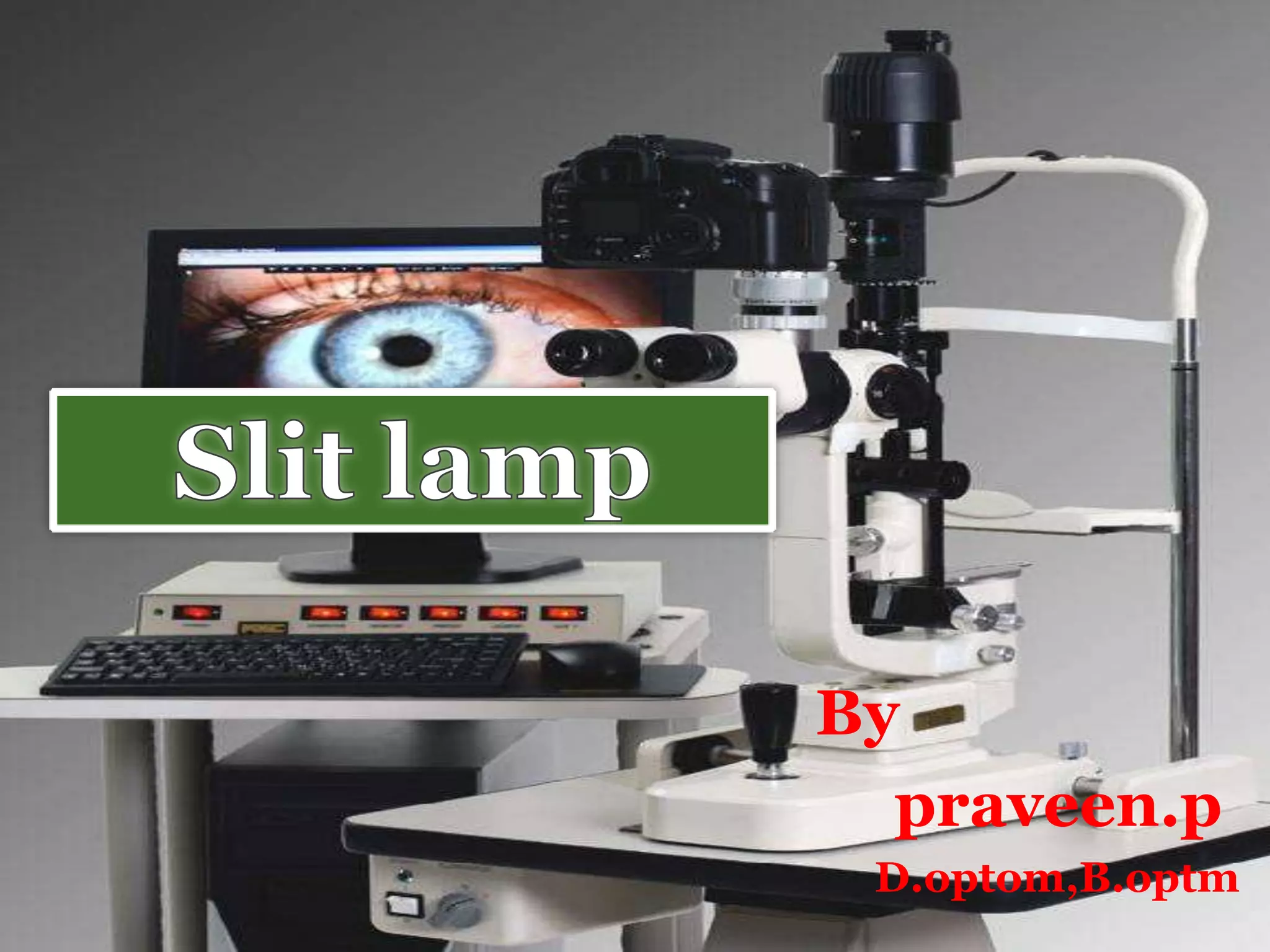
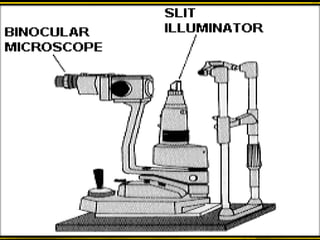
Slit lamps are used to carefully examine the cornea, conjunctiva, and lids of the anterior eye segment. The microscope and light source are coupled so that the same part of the eye being examined is illuminated. This coupling facilitates examination. Main components include the slit lamp microscopes, illumination systems, and mechanical coupling. Attachments allow for fundus examination, gonioscopy, tonometry, laser photocoagulation, and measuring visual acuity in patients with hazy media. Magnification typically ranges from 6x to 40x.


