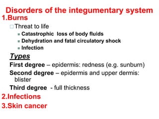
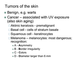
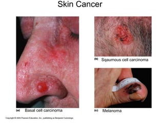
The Integumentary
System
Dr. Adanwali Hassan
Rn-Mw, Medical doctor (MD), Health Officer (HO) Gyn/Obest
The Integumentary System
An outer protective covering such as the
skin of an animal or a cuticle or seed coat or
rind or shell
Introduction
 Skin is the largest organ in the body and covers
the body's entire external surface.
 It is made up of three layers, the epidermis,
dermis, and the hypodermis, all three of which
vary significantly in their anatomy and function.
 The skin's structure is made up of an intricate
network which serves as the body’s initial
barrier against pathogens, UV light, and
chemicals, and mechanical injury.
Cont---
 It also regulates temperature and the
amount of water released into the
environment.
 This article discusses the relevant
anatomical structures of the skin’s
epidermal layer, its structure, function.
The Integumentary System
 Integument is/ means skin
 Skin and its appendages make up the
integumentary system
 A fatty layer (hypodermis) lies deep to it.
 Two distinct regions
Epidermis
Dermis
An external body part that projects from the
body
Functions of skin
 Protection:
Cushions and insulates (Separate in order to protect or prevent
interaction) and is waterproof
Protects from chemicals, heat, cold, bacteria
Screens UV
 Synthesizes vitamin D with UV
 Regulates body heat
 Prevents unnecessary water loss
 Sensory reception (nerve endings)
Epidermis
 Keratinized stratified squamous epithelium
 Four types of Cells in the Epidermis:
 Keratinocytes – deepest, produce
keratin (tough fibrous protein)
 Melanocytes - make dark skin
pigment melanin
 Merkel cells – associated with sensory
nerve endings
 Langerhans cells – macrophage-like
dendritic cells,
Keratin: A fibrous scleroprotein that occurs in the
outer layer of the skin and in horny tissues such
as hair, feathers, nails, and hooves
Layers (from deep to superficial)
1. Stratum basale or germinativum – single row
of cells attached to dermis; youngest cells
2. Stratum spinosum – spinyness is artifactual;
tonofilaments (bundles of protein) resist tension
3. Stratum granulosum – layers of flattened
keratinocytes producing keratin (hair and nails
made of it also)
4. Stratum lucidum (only on palms and soles)
5. Stratum corneum – horny layer (cells dead,
many layers thick)
Epithelium: layers (on left) and cell types (on right)
Remember…
 Four basic types of tissue
Epithelium – epidermis just discussed
Connective tissue - dermis
Muscle tissue
Nervous tissue
Dermis
 Strong, flexible connective tissue: your “hide”
Cells: fibroblasts, macrophages, mast cells, WBCs
Fiber types: collagen, elastic, reticular
 Rich supply of nerves and vessels
 Critical role in temperature regulation (the
vessels)
 Two layers (see next slides)
Papillary – areolar connective tissue;
includes dermal papillae
Reticular – “reticulum” (network) of
collagen and reticular fibers
*Dermis layers
*
*
*Dermal papillae
Epidermis and dermis of (a) thick skin and (b) thin skin
(which one makes the difference?)
Fingerprints, palmprints, footprints
 Dermal papillae lie atop dermal ridges
 Elevate the overlying epidermis into epidermal
ridges
 Are “sweat films” because of sweat pores
 Genetically determined
Flexion creases
 Deep dermis, from continual folding
Fibers
 Collagen: strength and resilience
 Elastic fibers: stretch-recoil
 Striae: stretch marks
 Tension lines (or lines of cleavage)
 The direction the bundles
of fibers are directed
The dermis is the receptive
site for the pigment of tattoos
Hypodermis
 “Hypodermis” (Gk) = below the skin
 “Subcutaneous” (Latin) = below the skin
 Also called “superficial fascia”
“fascia” (Latin) =band; in anatomy: sheet of
connective tissue
 Fatty tissue which stores fat and anchors
skin (areolar tissue and adipose cells)
 Different patterns of accumulation
(male/female)
Skin color
 Three skin pigments:
Melanin: the most important
Carotene: from carrots and yellow vegies
Hemoglobin: the pink of light skin
 Melanin in granules passes from
melanocytes (same number in all races) to
keratinocytes in stratum basale
Digested by lysosomes
Variations in color
Protection from UV light vs vitamin D?
Skin appendages
 Derived from epidermis but extend into
dermis
 Include
Hair and hair follicles
Sebaceous (oil) glands
Sweat (sudoiferous) glands
Nails
Nails
 Of hard keratin
 Corresponds to hooves and claws
 Grows from nail matrix
Hair and hair follicles: complex
Derived from epidermis and dermis
Everywhere but palms, soles, nipples, parts of genitalia
*“arrector pili” is smooth muscle
*
Hair papilla
is connective
tissue________________
Hair bulb:
epithelial cells
surrounding
papilla
Functions of hair
Warmth – less in man than other mammals
Sense light touch of the skin
Protection - scalp
 Parts
Root imbedded in skin
Shaft projecting above skin surface
 Make up of hair – hard keratin
 Three concentric layers
Medulla (core)
Cortex (surrounds medulla)
Cuticle (single layers, overlapping)
 Types of hair
 Vellus: fine, short hairs
 Intermediate hairs
 Terminal: longer, courser hair
 Hair growth: averages 2 mm/week
 Active: growing
 Resting phase then shed
 Hair loss
 Thinning – age related
 Male pattern baldness
 Hair color
 Amount of melanin for black or brown; distinct form
of melanin for red
 White: decreased melanin and air bubbles in the
medulla
 Genetically determined though influenced by
hormones and environment
Sebaceous (oil) glands
 Entire body except palms and soles
 Produce sebum by holocrine secretion
 Oils and lubricates
Sweat glands
 Entire skin surface
except nipples and
part of external
genitalia
 Prevent overheating
 500 cc to 12 l/day!
(is mostly water)
 Humans most
efficient (only
mammals have)
 Produced in
response to stress
as well as heat
Types of sweat glands
1.Eccrine or merocrine
 Most numerous
 True sweat: 99% water, some salts, traces of waste
 Open through pores
2.Apocrine
 Axillary, anal and genital areas only
 Ducts open into hair follices
 The organic molecules in it decompose with time - odor
3.Modified apocrine glands
 Ceruminous – secrete earwax
 Mammary – secrete milk
Disorders of the integumentary system
1.Burns
Threat to life
 Catastrophic loss of body fluids
 Dehydration and fatal circulatory shock
 Infection
Types
First degree – epidermis: redness (e.g. sunburn)
Second degree – epidermis and upper dermis:
blister
Third degree - full thickness
2.Infections
3.Skin cancer
Burns
First-degree
(epidermis only; redness)
Second-degree
(epidermis and dermis,
with blistering)
Third-degree
(full thickness, destroying
epidermis, dermis, often part
of hypodermis)
Critical burns
 Over 10% of the
body has third-
degree burns
 25 % of the body
has second-
degree burns
 Third-degree
burns on face,
hands, or feet
Estimate by “rule of 9’s”
Tumors of the skin
 Benign, e.g. warts
 Cancer – associated with UV exposure
(also skin aging)
Aktinic keratosis - premalignant
Basal cell - cells of stratum basale
Squamous cell - keratinocytes
Melanoma – melanocytes: most dangerous;
recognition:
 A - Asymmetry
 B - Border irregularity
 C - Colors
 D - Diameter larger than 6 mm
Basal cell carcinoma
Sqaumous cell carcinoma
Melanoma
Skin Cancer
Con…
Disadvantage
 The drugs prepared for IV administration is
expensive.
 Limited to highly soluble medications.
 Drug distribution is inhibited by poor
circulation.

1.The Integumentary System.ppt

  • 1.
    The Integumentary System Dr. AdanwaliHassan Rn-Mw, Medical doctor (MD), Health Officer (HO) Gyn/Obest
  • 2.
    The Integumentary System Anouter protective covering such as the skin of an animal or a cuticle or seed coat or rind or shell
  • 3.
    Introduction  Skin isthe largest organ in the body and covers the body's entire external surface.  It is made up of three layers, the epidermis, dermis, and the hypodermis, all three of which vary significantly in their anatomy and function.  The skin's structure is made up of an intricate network which serves as the body’s initial barrier against pathogens, UV light, and chemicals, and mechanical injury.
  • 4.
    Cont---  It alsoregulates temperature and the amount of water released into the environment.  This article discusses the relevant anatomical structures of the skin’s epidermal layer, its structure, function.
  • 5.
    The Integumentary System Integument is/ means skin  Skin and its appendages make up the integumentary system  A fatty layer (hypodermis) lies deep to it.  Two distinct regions Epidermis Dermis An external body part that projects from the body
  • 6.
    Functions of skin Protection: Cushions and insulates (Separate in order to protect or prevent interaction) and is waterproof Protects from chemicals, heat, cold, bacteria Screens UV  Synthesizes vitamin D with UV  Regulates body heat  Prevents unnecessary water loss  Sensory reception (nerve endings)
  • 7.
    Epidermis  Keratinized stratifiedsquamous epithelium  Four types of Cells in the Epidermis:  Keratinocytes – deepest, produce keratin (tough fibrous protein)  Melanocytes - make dark skin pigment melanin  Merkel cells – associated with sensory nerve endings  Langerhans cells – macrophage-like dendritic cells, Keratin: A fibrous scleroprotein that occurs in the outer layer of the skin and in horny tissues such as hair, feathers, nails, and hooves
  • 8.
    Layers (from deepto superficial) 1. Stratum basale or germinativum – single row of cells attached to dermis; youngest cells 2. Stratum spinosum – spinyness is artifactual; tonofilaments (bundles of protein) resist tension 3. Stratum granulosum – layers of flattened keratinocytes producing keratin (hair and nails made of it also) 4. Stratum lucidum (only on palms and soles) 5. Stratum corneum – horny layer (cells dead, many layers thick)
  • 9.
    Epithelium: layers (onleft) and cell types (on right)
  • 10.
    Remember…  Four basictypes of tissue Epithelium – epidermis just discussed Connective tissue - dermis Muscle tissue Nervous tissue
  • 11.
    Dermis  Strong, flexibleconnective tissue: your “hide” Cells: fibroblasts, macrophages, mast cells, WBCs Fiber types: collagen, elastic, reticular  Rich supply of nerves and vessels  Critical role in temperature regulation (the vessels)  Two layers (see next slides) Papillary – areolar connective tissue; includes dermal papillae Reticular – “reticulum” (network) of collagen and reticular fibers
  • 12.
  • 13.
    Epidermis and dermisof (a) thick skin and (b) thin skin (which one makes the difference?)
  • 14.
    Fingerprints, palmprints, footprints Dermal papillae lie atop dermal ridges  Elevate the overlying epidermis into epidermal ridges  Are “sweat films” because of sweat pores  Genetically determined Flexion creases  Deep dermis, from continual folding Fibers  Collagen: strength and resilience  Elastic fibers: stretch-recoil  Striae: stretch marks  Tension lines (or lines of cleavage)  The direction the bundles of fibers are directed The dermis is the receptive site for the pigment of tattoos
  • 15.
    Hypodermis  “Hypodermis” (Gk)= below the skin  “Subcutaneous” (Latin) = below the skin  Also called “superficial fascia” “fascia” (Latin) =band; in anatomy: sheet of connective tissue  Fatty tissue which stores fat and anchors skin (areolar tissue and adipose cells)  Different patterns of accumulation (male/female)
  • 16.
    Skin color  Threeskin pigments: Melanin: the most important Carotene: from carrots and yellow vegies Hemoglobin: the pink of light skin  Melanin in granules passes from melanocytes (same number in all races) to keratinocytes in stratum basale Digested by lysosomes Variations in color Protection from UV light vs vitamin D?
  • 17.
    Skin appendages  Derivedfrom epidermis but extend into dermis  Include Hair and hair follicles Sebaceous (oil) glands Sweat (sudoiferous) glands Nails
  • 19.
    Nails  Of hardkeratin  Corresponds to hooves and claws  Grows from nail matrix
  • 20.
    Hair and hairfollicles: complex Derived from epidermis and dermis Everywhere but palms, soles, nipples, parts of genitalia *“arrector pili” is smooth muscle * Hair papilla is connective tissue________________ Hair bulb: epithelial cells surrounding papilla
  • 21.
    Functions of hair Warmth– less in man than other mammals Sense light touch of the skin Protection - scalp  Parts Root imbedded in skin Shaft projecting above skin surface  Make up of hair – hard keratin  Three concentric layers Medulla (core) Cortex (surrounds medulla) Cuticle (single layers, overlapping)
  • 22.
     Types ofhair  Vellus: fine, short hairs  Intermediate hairs  Terminal: longer, courser hair  Hair growth: averages 2 mm/week  Active: growing  Resting phase then shed  Hair loss  Thinning – age related  Male pattern baldness  Hair color  Amount of melanin for black or brown; distinct form of melanin for red  White: decreased melanin and air bubbles in the medulla  Genetically determined though influenced by hormones and environment
  • 23.
    Sebaceous (oil) glands Entire body except palms and soles  Produce sebum by holocrine secretion  Oils and lubricates
  • 24.
    Sweat glands  Entireskin surface except nipples and part of external genitalia  Prevent overheating  500 cc to 12 l/day! (is mostly water)  Humans most efficient (only mammals have)  Produced in response to stress as well as heat
  • 25.
    Types of sweatglands 1.Eccrine or merocrine  Most numerous  True sweat: 99% water, some salts, traces of waste  Open through pores 2.Apocrine  Axillary, anal and genital areas only  Ducts open into hair follices  The organic molecules in it decompose with time - odor 3.Modified apocrine glands  Ceruminous – secrete earwax  Mammary – secrete milk
  • 26.
    Disorders of theintegumentary system 1.Burns Threat to life  Catastrophic loss of body fluids  Dehydration and fatal circulatory shock  Infection Types First degree – epidermis: redness (e.g. sunburn) Second degree – epidermis and upper dermis: blister Third degree - full thickness 2.Infections 3.Skin cancer
  • 27.
    Burns First-degree (epidermis only; redness) Second-degree (epidermisand dermis, with blistering) Third-degree (full thickness, destroying epidermis, dermis, often part of hypodermis)
  • 28.
    Critical burns  Over10% of the body has third- degree burns  25 % of the body has second- degree burns  Third-degree burns on face, hands, or feet Estimate by “rule of 9’s”
  • 29.
    Tumors of theskin  Benign, e.g. warts  Cancer – associated with UV exposure (also skin aging) Aktinic keratosis - premalignant Basal cell - cells of stratum basale Squamous cell - keratinocytes Melanoma – melanocytes: most dangerous; recognition:  A - Asymmetry  B - Border irregularity  C - Colors  D - Diameter larger than 6 mm
  • 30.
    Basal cell carcinoma Sqaumouscell carcinoma Melanoma Skin Cancer
  • 31.
    Con… Disadvantage  The drugsprepared for IV administration is expensive.  Limited to highly soluble medications.  Drug distribution is inhibited by poor circulation.

Editor's Notes

  • #8 Dendritic: Short fiber that conducts toward the cell body of the neuron