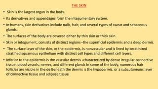
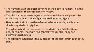
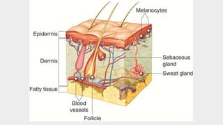
Presentation OF
The Skin
•‫عباس‬ ‫امني‬
THE SKIN
Skin Cells
Skin color:
THE SKIN
• Skin is the largest organ in the body.
• Its derivatives and appendages form the integumentary system.
• In humans, skin derivatives include nails, hair, and several types of sweat and sebaceous
glands.
• The surfaces of the body are covered either by thin skin or thick skin.
• Skin or integument, consists of distinct regions--the superficial epidermis and a deep dermis.
• The surface layer of the skin, or the epidermis, is nonvascular and is lined by keratinized
stratified squamous epithelium with distinct cell types and different cell layers.
• Inferior to the epidermis is the vascular dermis ,characterized by dense irregular connective
tissue, blood vessels, nerves, and different glands In some of the body, numerous hair
follicles are visible in the de Beneath the dermis is the hypodermis, or a subcutaneous layer
of connective tissue and adipose tissue
• The human skin is the outer covering of the body. In humans, it is the
largest organ of the integumentary system.
• The skin has up to seven layers of ectodermal tissue and guards the
underlying muscles, bones, ligamentsand internal organs.
• Human skin is similar to that of most other mammals, and human
skin is very similar to pigskin.
• Though nearly all human skin is covered with hair follicles, it can
appear hairless. There are two general types of skin, hairy and
glabrous skin (hairless).
• The adjective cutaneous literally means "of the skin" (from Latin cutis,
skin).
Epidermal Cell Layers
• There are four cell types in the epidermis of the skin, with the
keratinocytes being the most dominant cells. Keratinocytes
divide, grow, migrate up, undergo keratinization, or cornification,
and form the protective epidermal and surface layer for the skin.
The epidermis is composed of stratified keratinized squamous
epithelium. There are other less abundant cell types in the
epidermis. These are the melanocytes, Langerhans cells, and
Merkel cells, which are interspersed among the keratinocytes in
the epidermis. identified In epidermis of thick skin, five distinct
and recognizable cell layers can be
1. Stratum Basale (Germinativum): The First Layer
• Deepest or basal single layer of columnar to cuboidal cells that
rests on the basement membrane. Cells attached by
desmosomes and by hemidesmosomes to the basement
membrane. Cells serve as stem cells for the epidermis and
show increased mitotic activity. Cells mature and migrate
upward in the epidermis and produce intermediate keratin
filaments
2. Stratum Spin osum: The Second Layer
• Is the layer above the stratum basale that consists of four to
six rows of cell
• During histologic preparation, cells shrink and intercellular
spaces appear as spines
• Cells synthesize keratin filaments.
• Spines represent sites of desmosome attachments to keratin
filaments.
3. Stratum Granulosum: The Third Layer
• Cells above the stratum spinosum and consists of 3 to 5 cell
layers of flattened cells. Cells filled with dense keratohyalin
granules and membrane bound lamellar granules.
• Keratohyalin granules consist of the protein filaggrin that
cross-links with keratin filaments
• Combination of keratinfilaments with keratohyalin granules
produces soft keratin(keratinization)
• Lamellar granules discharge lipid material between cells and
waterproof the skin.
4. Stratum Lucidum: The Fourth Layer
• Lies superior to the stratum granulosum, found in thick skin
only; translucent and barely visible The tightly packed cells
lack nuclei or organelles and are dead. The flattened cells
contain densely packed keratin filaments.
5. Stratum Corneum: The Fifth Layer
• Most superficial layer and consists of flat, dead cells filled
with soft keratin ratinized cells continually shed or
desquamated from the surface and replaced by new During
keratinization, hydrolytic enzymes eliminate the nucleus and
organelles.
Skin Cells
1. Keratinocytes-produce keratin; predominant cell type in the
epidermis.
2. Melanocytes
• located between the stratum basale and stratum spinosum
• Long irregular cytoplasmic or dendritic extensions branch into the
epidermis
• Synthesize a dark brown pigment: melanin
• Melanin transferred from cytoplasmic extensions through to
keratinocytes in basal cell layers.
• Melanin darkens skin color and protects it from ultraviolet Radiation.
3. Langerhans Cells
• Dendritic-type cells originate from the bone marrow and
migrate via the blood to the skin.
• Reside primarily in the stratum spinosum and are part of the
immune system of the skin (Function as macrophages)
4. Merkel Cells
• Present in the basal layer of the epidermis and function as
mechanoreceptors for sensation.
Dermis: Papillary and Reticular Layers
• Dermis is the inferior connective tissue layer that binds to the
epidermis.
• A distinct basement membrane separates the epidermis from the
dermis.
• In addition, the dermis contains epidermal derivatives, such as the
sweat glands, sebaceous glands, and hair follicles.
• The junction of the dermis with the epidermis is irregular
• The superficial layer of the dermis forms numerous raised projections
called dermal papillae which with of the epidermis, called region of
the skin is the papillary layer of the dermis.
• It contains loose irregular connective tissue fibers, capillaries, blood
vessels, fibroblasts, macrophages, and other loose connective tissue
cells
• The deeper layer of the dermis is called the reticular layer.
• This layer is thicker and is characterized by dense irregular connective
tissue fibers (mainly type I collagen) and is less cellular than the papillary
layer.
• Also, this layer of the dermis can withstand more mechanical stresses
and can provide support for nerves, blood vessels, hair follicles, and all
the sweat glands.
• There is no distinct boundary between the two dermal layers, and the
papillary layer blends with the reticular layer Certain regions of the skin
exhibit arteriovenous anastomoses used for temperature regulation.
• Here, blood passes directly from the arteries into the veins.
• In addition, the dermis contains numerous sensory receptors (Meissner
corpuscles and Pacinian corpuscles)
Epidermis: Thick Versus Thin Skin
• Palms and soles, because of wear and tear, are covered by
thick skin
• Thick skin contains sweat glands but lacks hair, sebaceous
glands, and smooth muscle.
• Thin skin contains ous glands, hair, sweat glands, and
arrector pili smooth muscle
Major Skin Functions:
1.Protection through the keratinized epidermis from abrasion and the
entrance of pathogens
2. Impermeable to water, owing to lipid layer in the epidermis
3. Body temperature regulation as a result of sweating and changes in
vessel diameters
4. Sensory perception of touch, pain, pressure, and temperature
changes because of nerve endings
5. Excretions through sweat of water, sodium salts and nitrogenous
waste
6. Formation of vitamin D from precursor molecules produced in the
epidermis when exposed to the sun.
• Protection of the human body Sensation i.e. transmitting to the brain
information about surroundings Temperature regulation Immunity i.e.
the role of the skin within the immune system Enables movement
and growth without injury Excretion from the body of certain types of
waste materials Endocrine function e.g. re. Vitamin D It is useful to be
able to describe each of the above functions of the skin in further
detail with examples and explanations of the mechanisms that apply
in each case.
1. Protection
• Example(s) include protection of the body from:
• ultraviolet (UV) radiation e.g. sun damage
• dehydration
• microorganisms e.g. bacterial invasion
• mechanical trauma / physical injuries
How does the skin perform this function?
• The physical/mechanical barrier formed by the surface (stratum corneum
layer) of the skin.
• Mechanical strength of the tissues that form the skin.
• Keratin - a type of protein that is found in the skin.
• Melanin - a dark-coloured light-sensitive pigment that is found in the skin
and that protects against excessive amounts of ultraviolet (UV) radition,
usually coming from the sun.
2. Sensation
• Example:
• pressure / touch
• heat / cold
• pain
How does the skin perform this function?
• Somatic sensory receptors
3. Temperature Regulation
Example:
include retention or release of heat - depending on outide of body
temperature
How does the skin perform this function?
• Release of sweat from sweat glands followed by evapouration of sweat
from the surface of the skin (body)
• Regulation of blood flow to regions of skin, especially the extremities of
the body (i.e. limbs / appendicular skeleton)
Skin color:
a. Melanin ranges in color from yellow to reddish-brown to
black: Melanocytes are stimulated by sunlight and secreted
melanin transfer to keratinocytes to protects DNA from uv
light.
b. Carotene: yellow to orange: Accumulates in fatty tissue of
hypodermis.
c. Hemoglobin: in RBCs: oxyhemoglobin give reddish color to
skin
• Human skin color ranges in variety from the darkest brown to the lightesthues.
An individual's skin pigmentationis the result of genetics, being the product of
both of the individual's biological parents' genetic makeup.
• In evolution, skin pigmentation in human beings evolved by a process of
natural selection primarily to regulate the amount of ultraviolet
radiationpenetrating the skin, controlling its biochemical effects.
• The actual skin color of different humans is affected by many substances,
although the single most important substance is the pigment melanin. Melanin
is produced within the skin in cells called melanocytes and it is the main
determinant of the skin color of darker-skinned humans.
• The skin color of people with light skin is determined mainly by the bluish-
white connective tissue under the dermis and by the hemoglobin circulating in
the veins of the dermis.
• The red color underlying the skin becomes more visible, especially in the face,
when, as consequence of physical exercise or the stimulation of the nervous
system(anger, fear), arterioles dilate.
• Color is not entirely uniform across an individual's skin; for example, the skin of
the palm and the sole is lighter than most other skin, and this is especially
noticeable in darker-skinned people.
• There is a direct correlation between the geographic distribution of
ultraviolet radiation (UVR) and the distribution of indigenous skin
pigmentation around the world.
• Areas that receive higher amounts of UVR, generally located closer to the
equator, tend to have darker-skinned populations.
• Areas that are far from the tropics and closer to the poles have lower
intensity of UVR, which is reflected in lighter-skinned populations.
• Researchers suggest that human populations over the past 50,000 years have
changed from dark-skinned to light-skinned and vice versa as they migrated
to different UV zones, and that such major changes in pigmentation may have
happened in as little as 100 generations (≈2,500 years) through selective
sweeps.
• Natural skin color can also darken as a result of tanning due to exposure
to sunlight.
• The leading theory is that skin color adapts to intense sunlight irradiation
to provide partial protection against the ultraviolet fraction that
produces damage and thus mutations in the DNA of the skin cells.
• In addition, it has been observed that adult human females on average
are significantly lighter in skin pigmentation than males.
• Females need more calcium during pregnancy and lactation.
• The body synthesizes vitamin D from sunlight, which helps it absorb
calcium.
• Females evolved to have lighter skin so their bodies absorb more
calcium.
• The social significance of differences in skin color has varied across
cultures and over time, as demonstrated with regard to social status and
discrimination

Pdf |Histology| - The skin -

  • 1.
    Presentation OF The Skin •‫عباس‬‫امني‬ THE SKIN Skin Cells Skin color:
  • 2.
    THE SKIN • Skinis the largest organ in the body. • Its derivatives and appendages form the integumentary system. • In humans, skin derivatives include nails, hair, and several types of sweat and sebaceous glands. • The surfaces of the body are covered either by thin skin or thick skin. • Skin or integument, consists of distinct regions--the superficial epidermis and a deep dermis. • The surface layer of the skin, or the epidermis, is nonvascular and is lined by keratinized stratified squamous epithelium with distinct cell types and different cell layers. • Inferior to the epidermis is the vascular dermis ,characterized by dense irregular connective tissue, blood vessels, nerves, and different glands In some of the body, numerous hair follicles are visible in the de Beneath the dermis is the hypodermis, or a subcutaneous layer of connective tissue and adipose tissue
  • 3.
    • The humanskin is the outer covering of the body. In humans, it is the largest organ of the integumentary system. • The skin has up to seven layers of ectodermal tissue and guards the underlying muscles, bones, ligamentsand internal organs. • Human skin is similar to that of most other mammals, and human skin is very similar to pigskin. • Though nearly all human skin is covered with hair follicles, it can appear hairless. There are two general types of skin, hairy and glabrous skin (hairless). • The adjective cutaneous literally means "of the skin" (from Latin cutis, skin).
  • 5.
    Epidermal Cell Layers •There are four cell types in the epidermis of the skin, with the keratinocytes being the most dominant cells. Keratinocytes divide, grow, migrate up, undergo keratinization, or cornification, and form the protective epidermal and surface layer for the skin. The epidermis is composed of stratified keratinized squamous epithelium. There are other less abundant cell types in the epidermis. These are the melanocytes, Langerhans cells, and Merkel cells, which are interspersed among the keratinocytes in the epidermis. identified In epidermis of thick skin, five distinct and recognizable cell layers can be
  • 6.
    1. Stratum Basale(Germinativum): The First Layer • Deepest or basal single layer of columnar to cuboidal cells that rests on the basement membrane. Cells attached by desmosomes and by hemidesmosomes to the basement membrane. Cells serve as stem cells for the epidermis and show increased mitotic activity. Cells mature and migrate upward in the epidermis and produce intermediate keratin filaments
  • 7.
    2. Stratum Spinosum: The Second Layer • Is the layer above the stratum basale that consists of four to six rows of cell • During histologic preparation, cells shrink and intercellular spaces appear as spines • Cells synthesize keratin filaments. • Spines represent sites of desmosome attachments to keratin filaments.
  • 8.
    3. Stratum Granulosum:The Third Layer • Cells above the stratum spinosum and consists of 3 to 5 cell layers of flattened cells. Cells filled with dense keratohyalin granules and membrane bound lamellar granules. • Keratohyalin granules consist of the protein filaggrin that cross-links with keratin filaments • Combination of keratinfilaments with keratohyalin granules produces soft keratin(keratinization) • Lamellar granules discharge lipid material between cells and waterproof the skin.
  • 9.
    4. Stratum Lucidum:The Fourth Layer • Lies superior to the stratum granulosum, found in thick skin only; translucent and barely visible The tightly packed cells lack nuclei or organelles and are dead. The flattened cells contain densely packed keratin filaments.
  • 10.
    5. Stratum Corneum:The Fifth Layer • Most superficial layer and consists of flat, dead cells filled with soft keratin ratinized cells continually shed or desquamated from the surface and replaced by new During keratinization, hydrolytic enzymes eliminate the nucleus and organelles.
  • 11.
    Skin Cells 1. Keratinocytes-producekeratin; predominant cell type in the epidermis. 2. Melanocytes • located between the stratum basale and stratum spinosum • Long irregular cytoplasmic or dendritic extensions branch into the epidermis • Synthesize a dark brown pigment: melanin • Melanin transferred from cytoplasmic extensions through to keratinocytes in basal cell layers. • Melanin darkens skin color and protects it from ultraviolet Radiation.
  • 12.
    3. Langerhans Cells •Dendritic-type cells originate from the bone marrow and migrate via the blood to the skin. • Reside primarily in the stratum spinosum and are part of the immune system of the skin (Function as macrophages) 4. Merkel Cells • Present in the basal layer of the epidermis and function as mechanoreceptors for sensation.
  • 13.
    Dermis: Papillary andReticular Layers • Dermis is the inferior connective tissue layer that binds to the epidermis. • A distinct basement membrane separates the epidermis from the dermis. • In addition, the dermis contains epidermal derivatives, such as the sweat glands, sebaceous glands, and hair follicles. • The junction of the dermis with the epidermis is irregular • The superficial layer of the dermis forms numerous raised projections called dermal papillae which with of the epidermis, called region of the skin is the papillary layer of the dermis. • It contains loose irregular connective tissue fibers, capillaries, blood vessels, fibroblasts, macrophages, and other loose connective tissue cells
  • 14.
    • The deeperlayer of the dermis is called the reticular layer. • This layer is thicker and is characterized by dense irregular connective tissue fibers (mainly type I collagen) and is less cellular than the papillary layer. • Also, this layer of the dermis can withstand more mechanical stresses and can provide support for nerves, blood vessels, hair follicles, and all the sweat glands. • There is no distinct boundary between the two dermal layers, and the papillary layer blends with the reticular layer Certain regions of the skin exhibit arteriovenous anastomoses used for temperature regulation. • Here, blood passes directly from the arteries into the veins. • In addition, the dermis contains numerous sensory receptors (Meissner corpuscles and Pacinian corpuscles)
  • 15.
    Epidermis: Thick VersusThin Skin • Palms and soles, because of wear and tear, are covered by thick skin • Thick skin contains sweat glands but lacks hair, sebaceous glands, and smooth muscle. • Thin skin contains ous glands, hair, sweat glands, and arrector pili smooth muscle
  • 16.
    Major Skin Functions: 1.Protectionthrough the keratinized epidermis from abrasion and the entrance of pathogens 2. Impermeable to water, owing to lipid layer in the epidermis 3. Body temperature regulation as a result of sweating and changes in vessel diameters 4. Sensory perception of touch, pain, pressure, and temperature changes because of nerve endings 5. Excretions through sweat of water, sodium salts and nitrogenous waste 6. Formation of vitamin D from precursor molecules produced in the epidermis when exposed to the sun.
  • 17.
    • Protection ofthe human body Sensation i.e. transmitting to the brain information about surroundings Temperature regulation Immunity i.e. the role of the skin within the immune system Enables movement and growth without injury Excretion from the body of certain types of waste materials Endocrine function e.g. re. Vitamin D It is useful to be able to describe each of the above functions of the skin in further detail with examples and explanations of the mechanisms that apply in each case.
  • 18.
    1. Protection • Example(s)include protection of the body from: • ultraviolet (UV) radiation e.g. sun damage • dehydration • microorganisms e.g. bacterial invasion • mechanical trauma / physical injuries How does the skin perform this function? • The physical/mechanical barrier formed by the surface (stratum corneum layer) of the skin. • Mechanical strength of the tissues that form the skin. • Keratin - a type of protein that is found in the skin. • Melanin - a dark-coloured light-sensitive pigment that is found in the skin and that protects against excessive amounts of ultraviolet (UV) radition, usually coming from the sun.
  • 19.
    2. Sensation • Example: •pressure / touch • heat / cold • pain How does the skin perform this function? • Somatic sensory receptors
  • 20.
    3. Temperature Regulation Example: includeretention or release of heat - depending on outide of body temperature How does the skin perform this function? • Release of sweat from sweat glands followed by evapouration of sweat from the surface of the skin (body) • Regulation of blood flow to regions of skin, especially the extremities of the body (i.e. limbs / appendicular skeleton)
  • 21.
    Skin color: a. Melaninranges in color from yellow to reddish-brown to black: Melanocytes are stimulated by sunlight and secreted melanin transfer to keratinocytes to protects DNA from uv light. b. Carotene: yellow to orange: Accumulates in fatty tissue of hypodermis. c. Hemoglobin: in RBCs: oxyhemoglobin give reddish color to skin
  • 22.
    • Human skincolor ranges in variety from the darkest brown to the lightesthues. An individual's skin pigmentationis the result of genetics, being the product of both of the individual's biological parents' genetic makeup. • In evolution, skin pigmentation in human beings evolved by a process of natural selection primarily to regulate the amount of ultraviolet radiationpenetrating the skin, controlling its biochemical effects. • The actual skin color of different humans is affected by many substances, although the single most important substance is the pigment melanin. Melanin is produced within the skin in cells called melanocytes and it is the main determinant of the skin color of darker-skinned humans. • The skin color of people with light skin is determined mainly by the bluish- white connective tissue under the dermis and by the hemoglobin circulating in the veins of the dermis. • The red color underlying the skin becomes more visible, especially in the face, when, as consequence of physical exercise or the stimulation of the nervous system(anger, fear), arterioles dilate. • Color is not entirely uniform across an individual's skin; for example, the skin of the palm and the sole is lighter than most other skin, and this is especially noticeable in darker-skinned people.
  • 23.
    • There isa direct correlation between the geographic distribution of ultraviolet radiation (UVR) and the distribution of indigenous skin pigmentation around the world. • Areas that receive higher amounts of UVR, generally located closer to the equator, tend to have darker-skinned populations. • Areas that are far from the tropics and closer to the poles have lower intensity of UVR, which is reflected in lighter-skinned populations. • Researchers suggest that human populations over the past 50,000 years have changed from dark-skinned to light-skinned and vice versa as they migrated to different UV zones, and that such major changes in pigmentation may have happened in as little as 100 generations (≈2,500 years) through selective sweeps.
  • 24.
    • Natural skincolor can also darken as a result of tanning due to exposure to sunlight. • The leading theory is that skin color adapts to intense sunlight irradiation to provide partial protection against the ultraviolet fraction that produces damage and thus mutations in the DNA of the skin cells. • In addition, it has been observed that adult human females on average are significantly lighter in skin pigmentation than males. • Females need more calcium during pregnancy and lactation. • The body synthesizes vitamin D from sunlight, which helps it absorb calcium. • Females evolved to have lighter skin so their bodies absorb more calcium. • The social significance of differences in skin color has varied across cultures and over time, as demonstrated with regard to social status and discrimination