Anatomy and Histology of Skin(Dermis & Epidermis).pptx
•Download as PPTX, PDF•
0 likes•89 views

Deep to the epidermis lies the dermis. It is a thick layer of connective tissue consisting of collagen and elastin which allows for skin's strength and flexibility, respectively. The dermis also contains nerve endings, blood vessels, and adnexal structures such as hair shafts, sweat glands, and sebaceous glands.

Recommended
More Related Content
Similar to Anatomy and Histology of Skin(Dermis & Epidermis).pptx
Similar to Anatomy and Histology of Skin(Dermis & Epidermis).pptx (20)
More from Mathew Joseph
More from Mathew Joseph (20)
Recently uploaded
Recently uploaded (20)
Anatomy and Histology of Skin(Dermis & Epidermis).pptx
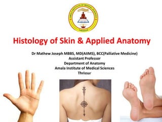
- 1. Histology of Skin & Applied Anatomy Dr Mathew Joseph MBBS, MD(AIIMS), BCC(Palliative Medicine) Assistant Professor Department of Anatomy Amala Institute of Medical Sciences Thrissur
- 3. Skin Facts • The human skin (cutis) - outer covering of body & is continuous with mucous membrane in the region of mouth, nose, urogenital organs & anus. • The mass of skin an adult accounts for approx 5 % while together with subcutaneous fat for about 16 to 18 per cent of the total body mass.
- 4. • Largest sensory organ • Weighs 4 kg • Thickness – 0.3 – 3 mm • Integumentary system • Rule of nine • Surface area: 1.5-2.0 sq meters • Growth rate of nail: 0.1mm per day • Growth rate of hair: 1.5-2.2 mm per week • Life span of hair: Eyelashes, axilla- 4 months Scalp – 4 years
- 5. •The skin surface is covered with hairs over a great area. •The areas devoid of hairs are lips (vermilion border), palms & soles, palmar surface of fingers & plantar surface of toes, glans penis, inner surface of prepuce, & inner surface of anus.
- 7. Layers of Skin Epidermis: Composed of keratinized stratified squamous epithelium. Dermis: Papillary region composed of loose connective tissue. Reticular region composed of dense irregular connective tissue. Hypodermis: Composed of areolar tissue with abundant adipocytes.
- 8. Epidermis
- 10. Germinative layer (stratum basale or stratum germinativum) • It consists of a single layer of columnar cells arranged like a palisade. • Between these cells slit-like spaces called intercellular bridges. • Among the cells of germinative layer localize melanocytes, which produce melanin. • Skin color straightly depends on the amount of melanin. • This layer presents stem cells conerned to mitosis.
- 15. Prickle cell layer (stratum spinosum) • Consists of 5 - 10 rows of cells , cuboid in deep parts of layer but become flatter gradually as they approach next layer, the granular layer • The cells of the prickle-cell layer are marked by presence of specific tonofibrils in their cytoplasm. • Special Langerhan's cells are demonstrated in this layer, which carry immunological function.
- 17. Granular layer (stratum granulosum) • Contains 1-5 rows of cells elongated parallel to epidermis • It was considered previously that they were formed of a special substance called keratohyalin • The presence of the keratohyalin granules is the first visible stage, of the beginning of the process of keratinization of the epidermal cells. •Serve as water-proof layer.
- 18. The epidermal germinative, prickle-cell, and granular layers are sometimes embraced under the name of Malpighian layer.
- 19. Lucid layer (stratum lucidum) • Composed of elongated cells containing a special protein substance which refracts light strongly • This substance resembles drops of oil and is called eleidin. • Besides its main component, eleidin, the stratum lucidum contains glycogen and fatty substances (lipoids, oleic acid)
- 20. Horny layer (stratum corneum) • It is composed of fine, anuclear keratinized elongated cells • They are firmly attached to one another and are filled with a horny substance (keratin) the chemical structure of which has still not been finally determined • It is believed that this is an albuminoid substance poor in water and rich in sulphur and contains fats and polysaccharides. • The outer part of stratum corneum is less compact and occasional lamina separate from the main bulk, i.e. the process of physiological desquamation occurs
- 22. Dermis Papillary layer - consists of thin bundles of astructural amorphous interstitial substance, collagen fibres & many fine elastic fibres Reticular layer - consists of collagen bundles are more compact and thick and intertwine into a thick network of loops The reticular and particularly the papillary layer of normal skin have a small number of various cell elements: fibroblasts, histiocytes, lymphocytes, mast, plasma cells & peculiar pigment cells Hairs, glands (epithelial appendages of the skin), muscles, vessels, nerves and nerve endings are located in the dermis
- 26. Hypodermis Consists of thick bundles of collagen and elastic fibres stretching from the reticular dermal layer and forming a wide-loop reticulum in which accumulations of large fat cells, lobules of fatty tissue, are lodged The thickness of the hypoderm varies from 2mm till 10cm and more, and in some areas there is no hypoderm at all (eyelids, prepuce, small pudendal lips, scrotum)
- 28. Skin Microscopy
- 30. Functions of the Skin Immunological Secretory Thermoregulation Receptory Excretory Protective
- 33. Receptors
- 34. Receptors
- 35. Cells of skin • Melanocytes • Keratinocytes • Langenhan cells • Merkel’s cells
- 36. Appendages of skin • Hair • Sebaceous gland • Sweat glands • Nail
- 38. Embryology ▪ The skin develops from two germinative zones: ▪ Ectoderm (the outermost embryonal layer) which is represented by the epidermis (the most superficial skin layer) ▪ Mesoderm (the middle embryonal layer) represented by two-layers, namely the true skin, or dermis (the middle layer) & the subcutaneous fat, or hypoderm (the deepest skin layer).
- 39. Summary Stratum Basale (Germinal/ Malpighian layer): Single layer of cuboidal cells resting on basement membrane. High mitotic activity. Stratum Spinosum: Several layers of polygonal cells. Cells are held together by desmosomes. Stratum Granulosum: 3-5 layers of flattened polygonal cells. Cells contain keratohyaline granules.
- 40. contd…. Stratum Lucidum: Seen only in non-hairy or thick skin. Cells are flattened, translucent, eosinophilic with indistinct boundaries & nucleus. Contains a product of keratohyaline i.e. eleidin. Stratum Corneum: Composed of structureless dehydrated dead cells. Flattened & scale-like. Filled with keratin. Superficial layer is continuosly sloughed off.
- 41. Stratum Basale
- 42. Stratum Spinosum
- 44. Stratum Lucidum
- 45. Stratum Corneum
- 46. TYPES OF SKIN Thin Skin Thick Skin Layers of epidermis St. corneum & spinosum are thin while lucidum is absent. St. corneum & spinosum are thick while lucidum is present. Thickness of epidermis 0.10-0.15 mm 0.6-4.5 mm Epidermal ridges Absent Present (well developed dermal paplillae) Hair follicles, arrector pili muscle & sebaceous gland Present Absent Sweat glands Few Many Sensory receptors Less More Distribution Covers all parts of body except palms & soles Present in palms, palmar surface of digits & soles
- 47. Self Assesment Q1. Which layer is present only in thick skin: a. Stratum basale b. Stratum spinosum c. Stratum granulosum d. Stratum lucidum
- 48. Q2. The characteristic feature of reticular layer of dermis is: a. High mitotic activity b. Contains keratin granules c. Dense irregular connective tissue d. Finger like processes
- 49. Q3. Secretion of sebaceous glands is aided by contraction of: a. Arrector pilorum muscle b. Myoepithelial cells c. Papillary layer of dermis d. Reticular layer of dermis
- 50. Q4. Langerhans cells are present in: a. Stratum basale b. Stratum spinosum c. Stratum granulosum d. Stratum lucidum
- 51. Q5. The sensory cells of epidermis are: a. Melanocytes b. Keratinocytes c. Langerhans cells d. Merkel cells