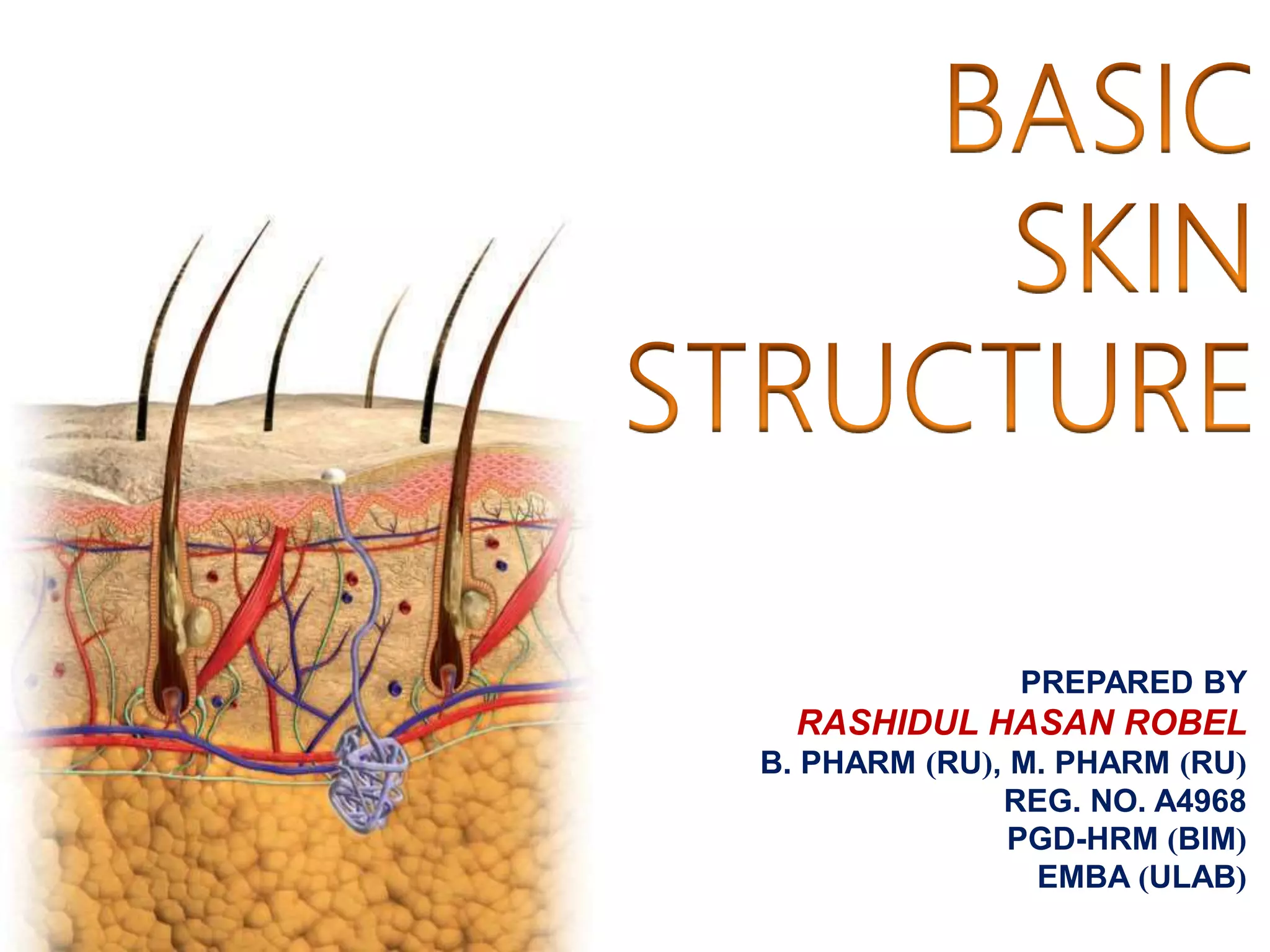
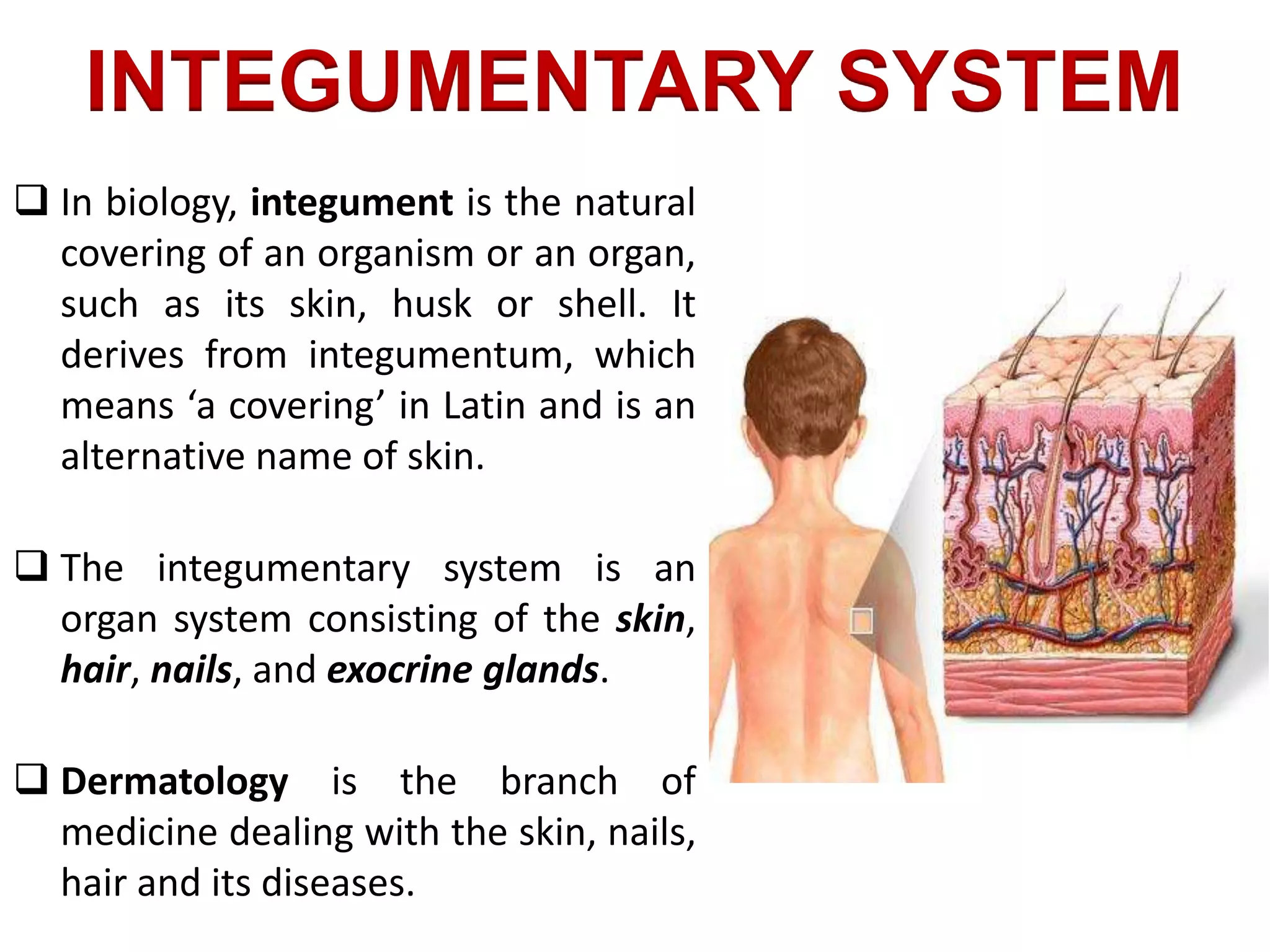
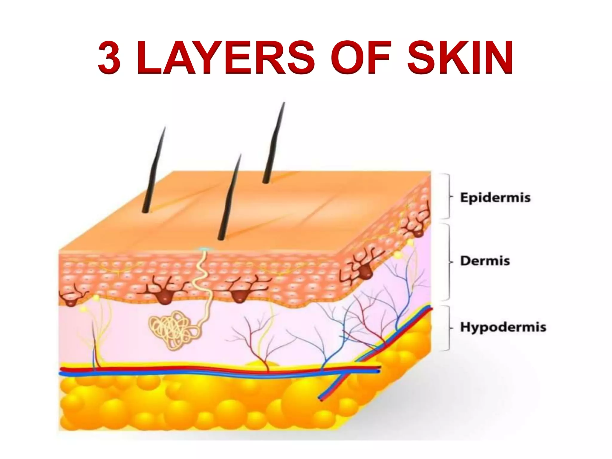
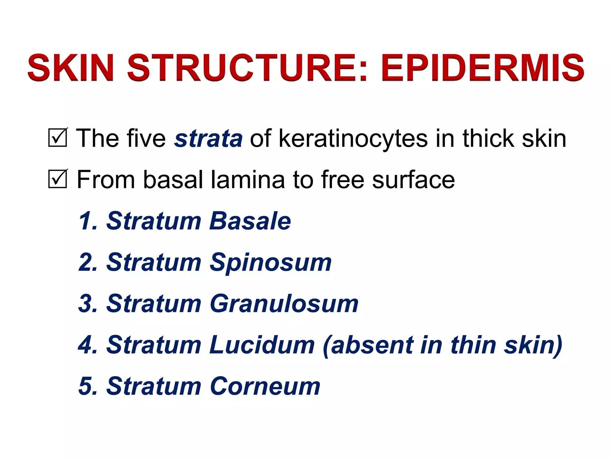
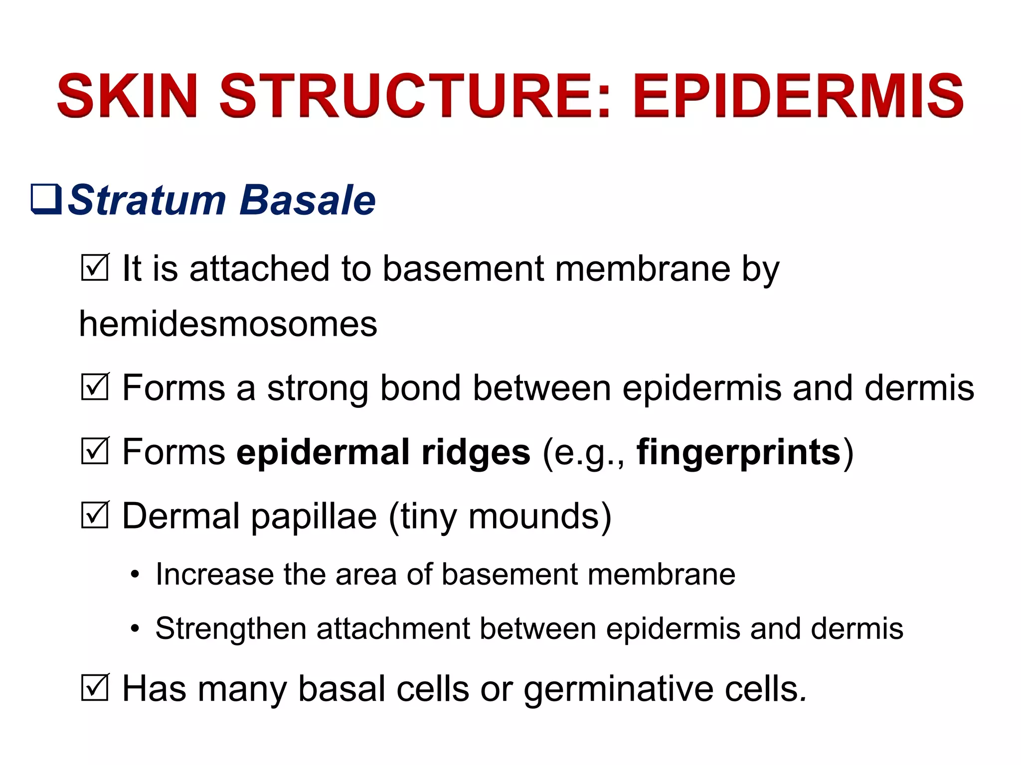
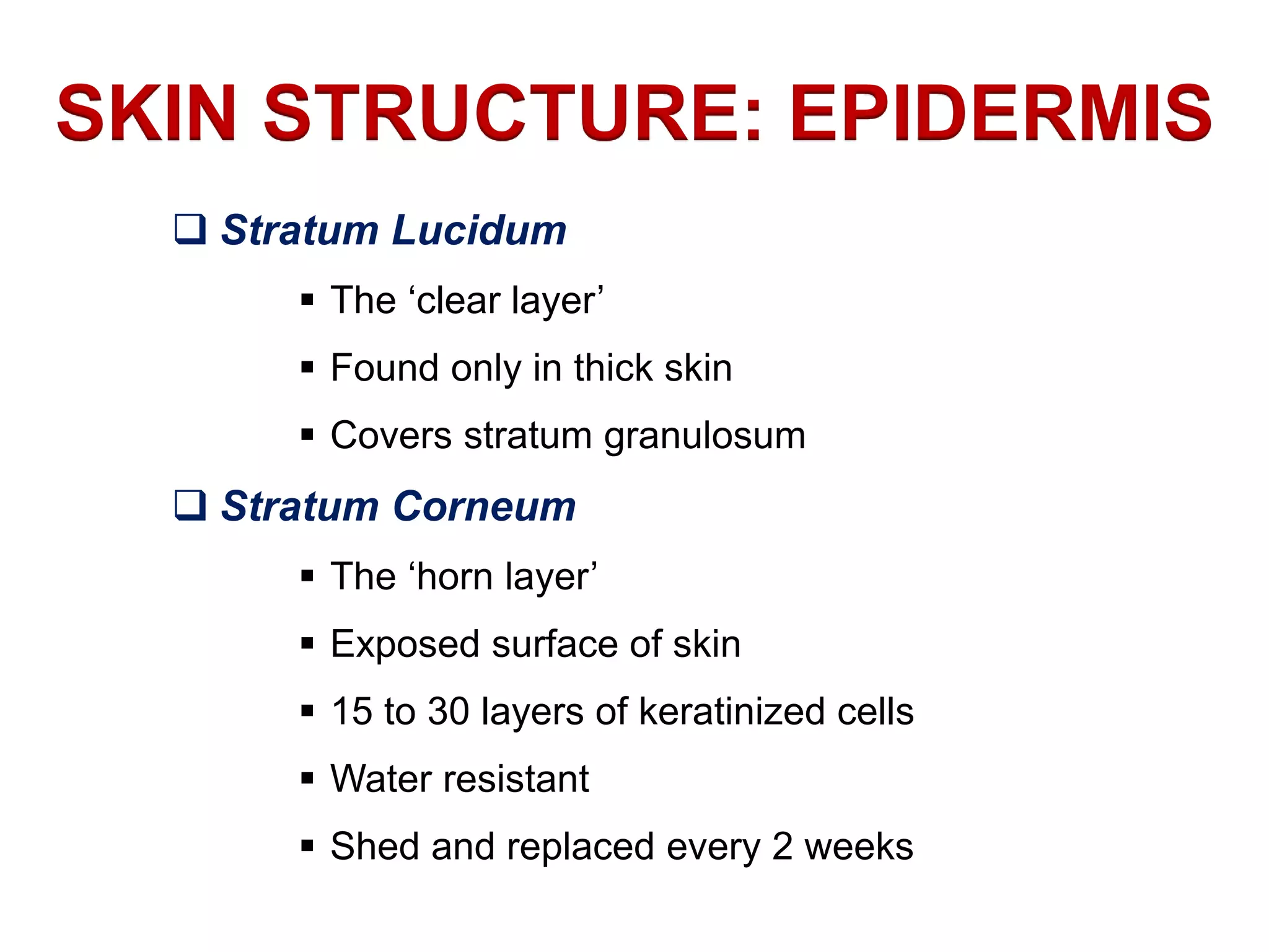
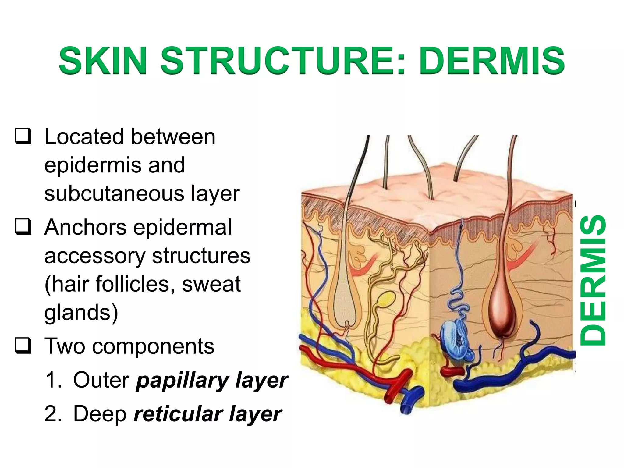
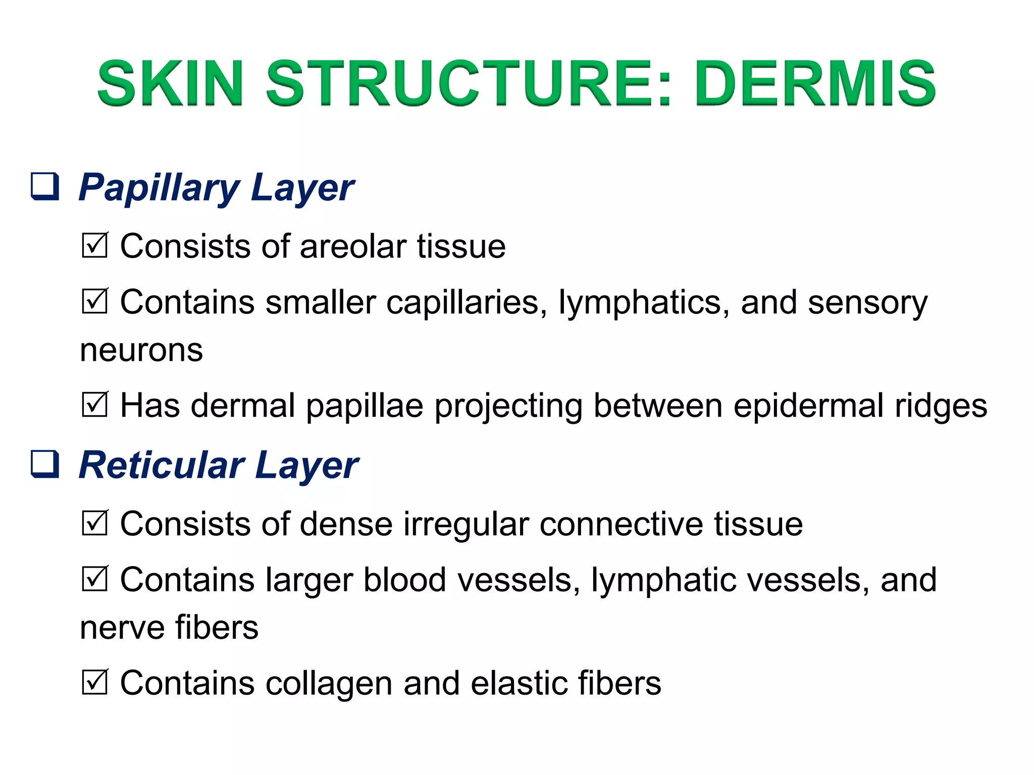
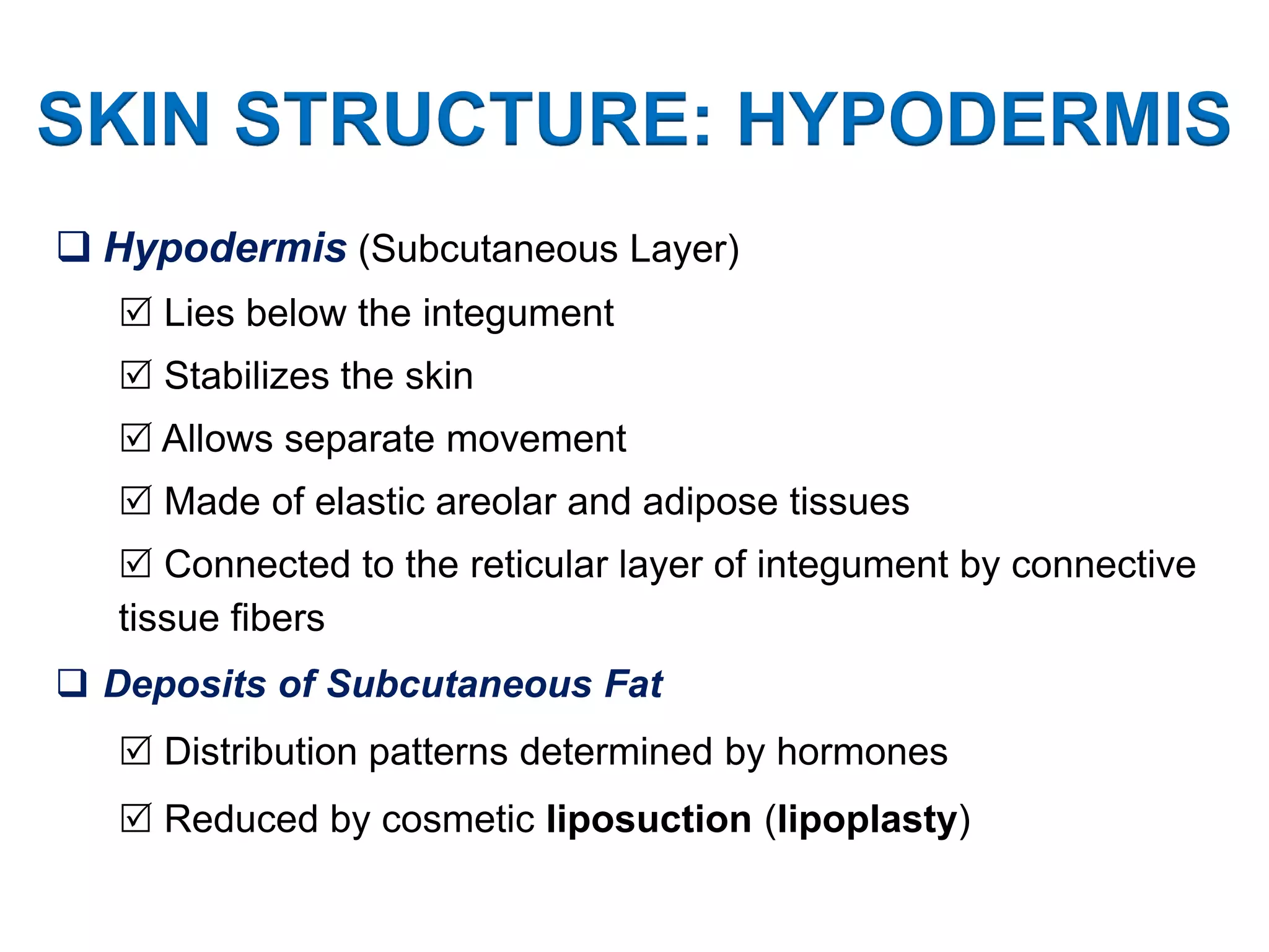
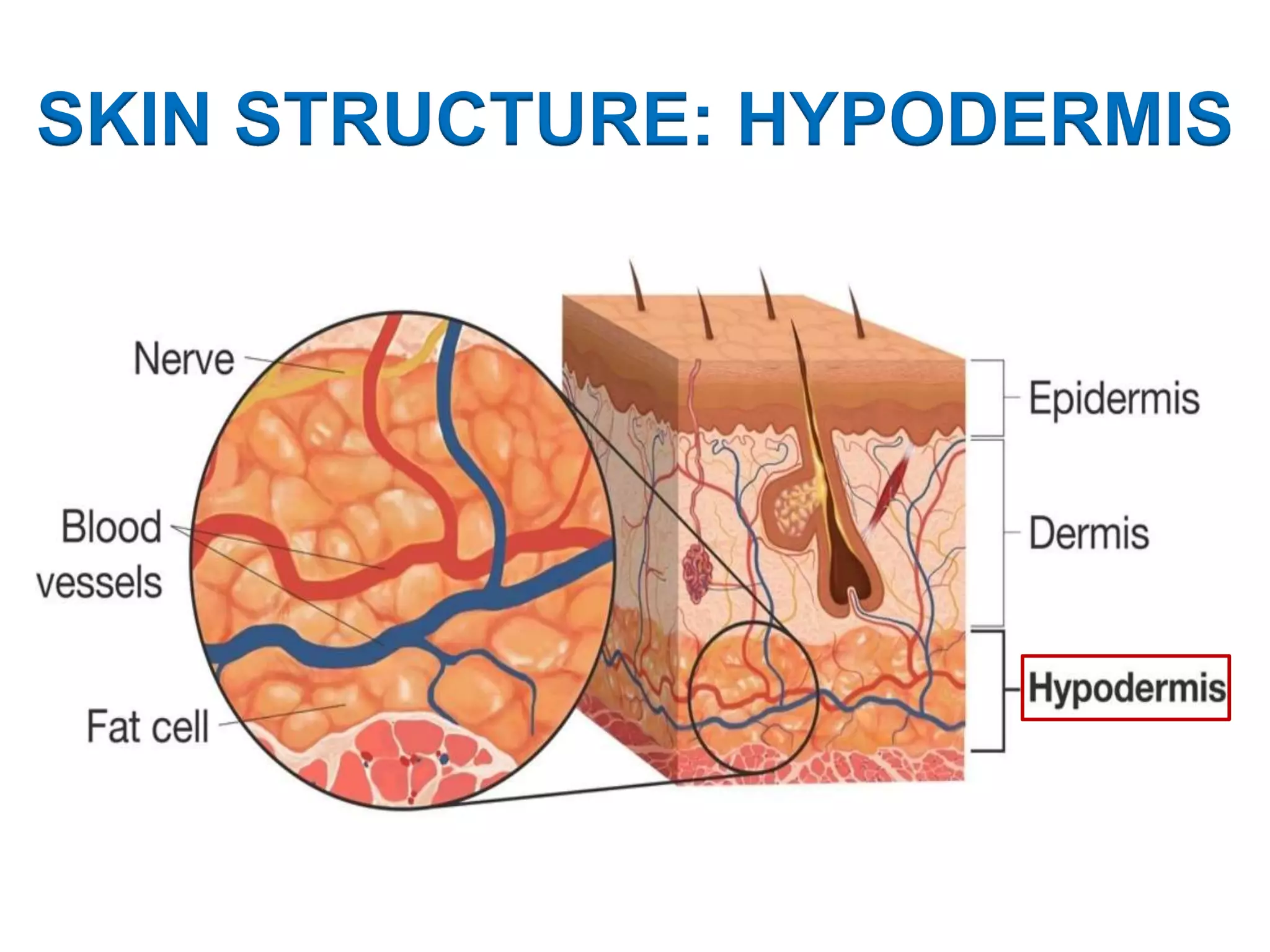
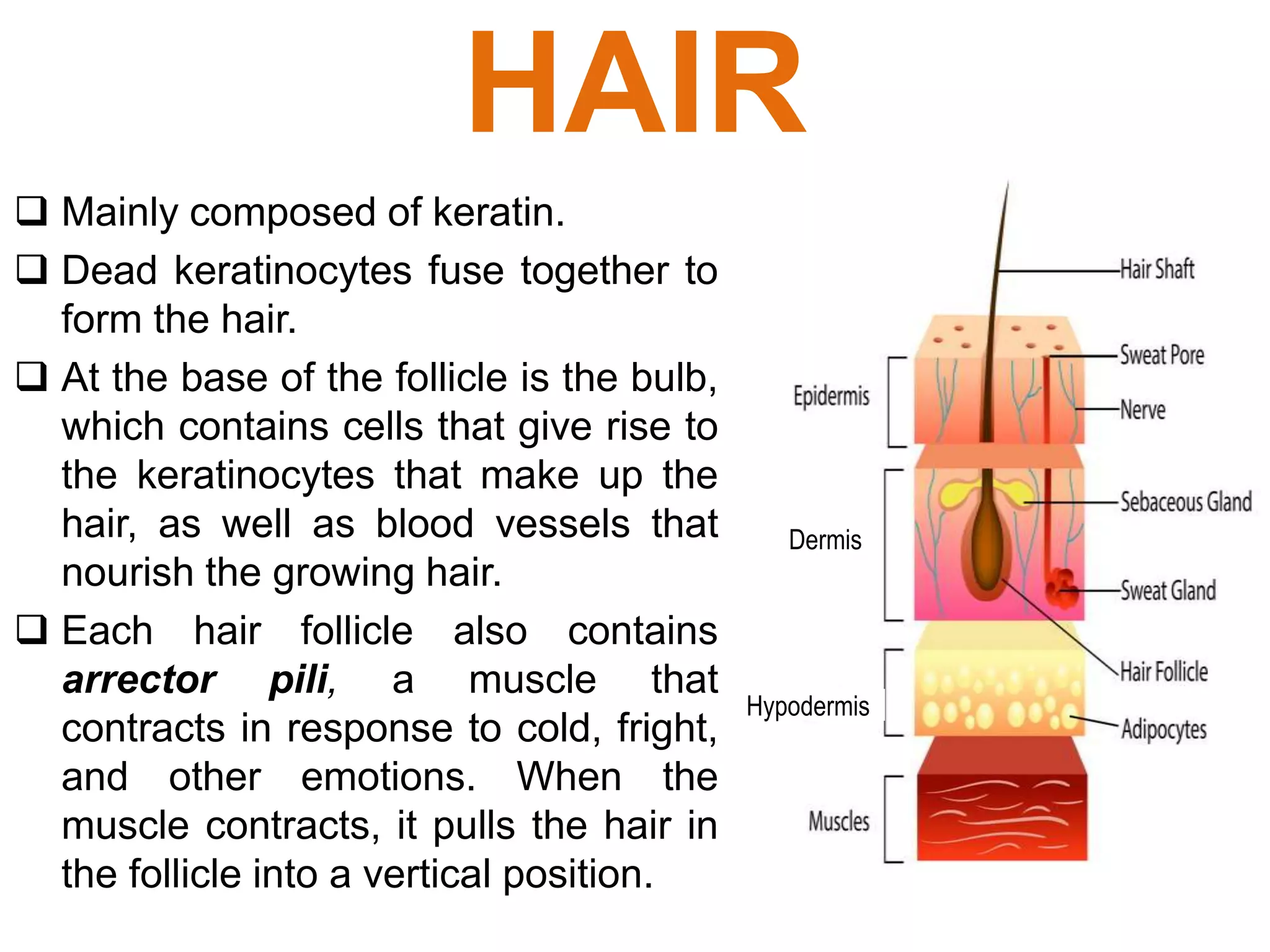
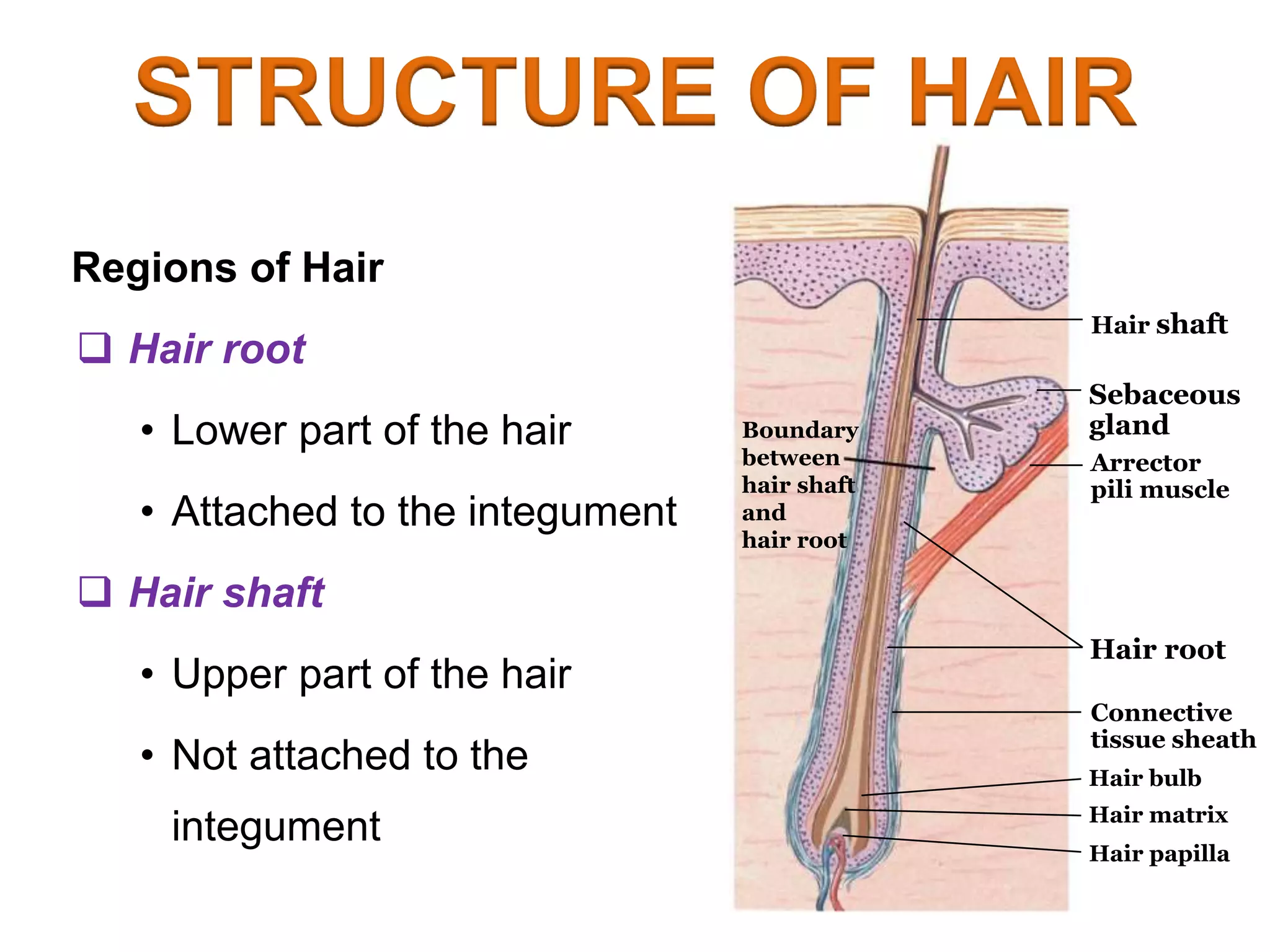
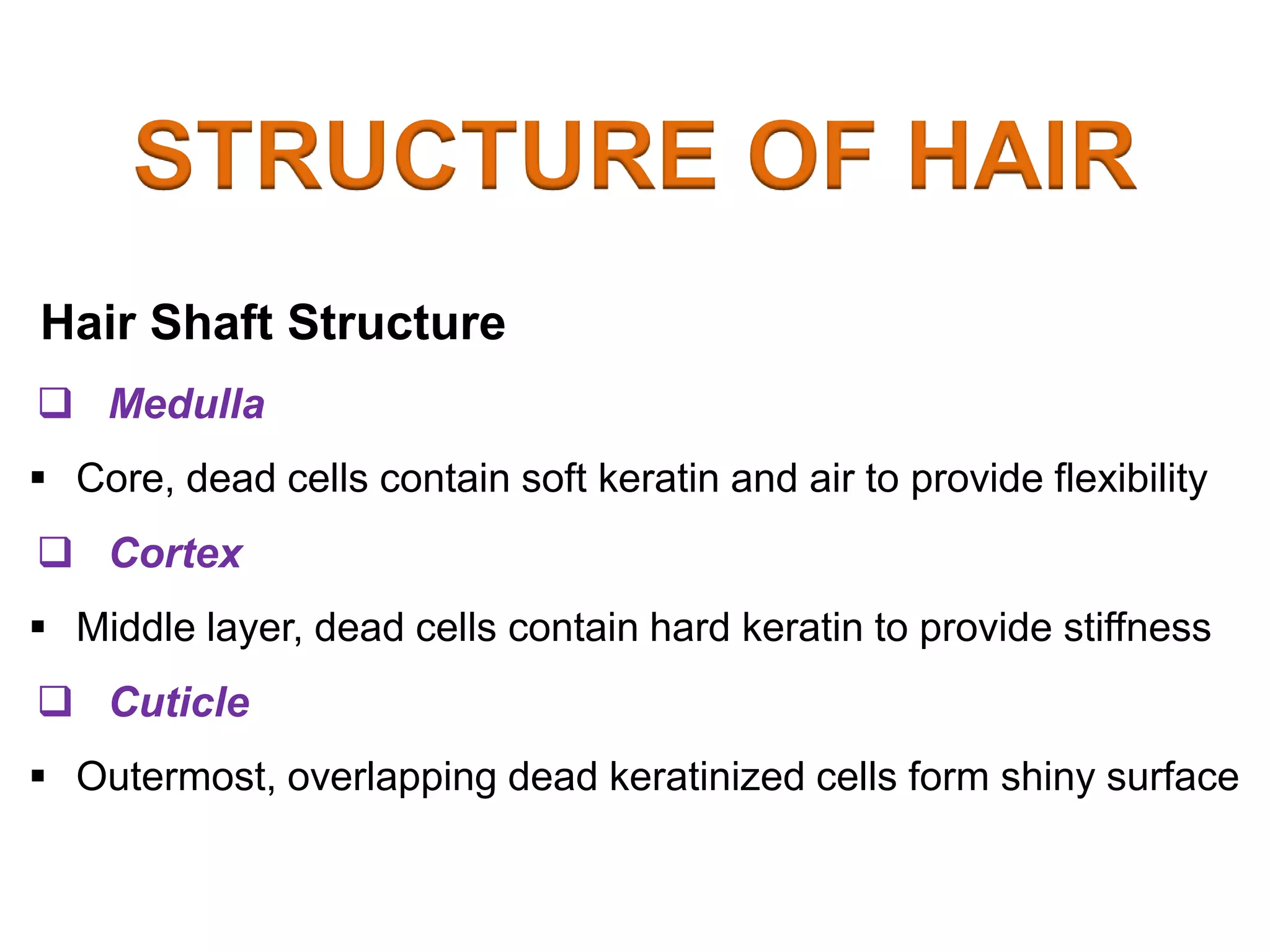
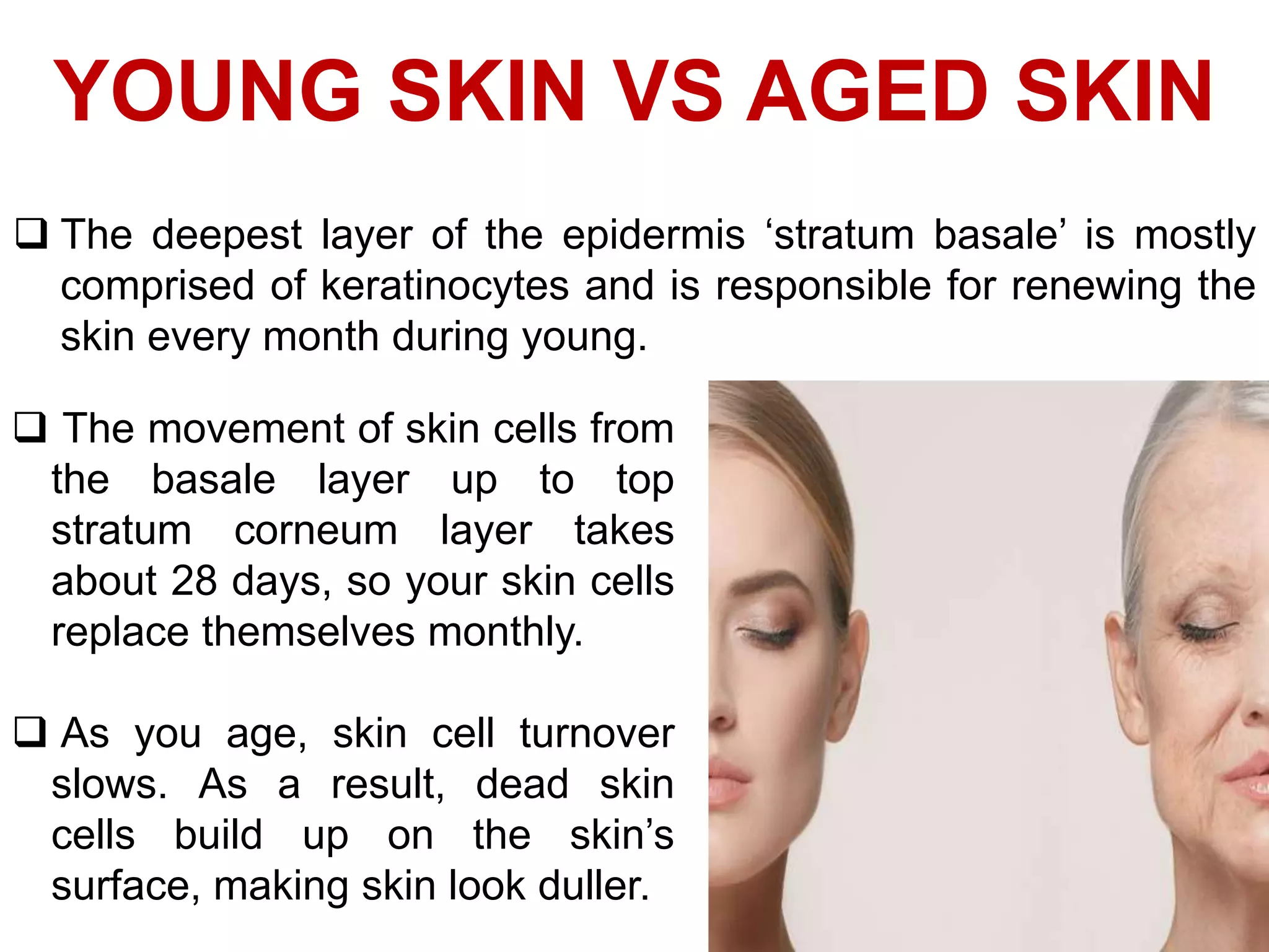
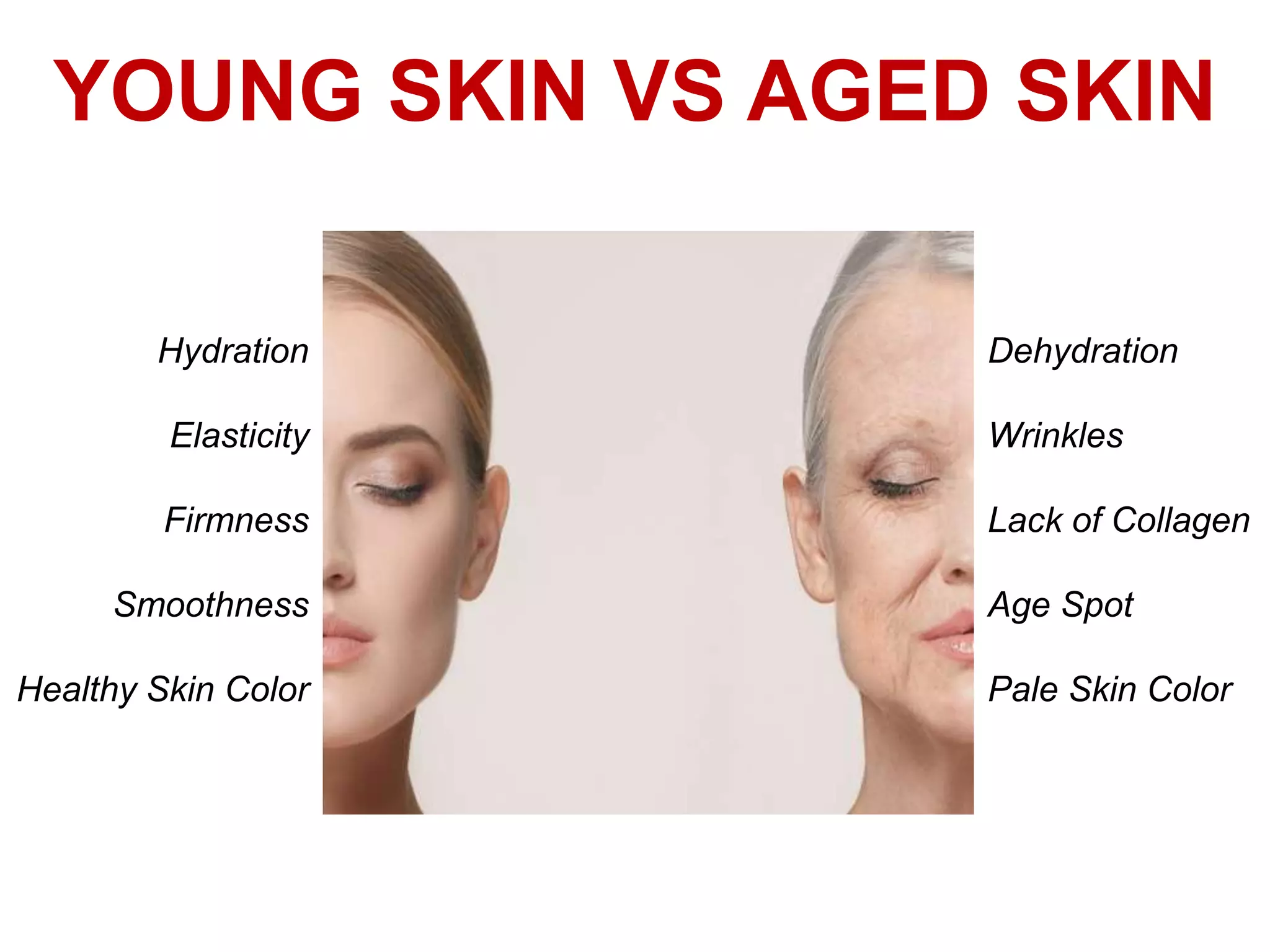
The skin is the largest organ of the body, with a total area of about 20 square feet. ... Skin has three layers: The epidermis, the outermost layer of skin, provides a waterproof barrier and creates our skin tone. The dermis, beneath the epidermis, contains tough connective tissue, hair follicles, and sweat glands.
The integumentary system is the organ system that protects the body from various kinds of damage, such as loss of water or abrasion from outside. The system comprises the skin and its appendages (including hair, scales, feathers, hooves, and nails). The integumentary system has a variety of functions; it may serve to waterproof, cushion, and protect the deeper tissues, excrete wastes, and regulate temperature, and is the attachment site for sensory receptors to detect pain, sensation, pressure, and temperature. In most terrestrial vertebrates with significant exposure to sunlight, the integumentary system also provides for vitamin D synthesis.
The integumentary system is an organ system consisting of the skin, hair, nails, and exocrine glands. The skin is only a few millimeters thick yet is by far the largest organ in the body. The average person's skin weighs 10 pounds and has a surface area of almost 20 square feet.
The integumentary system is the physical system that forms the barrier between the external environment and the internal systems of the body. In humans, this system consists of skin, hair, nails, and related glands.
Altogether, the integumentary system forms the largest organ in the body. The main function of this system is to protect bones, organs, and other internal structures from harm. In addition, the integumentary system performs important immune functions, cell fluid maintenance, synthesis of Vitamin D, body temperature regulation, and detection of stimuli.
Skin and nail ( the integumentary system)Ravish Yadav
The all the content in this profile is completed by the teachers, students as well as other health care peoples.
thank you, all the respected peoples, for giving the information to complete this presentation.
this information is free to use by anyone.
The branch of science concerned with the bodily structure of humans, animals, and other living organisms, especially as revealed by dissection and the separation of parts.
The integumentary system comprises the skin and its appendages. Skin + derivatives= Integument.
It aims to protect the body from various kinds of damage, such as loss of water or damages from outside.
The integumentary system in chordates includes hair, scales, feathers, hooves, and nails.
It may serve to water proof, and protect the deeper tissues.
Excrete wastes, and regulate body temperature.
It is the attachment site for sensory receptors to detect pain, sensation, pressure, and temperature.
Skin = integument
Skin+ accessory organs = integumentary system.
The integumentary system comprises the skin and its appendages acting to protect the body from various kinds of damage, such as loss of water or damages from outside.
It includes hair, glands and nails.
It has a variety of additional functions; it may serve to waterproof, and protect the deeper tissues, excrete wastes, and regulate body temperature, and is the attachment site for sensory receptors to detect pain, sensation, pressure, and temperature,vitamin D synthesis.
skin and its layuer
Epidermis
Dermis
Hypodermis ( subcutaneous layer)
Epidermis
Most superficial layer of the skin
Approx 10 to 30 cells thick ( epithelial)
Cell types
Keratinocytes -90%
Melanocytes
Merkel cells 10%
Langerhens
stratum basale
Stratum germinativum
Deepest epidermal layer
Attaches to basal lamina
Cells bond to dermis via collagen fibres
Finger like projection called dermal papillae in dermis
Helps in stronger connection
Cells- cuboidal shaped keratinocytes
Grows constantly ,mitosis
Pushed up old cells
Applied aspects
Cells – merkel cell – function as receptor- stimulating sensory nerve fibres
Found in hairless skin.
Abundant on surface of hand and feet.
Melanocytes- produces pigment melanin
- gives hair and skin its color
-protect from u rays
Note: skin color influenced by ,melanin and carotene – carrot, oxygenated haemoglobin
Fingerprints-epidermal ridges
Stratum spinosum
Spiny in appearance
Desmosomsis protruding cell
Interlock between cells
Composed of 8to 10 layers of keratinocytes
Applied aspects
Langerhans cell –as a macrophage by engulfing bacteria and foreign body and damaged cell.
Keratinocytes synthesise keratin and prevent water loss
Stratum granulosam
Has a grainy appearance
Cells become flattened,and cell membrane thicken, and generate large amount of protein keratin and keratohyalin
After cell dies , keratin ,keratohyalin and cell membrane forms stratum lucidum
And accessory structures of hair and nail
Stratum lucidum
Smooth translucent layer
It is found only in thick skin of palms ,soles,digits
Cells are densely packed with eleidin , a clear protein rich in lipids,
Transparent appearance
Stratum corneam
Most superficial layer of epidermis
The increased keratinization of cell
It has 15 to 30 layers of cells
Water is lost from skin in 2 ways
Insensible perspiration
water diffuses from stratum corneum and evaporates from skin
-500 ml per day
Sensible perspiration
Water excreted by sweat glands.
clinical application
Some medications are toxic if swallowed, but safe if used topically (applied to surface of skin)
• Certain topical antibiotics are fairly toxic if taken by mouth, butcan be applied to skin with minimal risk of systemic absorption;they are polar molecules that cannot pass through epidermis toreach blood vessels in dermis; allows for local effect only
• Nonpolar substances cross epidermis much more easily;provides a c
Acetabularia Information For Class 9 .docxvaibhavrinwa19
Acetabularia acetabulum is a single-celled green alga that in its vegetative state is morphologically differentiated into a basal rhizoid and an axially elongated stalk, which bears whorls of branching hairs. The single diploid nucleus resides in the rhizoid.
Introduction to AI for Nonprofits with Tapp NetworkTechSoup
Dive into the world of AI! Experts Jon Hill and Tareq Monaur will guide you through AI's role in enhancing nonprofit websites and basic marketing strategies, making it easy to understand and apply.
The Roman Empire A Historical Colossus.pdfkaushalkr1407
The Roman Empire, a vast and enduring power, stands as one of history's most remarkable civilizations, leaving an indelible imprint on the world. It emerged from the Roman Republic, transitioning into an imperial powerhouse under the leadership of Augustus Caesar in 27 BCE. This transformation marked the beginning of an era defined by unprecedented territorial expansion, architectural marvels, and profound cultural influence.
The empire's roots lie in the city of Rome, founded, according to legend, by Romulus in 753 BCE. Over centuries, Rome evolved from a small settlement to a formidable republic, characterized by a complex political system with elected officials and checks on power. However, internal strife, class conflicts, and military ambitions paved the way for the end of the Republic. Julius Caesar’s dictatorship and subsequent assassination in 44 BCE created a power vacuum, leading to a civil war. Octavian, later Augustus, emerged victorious, heralding the Roman Empire’s birth.
Under Augustus, the empire experienced the Pax Romana, a 200-year period of relative peace and stability. Augustus reformed the military, established efficient administrative systems, and initiated grand construction projects. The empire's borders expanded, encompassing territories from Britain to Egypt and from Spain to the Euphrates. Roman legions, renowned for their discipline and engineering prowess, secured and maintained these vast territories, building roads, fortifications, and cities that facilitated control and integration.
The Roman Empire’s society was hierarchical, with a rigid class system. At the top were the patricians, wealthy elites who held significant political power. Below them were the plebeians, free citizens with limited political influence, and the vast numbers of slaves who formed the backbone of the economy. The family unit was central, governed by the paterfamilias, the male head who held absolute authority.
Culturally, the Romans were eclectic, absorbing and adapting elements from the civilizations they encountered, particularly the Greeks. Roman art, literature, and philosophy reflected this synthesis, creating a rich cultural tapestry. Latin, the Roman language, became the lingua franca of the Western world, influencing numerous modern languages.
Roman architecture and engineering achievements were monumental. They perfected the arch, vault, and dome, constructing enduring structures like the Colosseum, Pantheon, and aqueducts. These engineering marvels not only showcased Roman ingenuity but also served practical purposes, from public entertainment to water supply.
Francesca Gottschalk - How can education support child empowerment.pptxEduSkills OECD
Francesca Gottschalk from the OECD’s Centre for Educational Research and Innovation presents at the Ask an Expert Webinar: How can education support child empowerment?
Unit 8 - Information and Communication Technology (Paper I).pdfThiyagu K
This slides describes the basic concepts of ICT, basics of Email, Emerging Technology and Digital Initiatives in Education. This presentations aligns with the UGC Paper I syllabus.
Palestine last event orientationfvgnh .pptxRaedMohamed3
An EFL lesson about the current events in Palestine. It is intended to be for intermediate students who wish to increase their listening skills through a short lesson in power point.
Macroeconomics- Movie Location
This will be used as part of your Personal Professional Portfolio once graded.
Objective:
Prepare a presentation or a paper using research, basic comparative analysis, data organization and application of economic information. You will make an informed assessment of an economic climate outside of the United States to accomplish an entertainment industry objective.
How to Make a Field invisible in Odoo 17Celine George
It is possible to hide or invisible some fields in odoo. Commonly using “invisible” attribute in the field definition to invisible the fields. This slide will show how to make a field invisible in odoo 17.
Read| The latest issue of The Challenger is here! We are thrilled to announce that our school paper has qualified for the NATIONAL SCHOOLS PRESS CONFERENCE (NSPC) 2024. Thank you for your unwavering support and trust. Dive into the stories that made us stand out!
2024.06.01 Introducing a competency framework for languag learning materials ...Sandy Millin
http://sandymillin.wordpress.com/iateflwebinar2024
Published classroom materials form the basis of syllabuses, drive teacher professional development, and have a potentially huge influence on learners, teachers and education systems. All teachers also create their own materials, whether a few sentences on a blackboard, a highly-structured fully-realised online course, or anything in between. Despite this, the knowledge and skills needed to create effective language learning materials are rarely part of teacher training, and are mostly learnt by trial and error.
Knowledge and skills frameworks, generally called competency frameworks, for ELT teachers, trainers and managers have existed for a few years now. However, until I created one for my MA dissertation, there wasn’t one drawing together what we need to know and do to be able to effectively produce language learning materials.
This webinar will introduce you to my framework, highlighting the key competencies I identified from my research. It will also show how anybody involved in language teaching (any language, not just English!), teacher training, managing schools or developing language learning materials can benefit from using the framework.
2. INTEGUMENTARY SYSTEM
In biology, integument is the natural
covering of an organism or an organ,
such as its skin, husk or shell. It
derives from integumentum, which
means ‘a covering’ in Latin and is an
alternative name of skin.
The integumentary system is an
organ system consisting of the skin,
hair, nails, and exocrine glands.
Dermatology is the branch of
medicine dealing with the skin, nails,
hair and its diseases.
3. SKIN
• Largest organ of the body
• Completely covers the body
• Average thickness: 1-2 mm. (0.5mm eyelids, 6mm palms and soles)
• pH: 4 - 5.6
• Renewal of skin takes place in 28-50 days by
shedding of the outer layer.
The thin layer of tissue forming the natural outer
covering of the body of a person or animal. Skin is an
organ because it consists of different tissues that are
joined to perform a specific function.
4. FUNCTIONS OF SKIN
1. Protection
- Physical barrier that protects underlying tissues from injury,
UV light and bacterial invasion.
- Mechanical barrier is part non specific immunity (skin,
tears and saliva).
2. Regulation of Body Temperature
- High temperature or strenuous exercise; sweat is evaporated
from the skin surface to cool it down.
- vasodilation (increases blood flow) and vasoconstriction
(decrease in blood flow) regulates body temperature.
5. 3. Sensation
- Nerve endings and receptor cells that detect stimuli to
temperature, pain, pressure and touch.
4. Excretion
- Sweat removes water and small amounts of salt, uric acid
and ammonia from the body surface
5. Blood Reservoir
- Dermis houses an extensive network of blood vessels
carrying 8-10% of total blood flow in a resting adult.
6. Synthesis of Vitamin D (cholecalciferol)
- UV rays in sunlight stimulate the production of Vit-D.
Enzymes in the kidney and liver modify and convert to final
form; calcitriol (most active form of Vit-D.) Calcitriol aids
in absorption of calcium from foods and is considered a
hormone.
8. EPIDERMIS
Epidermis
It is avascular stratified squamous epithelium
Nutrients and oxygen diffuse from capillaries in the dermis
Cells of Epidermis
Keratinocytes (90%) : Waterproofs & protects skin, nails, hair,
stratum corneum and also provides rigidity by producing tough,
triple-helix-shaped protein strand called Keratin.
Melanocytes (8%) : Produce melanin that gives skin color
Merkel Cells : Slowly adapting mechanoreceptors that
mediate the senses of touch and hair movement.
Langerhans’ Cells : Immunological defense
9. EPIDERMIS
The structural relationship and
interface between the epidermis
and underlying dermis. The
proportions of the various layers
differ with the location sampled.
Dermis
Epidermis
Epidermal
ridge
Dermal
papilla
10. SKIN STRUCTURE: EPIDERMIS
Thin Skin
Covers most of the body
Has four layers of keratinocytes
Thick Skin
Covers the palms of the hands and soles of the feet
Has five layers of keratinocytes
11. SKIN STRUCTURE: EPIDERMIS
The five strata of keratinocytes in thick skin
From basal lamina to free surface
1. Stratum Basale
2. Stratum Spinosum
3. Stratum Granulosum
4. Stratum Lucidum (absent in thin skin)
5. Stratum Corneum
13. SKIN STRUCTURE: EPIDERMIS
Stratum Basale
It is attached to basement membrane by
hemidesmosomes
Forms a strong bond between epidermis and dermis
Forms epidermal ridges (e.g., fingerprints)
Dermal papillae (tiny mounds)
• Increase the area of basement membrane
• Strengthen attachment between epidermis and dermis
Has many basal cells or germinative cells.
14. Thick skin SEM 25
Epidermal
ridge
Pores of sweat
gland ducts
Epidermal ridges
15. SKIN STRUCTURE: EPIDERMIS
Stratum Spinosum
the ‘spiny layer’
Produced by division of stratum basale
8-10 layers of keratinocytes bound by desmosomes
Cells shrink until cytoskeletons stick out (spiny)
Continue to divide, increasing thickness of epithelium
Contain dendritic (Langerhans) cells, active in immune
response
16. SKIN STRUCTURE: EPIDERMIS
Stratum Granulosum
The ‘grainular layer’
Stops dividing, starts producing keratin & keratohyalin
• Keratin
a) A tough, fibrous protein
b) Makes up hair and nails
• Keratohyalin
a) Dense granules
b) Cross-link keratin fibers
17. SKIN STRUCTURE: EPIDERMIS
Stratum Lucidum
The ‘clear layer’
Found only in thick skin
Covers stratum granulosum
Stratum Corneum
The ‘horn layer’
Exposed surface of skin
15 to 30 layers of keratinized cells
Water resistant
Shed and replaced every 2 weeks
18. Located between
epidermis and
subcutaneous layer
Anchors epidermal
accessory structures
(hair follicles, sweat
glands)
Two components
1. Outer papillary layer
2. Deep reticular layer
SKIN STRUCTURE: DERMIS
DERMIS
19. SKIN STRUCTURE: DERMIS
Papillary Layer
Consists of areolar tissue
Contains smaller capillaries, lymphatics, and sensory
neurons
Has dermal papillae projecting between epidermal ridges
Reticular Layer
Consists of dense irregular connective tissue
Contains larger blood vessels, lymphatic vessels, and
nerve fibers
Contains collagen and elastic fibers
21. SKIN STRUCTURE: HYPODERMIS
Hypodermis (Subcutaneous Layer)
Lies below the integument
Stabilizes the skin
Allows separate movement
Made of elastic areolar and adipose tissues
Connected to the reticular layer of integument by connective
tissue fibers
Deposits of Subcutaneous Fat
Distribution patterns determined by hormones
Reduced by cosmetic liposuction (lipoplasty)
24. HAIR
Mainly composed of keratin.
Dead keratinocytes fuse together to
form the hair.
At the base of the follicle is the bulb,
which contains cells that give rise to
the keratinocytes that make up the
hair, as well as blood vessels that
nourish the growing hair.
Each hair follicle also contains
arrector pili, a muscle that
contracts in response to cold, fright,
and other emotions. When the
muscle contracts, it pulls the hair in
the follicle into a vertical position.
Dermis
Hypodermis
25. This response may help
some mammals keep
warm or look bigger to
frighten or intimidate
their enemies.
But in humans, because
of our sparse coat of
body hair, it merely
produces ‘goose bumps’.
26. STRUCTURE OF HAIR
Hair Follicle
Hair follicles are the organs that form the hairs
Located deep in dermis
Produces nonliving hairs
Wrapped in a dense connective tissue sheath
Base is surrounded by sensory nerves (root hair plexus)
Control bacteria
28. Regions of Hair
Hair root
• Lower part of the hair
• Attached to the integument
Hair shaft
• Upper part of the hair
• Not attached to the
integument
Boundary
between
hair shaft
and
hair root
Hair shaft
Sebaceous
gland
Arrector
pili muscle
Hair root
Connective
tissue sheath
Hair bulb
Hair matrix
Hair papilla
STRUCTURE OF HAIR
29. Hair Shaft Structure
Medulla
Core, dead cells contain soft keratin and air to provide flexibility
Cortex
Middle layer, dead cells contain hard keratin to provide stiffness
Cuticle
Outermost, overlapping dead keratinized cells form shiny surface
STRUCTURE OF HAIR
30. Hair on Head
UV protection
Cushion from trauma
Insulation
Nostrils, Ear canals, Eyelashes
Prevent entry of foreign material
Body Hair
Sensory detection
Root Hair Plexus
Sensory nerves at base of hair follicle that detect slight
movement of hair
Arrector Pili Muscle
Attached to every hair follicle
Contract to stand hair perpendicular to skin surface
FUNCTIONS OF HAIR
31. STRUCTURE & FUNCTION OF NAIL
Nails
Protect fingers and toes
Made of dead cells packed with keratin
Metabolic disorders can change nail
structure
Nail Production
Occurs in a deep epidermal fold near
the bone called the nail root
Free edge
of Nail
Body of
Nail
Laternal
Nail fold
Lunula
Eponychium
(cuticle)
32. STRUCTURE & FUNCTION OF NAIL
Nail Structure
Nail body
The visible portion of the nail
Covers the nail bed
Lunula
The pale crescent at the base of the nail
Nails Sides
Lie in lateral nail grooves
Surrounded by lateral nail folds
35. SKIN GLANDS
An adult human has between 1.6 to 4
million sudoriferous glands, or sweat
glands.
Most common type called Eccrine
Sweat Glands, which are found almost
all over the surface of the body and are
most numerous on the palms and soles.
These glands begin deep in the dermis
and connect to the surface of the skin
by a coiled duct.
In addition, nerve fibers that encircle the
sweat glands stimulate the glands in
response to fear, excitement, or anxiety.
Sweat Glands
36. Other sweat glands, known as Apocrine Sweat Glands, are
much less numerous than eccrine sweat glands. They are also
anchored deep in the dermis, but open into hair follicles rather
than onto the surface of the skin.
Apocrine sweat glands are located mainly in the armpit, genital
area, and around the nipples of the breasts.
37. Oil or sebaceous glands are found all
over the body except on the palms, the
soles, and the top of the feet.
These are most numerous on the face
and scalp.
Most sebaceous glands open into hair
follicles.
But the glands also occur in some
hairless areas, such as the lips and
inside the mouth.
It produces an oily substance called
Sebum, which keeps the skin and hair
from drying out and inhibits the growth
of certain harmful bacteria.
Oil Glands
38. Wax or ceruminous
glands are located in
the ear canal.
They secrete a waxy
substance that helps
prevent foreign particles
from entering the ear.
These glands are
modified sweat glands.
Wax Glands
39. SKIN COLOR
(Sub-Saharan African, Indian, Southern European, and Northwest European)
Skin Color Reason: Melanin, Carotene & Hemoglobin
Melanin Pigments
Eumelanin
Pheomelanin
High Eumelanin & Low Pheomelanin - Darker skin and hair color
Low Eumelanin & High Pheomelanin - Lighter skin and hair color
40. YOUNG SKIN VS AGED SKIN
The deepest layer of the epidermis ‘stratum basale’ is mostly
comprised of keratinocytes and is responsible for renewing the
skin every month during young.
The movement of skin cells from
the basale layer up to top
stratum corneum layer takes
about 28 days, so your skin cells
replace themselves monthly.
As you age, skin cell turnover
slows. As a result, dead skin
cells build up on the skin’s
surface, making skin look duller.
41. When you age, collagen
production decreases
and your skin’s ability to
repair collagen and
elastin decreases,
resulting in wrinkles and
loss of elasticity.
Young skin has thick
bundles of tightly packed
collagen while older skin
has thin, loose bundles
of collagen.
YOUNG SKIN VS AGED SKIN
42. As you get older, there’s decreased blood flow
through the dermal capillaries.
Fewer nutrients reach the epidermis, leading to
thinning of the epidermis, slower cell turnover
rate and a reduction in the barrier function of the
epidermis.
These changes lead to increased water loss
and dry skin. Skin cells have 30 percent less
natural moisture.
YOUNG SKIN VS AGED SKIN