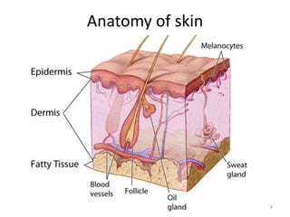
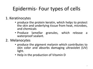
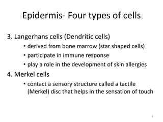
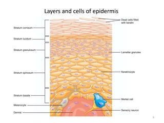
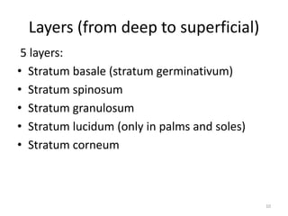
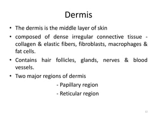
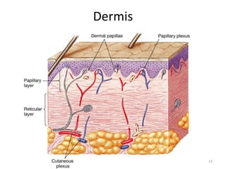
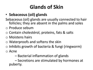
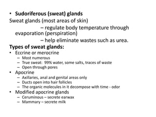
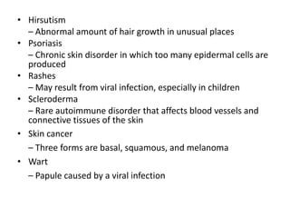
The Integumentary System
1
Integumentary system
• Integument is skin
• Integumentary system is the skin and the
organs derived from it (hair, glands, nails)
• One of the largest organs
– surface area of between - 1.5-2.0 square meters ,
– total average weight :10-11 lbs.
– Largest sense organ in the body
• The study of the skin is Dermatology
2
Functions of skin
1.Covers and protects the body from
– Pathogens
– Injury
– Ultra-violet radiation
2. Regulate body temperature through
– Sweating
– Dilatation/constriction of blood vessels
3. Excretes Waste as
- Urea
- sweat
4. Reduces water loss - Keeps the body from drying out
5. Houses sensory receptors
3
4
Anatomy of skin
Anatomy of skin
• Epidermis
• Dermis
• Subcutaneous layer or hypodermis
5
Epidermis
• Stratified squamous epithelium
– Avascular (contains no blood vessels)
– Most of the cells in the epidermis are
keratinocytes.
Inner part is composed of living cells
Outer part is composed of dead cells
6
Epidermis- Four types of cells
1. Keratinocytes
• produce the protein keratin, which helps to protect
the skin and underlying tissue from heat, microbes,
and chemicals
• Produce lamellar granules, which release a
waterproof sealant.
2. Melanocytes
• produce the pigment melanin which contributes to
skin color and absorbs damaging ultraviolet (UV)
light.
• Help in the production of Vitamin D
7
3. Langerhans cells (Dendritic cells)
• derived from bone marrow (star shaped cells)
• participate in immune response
• play a role in the development of skin allergies
4. Merkel cells
• contact a sensory structure called a tactile
(Merkel) disc that helps in the sensation of touch
8
Epidermis- Four types of cells
Layers and cells of epidermis
9
Layers (from deep to superficial)
5 layers:
• Stratum basale (stratum germinativum)
• Stratum spinosum
• Stratum granulosum
• Stratum lucidum (only in palms and soles)
• Stratum corneum
10
– Stratum basale or germinativum – single row of
cells attached to dermis; youngest cells
– Stratum spinosum – provides strength and
flexibility to the skin, 8 to 10 cell layers are held
together by desmosomes.
– Stratum granulosum – layers of flattened
keratinocytes, producing keratin (hair and nails
made of it also)
– Stratum lucidum (only on palms and soles)
– Stratum corneum – horny layer (cells dead, many
layers thick)
11
Layers (from deep to superficial)
Dermis
• The dermis is the middle layer of skin
• composed of dense irregular connective tissue -
collagen & elastic fibers, fibroblasts, macrophages &
fat cells.
• Contains hair follicles, glands, nerves & blood
vessels.
• Two major regions of dermis
- Papillary region
- Reticular region
12
13
Dermis
Dermis - Papillary Region
• Top 20% of dermis
• Finger like projections are called dermal papillae
– anchors epidermis to dermis
– contains capillaries that feed epidermis
– contains Meissner’s corpuscles (a sensory
nerve ending that is sensitive to mechanical
stimuli like touch) & free nerve endings for
sensations of heat, cold, pain, tickle, and itch
14
Dermis
Dermis - Reticular Region
• Dense irregular connective tissue
• Contains interlacing collagen and elastic fibers
• Packed with oil glands, sweat gland ducts, fat & hair follicles
• Provides strength, extensibility & elasticity to skin
– Stretch marks are dermal tears from extreme
stretching
• Epidermal ridges form in fetus
– Fingerprints are left by sweat glands open on ridges
– Increase grip of hand
Dermis
Hypodermis
• Hypodermis (Gk) = below the skin
• “Subcutaneous” (Latin) = below the skin
• Also called “superficial fascia”
“fascia” (Latin) =band; in anatomy: sheet of
connective tissue
• Fatty tissue which stores fat and anchors skin
• Attaches the reticular layer to the underlying organs
• It contains larger blood vessels and nerves than
those found in the dermis.
16
Hypodermis
Hypodermis
• It is essentially composed of a type of cells specialised
in accumulating and storing fats, known as adipocytes.
• These cells are grouped together in lobules separated
by connective tissue - collagen and elastin fibres
• The number of adipocytes varies among different areas
of the body, while their size varies according to the
body's nutritional state.
• It acts as energy reserve
• provides some minor thermoregulation via insulation
Skin Appendages or Accessory
Organs of Skin
• Derived from epidermis but extend into dermis
• Include
– Hair and hair follicles
– Sebaceous (oil) glands
– Sweat (sudoiferous) glands
– Nails
Skin Appendages
Hair
Hairs are present on most skin surfaces except the
palms and soles.
Hair consists of:
• Shaft -- visible
• Root -- below the surface
• Follicle surrounds root
– External root sheath
– Internal root sheath
– Base of follicle is bulb
• Blood vessels
Hair
Nails
• Tightly packed keratinized cells
• Nail body
– Visible portion pink due to underlying
capillaries
– Free edge appears white
• Nail root
– buried under skin layers
– lunula is white due to thickened stratum
basale
• Eponychium (cuticle)
Nails
Glands of Skin
• Sebaceous (oil) glands
Sebaceous (oil) glands are usually connected to hair
follicles; they are absent in the palms and soles
o Produce sebum
o Contain cholestérol, proteins, fats & salts
o Moistens hairs
o Waterproofs and softens the skin
o Inhibits growth of bacteria & fungi (ringworm)
o Acne
– Bacterial inflammation of glands
– Secretions are stimulated by hormones at
puberty.
Glands of the Skin
• Sudoriferous (sweat) glands
Sweat glands (most areas of skin)
– regulate body temperature through
evaporation (perspiration)
– help eliminate wastes such as urea.
Types of sweat glands:
• Eccrine or merocrine
– Most numerous
– True sweat: 99% water, some salts, traces of waste
– Open through pores
• Apocrine
– Axillaries, anal and genital areas only
– Ducts open into hair follicles
– The organic molecules in it decompose with time - odor
• Modified apocrine glands
– Ceruminous – secrete earwax
– Mammary – secrete milk
Disorders of the Integumentary System
• Acne vulgaris
– Caused by increased secretion of oil related to increased
hormones during puberty
• Albinism
– Inherited disorder in which melanin is not produced
• Alopecia
– Baldness
• Cellulitis
– Bacterial infection of the dermis and subcutaneous layer of
the skin
• Dandruff
– White flakes of dead skin cells from the scalp
• Eczema
– Group of disorders caused by allergic or irritant reactions
• Hirsutism
– Abnormal amount of hair growth in unusual places
• Psoriasis
– Chronic skin disorder in which too many epidermal cells are
produced
• Rashes
– May result from viral infection, especially in children
• Scleroderma
– Rare autoimmune disorder that affects blood vessels and
connective tissues of the skin
• Skin cancer
– Three forms are basal, squamous, and melanoma
• Wart
– Papule caused by a viral infection

03. Integumentary system_part-1.pptx

  • 1.
  • 2.
    Integumentary system • Integumentis skin • Integumentary system is the skin and the organs derived from it (hair, glands, nails) • One of the largest organs – surface area of between - 1.5-2.0 square meters , – total average weight :10-11 lbs. – Largest sense organ in the body • The study of the skin is Dermatology 2
  • 3.
    Functions of skin 1.Coversand protects the body from – Pathogens – Injury – Ultra-violet radiation 2. Regulate body temperature through – Sweating – Dilatation/constriction of blood vessels 3. Excretes Waste as - Urea - sweat 4. Reduces water loss - Keeps the body from drying out 5. Houses sensory receptors 3
  • 4.
  • 5.
    Anatomy of skin •Epidermis • Dermis • Subcutaneous layer or hypodermis 5
  • 6.
    Epidermis • Stratified squamousepithelium – Avascular (contains no blood vessels) – Most of the cells in the epidermis are keratinocytes. Inner part is composed of living cells Outer part is composed of dead cells 6
  • 7.
    Epidermis- Four typesof cells 1. Keratinocytes • produce the protein keratin, which helps to protect the skin and underlying tissue from heat, microbes, and chemicals • Produce lamellar granules, which release a waterproof sealant. 2. Melanocytes • produce the pigment melanin which contributes to skin color and absorbs damaging ultraviolet (UV) light. • Help in the production of Vitamin D 7
  • 8.
    3. Langerhans cells(Dendritic cells) • derived from bone marrow (star shaped cells) • participate in immune response • play a role in the development of skin allergies 4. Merkel cells • contact a sensory structure called a tactile (Merkel) disc that helps in the sensation of touch 8 Epidermis- Four types of cells
  • 9.
    Layers and cellsof epidermis 9
  • 10.
    Layers (from deepto superficial) 5 layers: • Stratum basale (stratum germinativum) • Stratum spinosum • Stratum granulosum • Stratum lucidum (only in palms and soles) • Stratum corneum 10
  • 11.
    – Stratum basaleor germinativum – single row of cells attached to dermis; youngest cells – Stratum spinosum – provides strength and flexibility to the skin, 8 to 10 cell layers are held together by desmosomes. – Stratum granulosum – layers of flattened keratinocytes, producing keratin (hair and nails made of it also) – Stratum lucidum (only on palms and soles) – Stratum corneum – horny layer (cells dead, many layers thick) 11 Layers (from deep to superficial)
  • 12.
    Dermis • The dermisis the middle layer of skin • composed of dense irregular connective tissue - collagen & elastic fibers, fibroblasts, macrophages & fat cells. • Contains hair follicles, glands, nerves & blood vessels. • Two major regions of dermis - Papillary region - Reticular region 12
  • 13.
  • 14.
    Dermis - PapillaryRegion • Top 20% of dermis • Finger like projections are called dermal papillae – anchors epidermis to dermis – contains capillaries that feed epidermis – contains Meissner’s corpuscles (a sensory nerve ending that is sensitive to mechanical stimuli like touch) & free nerve endings for sensations of heat, cold, pain, tickle, and itch 14 Dermis
  • 15.
    Dermis - ReticularRegion • Dense irregular connective tissue • Contains interlacing collagen and elastic fibers • Packed with oil glands, sweat gland ducts, fat & hair follicles • Provides strength, extensibility & elasticity to skin – Stretch marks are dermal tears from extreme stretching • Epidermal ridges form in fetus – Fingerprints are left by sweat glands open on ridges – Increase grip of hand Dermis
  • 16.
    Hypodermis • Hypodermis (Gk)= below the skin • “Subcutaneous” (Latin) = below the skin • Also called “superficial fascia” “fascia” (Latin) =band; in anatomy: sheet of connective tissue • Fatty tissue which stores fat and anchors skin • Attaches the reticular layer to the underlying organs • It contains larger blood vessels and nerves than those found in the dermis. 16
  • 17.
  • 18.
    Hypodermis • It isessentially composed of a type of cells specialised in accumulating and storing fats, known as adipocytes. • These cells are grouped together in lobules separated by connective tissue - collagen and elastin fibres • The number of adipocytes varies among different areas of the body, while their size varies according to the body's nutritional state. • It acts as energy reserve • provides some minor thermoregulation via insulation
  • 19.
    Skin Appendages orAccessory Organs of Skin
  • 20.
    • Derived fromepidermis but extend into dermis • Include – Hair and hair follicles – Sebaceous (oil) glands – Sweat (sudoiferous) glands – Nails Skin Appendages
  • 21.
    Hair Hairs are presenton most skin surfaces except the palms and soles. Hair consists of: • Shaft -- visible • Root -- below the surface • Follicle surrounds root – External root sheath – Internal root sheath – Base of follicle is bulb • Blood vessels
  • 22.
  • 23.
    Nails • Tightly packedkeratinized cells • Nail body – Visible portion pink due to underlying capillaries – Free edge appears white • Nail root – buried under skin layers – lunula is white due to thickened stratum basale • Eponychium (cuticle)
  • 24.
  • 25.
    Glands of Skin •Sebaceous (oil) glands Sebaceous (oil) glands are usually connected to hair follicles; they are absent in the palms and soles o Produce sebum o Contain cholestérol, proteins, fats & salts o Moistens hairs o Waterproofs and softens the skin o Inhibits growth of bacteria & fungi (ringworm) o Acne – Bacterial inflammation of glands – Secretions are stimulated by hormones at puberty.
  • 26.
  • 27.
    • Sudoriferous (sweat)glands Sweat glands (most areas of skin) – regulate body temperature through evaporation (perspiration) – help eliminate wastes such as urea. Types of sweat glands: • Eccrine or merocrine – Most numerous – True sweat: 99% water, some salts, traces of waste – Open through pores • Apocrine – Axillaries, anal and genital areas only – Ducts open into hair follicles – The organic molecules in it decompose with time - odor • Modified apocrine glands – Ceruminous – secrete earwax – Mammary – secrete milk
  • 28.
    Disorders of theIntegumentary System • Acne vulgaris – Caused by increased secretion of oil related to increased hormones during puberty • Albinism – Inherited disorder in which melanin is not produced • Alopecia – Baldness • Cellulitis – Bacterial infection of the dermis and subcutaneous layer of the skin • Dandruff – White flakes of dead skin cells from the scalp • Eczema – Group of disorders caused by allergic or irritant reactions
  • 29.
    • Hirsutism – Abnormalamount of hair growth in unusual places • Psoriasis – Chronic skin disorder in which too many epidermal cells are produced • Rashes – May result from viral infection, especially in children • Scleroderma – Rare autoimmune disorder that affects blood vessels and connective tissues of the skin • Skin cancer – Three forms are basal, squamous, and melanoma • Wart – Papule caused by a viral infection