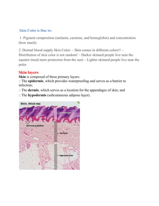
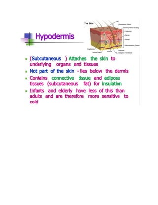
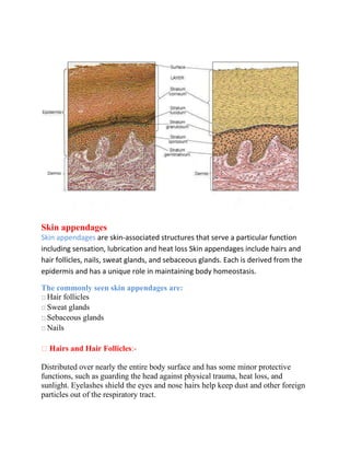
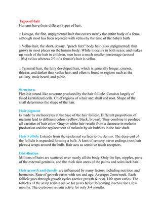
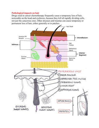
SlideShare a Scribd company logo
Integumentary system = skin and its derivatives
Dr.Raghda Alkhateeb, Cardiovascular Surgeon, Lecturer M.B.Ch.B, CABHS, JBCS
Nursing College of Sulaymaniyah
The integumentary system is the physical system that forms the barrier
between the external environment and the internal systems of the body. In
humans, this system consists of skin, hair, nails, and related glands.
Altogether, the integumentary system forms the largest organ in the body.
Human skin
The human skin is is the largest organ of the body making 15 % of the body
weight.
Embryology of the skin
Origin from ectoderm, mesoderm layers and the mesenchymal cells
during the fetal life, in the 1st
and 2nd
trimester of pregnancy.
What makes mesenchymal cells?
Mesenchymal stem cells (MSCs) are adult stem cells isolated from different sources that
can differentiate into other types of cells. In humans, these sources include; bone marrow,
fat (adipose tissue), umbilical cord tissue (Wharton's Jelly) or amniotic fluid (the fluid
surrounding a fetus).
Types of Stem Cells
 Embryonic stem cells.
 Tissue-specific stem cells.
 Mesenchymal stem cells.
 Induced pluripotent stem cells.

Functions
Skin performs the following functions:
1. Protection: an anatomical barrier from pathogens and damage between the
internal and external environment
2. Sensation: contains a variety of nerve endings that react to heat and cold, touch,
pressure, vibration, and tissue injury.
3. Heat regulation: the skin contains a blood supply far greater than its
requirements which allows precise control of energy loss by radiation, convection
and conduction.
4. Control of evaporation: the skin provides a relatively dry and semi-
impermeable barrier to fluid loss. Loss of this function contributes to the massive
fluid loss in burns.
5. Aesthetics and communication: others see our skin and can assess our mood,
physical state and attractiveness.
6. Storage and synthesis: acts as a storage center for lipids and water, as well as a
means of synthesis of vitamin D by action of UV on certain parts of the skin.
7. Excretion: sweating, getting rid of waste products.
Pigments
Melanin: It is brown in color
Melanoid: It resembles melanin
Keratin: This pigment is yellow to orange in color. It is present in the stratum
corneum and fat cells of dermis and superficial fascia.
Hemoglobin: It is found in blood and is not a pigment of the skin but develops a
purple color.
Oxyhemoglobin: It is also found in blood and is not a pigment of the skin. It
develops a red color.
Carotene
Skin Color is Due to:
1. Pigment composition (melanin, carotene, and hemoglobin) and concentration
(how much)
2. Dermal blood supply Skin Color: – Skin comes in different colors!! –
Distribution of skin color is not random! – Darker skinned people live near the
equator (need more protection from the sun) – Lighter skinned people live near the
poles
Skin layers
Skin is composed of three primary layers:
The epidermis, which provides waterproofing and serves as a barrier to
infection;
The dermis, which serves as a location for the appendages of skin; and
The hypodermis (subcutaneous adipose layer).
Epidermis: is the outermost layer of the skin. It forms the waterproof, protective
wrap over the body's surface and is made up of stratified (layers) squamous
epithelium with an underlying basal lamina.
Components
The epidermis contains no blood vessels, and is nourished by diffusion from the
dermis. The main cell types of the epidermis are keratinocytes, melanocytes,
Langerhans cells and Merkels cells. The epidermis helps the skin to regulate body
temperature.
4.
Epidermis Layers
Epidermis is divided into several layers where cells are formed through mitosis at
the innermost layers. They move up the strata changing shape and composition as
they differentiate and become filled with keratin. They eventually reach the top
layer are start with Stratum Corneum ,Stratum Lucidum ,Stratum Granulosum
,Stratum Mucosum and Stratum Germinativum.
The Corneum layer is the top layer of your skin. This is the layer you can see Here
the cells are dead and continually flake off the surface.
The Lucidum layer is only found on the palms of your hands and soles of your feet.
This is the layer that thickens to fight mechanical attack
The Granulosum layer is where the cells are found with small granules in them,
thought to make the skin tough. Your lips and skin under fingernails do
not have this layer in them.
The Mucosum layer is where tissue fluid is stored
The Germinativum layer is the bottom layer and here the cells are constantly
reproducing. The melanocyte cells are also located in this layer.
Skin appendages
Skin appendages are skin-associated structures that serve a particular function
including sensation, lubrication and heat loss Skin appendages include hairs and
hair follicles, nails, sweat glands, and sebaceous glands. Each is derived from the
epidermis and has a unique role in maintaining body homeostasis.
The commonly seen skin appendages are:
Hair follicles
Sweat glands
Sebaceous glands
Nails
Hairs and Hair Follicles:-
Distributed over nearly the entire body surface and has some minor protective
functions, such as guarding the head against physical trauma, heat loss, and
sunlight. Eyelashes shield the eyes and nose hairs help keep dust and other foreign
particles out of the respiratory tract.
Types of hair
Humans have three different types of hair:
Lanugo, the fine, unpigmented hair that covers nearly the entire body of a fetus,
although most has been replaced with vellus by the time of the baby's birth
Vellus hair, the short, downy, "peach fuzz" body hair (also unpigmented) that
grows in most places on the human body. While it occurs in both sexes, and makes
up much of the hair in children, men have a much smaller percentage (around
10%) vellus whereas 2/3 of a female's hair is vellus.
Terminal hair, the fully developed hair, which is generally longer, coarser,
thicker, and darker than vellus hair, and often is found in regions such as the
axillary, male beard, and pubic.
Structure:
Flexible strand-like structure produced by the hair follicle. Consists largely of
fused keratinized cells. Chief regions of a hair are: shaft and root. Shape of the
shaft determines the shape of the hair.
Hair pigment
Is made by melanocytes at the base of the hair follicle. Different proportions of
melanin lead to different colors (yellow, black, brown). They combine to produce
all varieties of hair color. Gray or white hair results from a decrease in melanin
production and the replacement of melanin by air bubbles in the hair shaft.
Hair Follicle Extends from the epidermal surface to the dermis. The deep end of
the follicle is expanded forming a bulb. A knot of sensory nerve endings (root hair
plexus) wraps around the bulb. Hair acts as sensitive touch receptors.
Distribution
Millions of hairs are scattered over nearly all the body. Only the lips, nipples, parts
of the external genitalia, and the thick skin areas of the palms and soles lack hair.
Hair growth and density are influenced by many factors including nutrition and
hormones. Rate of growth varies with sex and age. Averages 2mm/week. Each
follicle goes through growth cycles (active growth & rest). Life span varies. The
follicles of the scalp remain active for years before becoming inactive for a few
months. The eyebrows remain active for only 3-4 months.
Pathological impacts on hair
Drugs used in cancer chemotherapy frequently cause a temporary loss of hair,
noticeable on the head and eyebrows, because they kill all rapidly dividing cells,
not just the cancerous ones. Other diseases and traumas can cause temporary or
permanent loss of hair, either generally or in patches.
Sweat Glands
More than 2.5 million sweat glands are distributed over the entire body. Two types
of sweat glands: eccrine and apocrine.
Eccrine Sweat Glands
Eccrine sweat glands are found in nearly all body regions more numerous
especially on the palms, soles and forehead. They secrete a hypotonic solution
(sweat) derived from blood plasma. It is 99% water with some other substances in
it. Sweating is regulated by the sympathetic nervous system. And assists in
thermoregulation.
Human eccrine sweat is composed chiefly of water with various salts and organic
compounds in solution. It contains minute amounts of fatty materials, urea, and
other wastes. The concentration of sodium varies from 35–65 mmol/l and is lower
in people acclimatised to a hot environment. The sweat of other species generally
differ in composition.
Apocrine Sweat Glands
Apocrine glands are only found in largely confined to the axillary and perineal
regions. They are larger in size. The secretions are the same as eccrine with the
addition of fatty substances and proteins which make the secretions viscous. The
secretions are odorless until bacterial decomposition occurs. Precise function
unknown. Activated by the sympathetic nervous system during pain or stress.
Apocrine glands typically empty into hair follicles. Emotional stress increases the
production of sweat from the apocrine glands, or more precisely: the sweat
already
present in the tubule is squeezed out. Apocrine sweat glands essentially serve as
scent glands.
Sebaceous (oil) Glands
Simple glands found all over the body except on the palms and soles. They are
small on the body trunk and limbs and larger on the face, neck, and upper chest.
Secrete an oily secretion called sebum. The sebum is usually ducted into a hair
follicle. Sebum softens and lubricates the hair and skin and it has a bactericidal
action.
Sebum is odorless, but its bacterial breakdown can produce odors. Sebum is the
cause of some people experiencing "oily" hair if it is not washed for several days.
Earwax is partly sebum, as is mucopurulent discharge, the dry substance
accumulating in the corners of the eye after sleeping.
The activity of the sebaceous glands increases during puberty because of
heightened levels of androgens.
Sebaceous glands are involved in skin problems such as acne and keratosis pilaris.
A blocked sebaceous gland can result in a sebaceous cyst. The prescription drug
isotretinoin significantly reduces the amount of sebum produced by the sebaceous
glands, and is used to treat acne. The extreme use (up to 10 times doctor prescribed
amounts) of anabolic steroids by bodybuilders to prevent weight loss tend to
stimulate the sebaceous glands which can cause acne. Acne is an active
inflammation of the glands accompanied by "pimples." Acne is usually caused by
bacteria, especially staphylococcus
Nails
Scale-like modifications of the epidermis that forms a clear protective covering on
the dorsum of the distal part of a finger or toe. Each nail has a free edge, a body,
and a root embedded in the skin. The thickened proximal part of the nail bed - nail
matrix - is responsible for nail growth. Nail cells are heavily keratinized. Nail body
slides distally over the nail bed during growth.
Nail Diseases
Nail diseases are in a separate category from diseases of the skin. Although nails
are a skin appendage, they have their own signs and symptoms which may relate to
other medical conditions. Nail conditions that show signs of infection or
inflammation require medical assistance and cannot be treated at a beauty parlor.
Deformity or disease of the nails may be referred to as onychosis.
There are many diseases that can occur with the fingernails and toenails. The most
common of these diseases are ingrown nails and fungal infections.
Nutrition for healthy skin
Vitamin A, also known as retinoids, benefits the skin by normalizing
keratinization, downregulating sebum production which contributes to acne, and
reversing and treating photodamage, striae, and cellulite.
Vitamin D and analogs are used to downregulate the cutaneous immune system
and epithelial proliferation while promoting differentiation.
Vitamin C is an antioxidant that regulates collagen synthesis, forms barrier lipids,
regenerates vitamin E, and provides photoprotection.
Vitamin E is a membrane antioxidant that protects against oxidative damage and
also provides protection against harmful UV rays.
Wound Definition. A wound may be defined as any disruption
of the integrity of skin, mucous membrane or organ tissue.
exudative phase. resorptive phase. proliferative phase.
How does skin heal?
When skin is damaged, it attends to heal itself to continue to protect the body. This
regeneration process occurs naturally in healthy individuals.
There are four phases of wound healing:
1. Hemostasis: begins immediately following injury to the skin.
Special blood cells called platelets block off damaged blood vessels. Platelets also
secrete proteins called growth factors, which help initiate later healing steps.
2. Inflammation: swelling and warmth associated with the pain of the injury.
Inflammation usually lasts up to 4 days following the injury. Inflammation causes
blood vessels to leak plasma and microorganisms into surrounding tissue, which
helps prevent infection. During this stage, growth factors help the cell divide and
produce more cells. Specialized cells called macrophages can consume bacteria,
providing another line of defence against infection. These cells also help resolve the
inflammation at the end of this stage, causing transition into stage 3.
3. Proliferation (also known as granulation and contraction):
This involves replacement of dermal tissue (and subdermal tissue in deeper
wounds).
The proliferation stage begins about 4 days after injury to the skin, and usually
continues until day 21. Special cells called fibroblasts secrete collagen, which assists
with the regeneration of dermal tissue. This healing stage is also characterized by
contraction, during which the edges of the wound are pulled together.
4. Remodeling (also known as maturation): realignment of collagen.
Remodeling can take up to 2 years after injury to the skin. After remodeling occurs,
skin’s ability to stretch and return to shape will be only 70-80% as strong as before
the injury. Scar tissue forms and becomes thicker over time.
Inflammation is recognized as a set of complex changing responses to tissue injury primarily caused
by toxic chemicals, some environmental agents, trauma, overuse, or infection. Some of these responses
can be beneficial in wound healing and infection control or pathological as in many chronic disease
states. Inflammation is a “second-line” defense against infectious agents.
Or Inflammation is the immune system's response to harmful stimuli, such as pathogens,
damaged cells, toxic compounds, or irradiation, and acts by removing injurious stimuli and
initiating the healing process.
Skin flora:
Integumentary sytem .pdf

More Related Content

Similar to Integumentary sytem .pdf

Lecture 7 the integumentary system
Lecture 7 the integumentary systemLecture 7 the integumentary system
Lecture 7 the integumentary system
BilalHoushaymi
 
Module 3 Part 1
Module 3 Part 1Module 3 Part 1
Module 3 Part 1pamiepk
 
Module 3 integumentary system 1
Module 3   integumentary system 1Module 3   integumentary system 1
Module 3 integumentary system 1pamiepk
 
Integumentary system .ppsx
Integumentary system .ppsxIntegumentary system .ppsx
Integumentary system .ppsx
lumaGhaziALzamel
 
Skin structures and functions ppt Dr.Mumux
Skin structures and functions ppt Dr.MumuxSkin structures and functions ppt Dr.Mumux
Skin structures and functions ppt Dr.Mumux
Mumux Mirani
 
Anatomy of skin
Anatomy of skinAnatomy of skin
Anatomy of skin
A Y
 
3. Integumentary system (Skin).pptx
3. Integumentary system (Skin).pptx3. Integumentary system (Skin).pptx
3. Integumentary system (Skin).pptx
AbhiDabra
 
integmentary skin Dr Ahmed H Ahmed.pdf
integmentary skin Dr Ahmed H Ahmed.pdfintegmentary skin Dr Ahmed H Ahmed.pdf
integmentary skin Dr Ahmed H Ahmed.pdf
Salahaddin University
 
Skin
Skin Skin
INTEGUMENTARY SYSTEM BY EVA KIPTOO.pptx
INTEGUMENTARY SYSTEM BY EVA KIPTOO.pptxINTEGUMENTARY SYSTEM BY EVA KIPTOO.pptx
INTEGUMENTARY SYSTEM BY EVA KIPTOO.pptx
NabiswaboazWangila
 
Chapter 3 Presentation
Chapter 3 PresentationChapter 3 Presentation
Chapter 3 Presentationfrancine king
 
anatomy -skin
anatomy -skinanatomy -skin
anatomy -skin
Labeeb Al Maktary
 
TAM1
TAM1TAM1
Integumentary system
Integumentary systemIntegumentary system
Integumentary system
Ivy Janelle
 
EVA KIPTOO DENTAL PRESENTATION - Copy.pptx
EVA KIPTOO DENTAL PRESENTATION - Copy.pptxEVA KIPTOO DENTAL PRESENTATION - Copy.pptx
EVA KIPTOO DENTAL PRESENTATION - Copy.pptx
boaznabiswa
 
Skin structure and development
Skin structure and developmentSkin structure and development
Skin structure and development
NIKHILESH REDDY PURUMANDLA
 
The Sensory Organs ( Anatomy & Physiology)
The Sensory Organs ( Anatomy & Physiology)The Sensory Organs ( Anatomy & Physiology)
The Sensory Organs ( Anatomy & Physiology)
Nagamani Manjunath
 
Powerpoint of integumentary syytem
Powerpoint of  integumentary syytemPowerpoint of  integumentary syytem
Powerpoint of integumentary syytemnone
 
Powerpoint of integumentary system of vertebrates
Powerpoint of  integumentary system of vertebratesPowerpoint of  integumentary system of vertebrates
Powerpoint of integumentary system of vertebratesnone
 
Integumentary system
Integumentary systemIntegumentary system
Integumentary system
NeilfieOrit2
 

Similar to Integumentary sytem .pdf (20)

Lecture 7 the integumentary system
Lecture 7 the integumentary systemLecture 7 the integumentary system
Lecture 7 the integumentary system
 
Module 3 Part 1
Module 3 Part 1Module 3 Part 1
Module 3 Part 1
 
Module 3 integumentary system 1
Module 3   integumentary system 1Module 3   integumentary system 1
Module 3 integumentary system 1
 
Integumentary system .ppsx
Integumentary system .ppsxIntegumentary system .ppsx
Integumentary system .ppsx
 
Skin structures and functions ppt Dr.Mumux
Skin structures and functions ppt Dr.MumuxSkin structures and functions ppt Dr.Mumux
Skin structures and functions ppt Dr.Mumux
 
Anatomy of skin
Anatomy of skinAnatomy of skin
Anatomy of skin
 
3. Integumentary system (Skin).pptx
3. Integumentary system (Skin).pptx3. Integumentary system (Skin).pptx
3. Integumentary system (Skin).pptx
 
integmentary skin Dr Ahmed H Ahmed.pdf
integmentary skin Dr Ahmed H Ahmed.pdfintegmentary skin Dr Ahmed H Ahmed.pdf
integmentary skin Dr Ahmed H Ahmed.pdf
 
Skin
Skin Skin
Skin
 
INTEGUMENTARY SYSTEM BY EVA KIPTOO.pptx
INTEGUMENTARY SYSTEM BY EVA KIPTOO.pptxINTEGUMENTARY SYSTEM BY EVA KIPTOO.pptx
INTEGUMENTARY SYSTEM BY EVA KIPTOO.pptx
 
Chapter 3 Presentation
Chapter 3 PresentationChapter 3 Presentation
Chapter 3 Presentation
 
anatomy -skin
anatomy -skinanatomy -skin
anatomy -skin
 
TAM1
TAM1TAM1
TAM1
 
Integumentary system
Integumentary systemIntegumentary system
Integumentary system
 
EVA KIPTOO DENTAL PRESENTATION - Copy.pptx
EVA KIPTOO DENTAL PRESENTATION - Copy.pptxEVA KIPTOO DENTAL PRESENTATION - Copy.pptx
EVA KIPTOO DENTAL PRESENTATION - Copy.pptx
 
Skin structure and development
Skin structure and developmentSkin structure and development
Skin structure and development
 
The Sensory Organs ( Anatomy & Physiology)
The Sensory Organs ( Anatomy & Physiology)The Sensory Organs ( Anatomy & Physiology)
The Sensory Organs ( Anatomy & Physiology)
 
Powerpoint of integumentary syytem
Powerpoint of  integumentary syytemPowerpoint of  integumentary syytem
Powerpoint of integumentary syytem
 
Powerpoint of integumentary system of vertebrates
Powerpoint of  integumentary system of vertebratesPowerpoint of  integumentary system of vertebrates
Powerpoint of integumentary system of vertebrates
 
Integumentary system
Integumentary systemIntegumentary system
Integumentary system
 

More from Raghda ALKhateeb

MCQ CVS Physiology
MCQ CVS PhysiologyMCQ CVS Physiology
MCQ CVS Physiology
Raghda ALKhateeb
 
CNS AND PNS NURSING .pptx1-1.pptx
CNS AND PNS NURSING .pptx1-1.pptxCNS AND PNS NURSING .pptx1-1.pptx
CNS AND PNS NURSING .pptx1-1.pptx
Raghda ALKhateeb
 
Vital Signs Review .pptx1-1.pptx
Vital Signs Review .pptx1-1.pptxVital Signs Review .pptx1-1.pptx
Vital Signs Review .pptx1-1.pptx
Raghda ALKhateeb
 
Blood Physiology.pptx
Blood Physiology.pptxBlood Physiology.pptx
Blood Physiology.pptx
Raghda ALKhateeb
 
coronarycameralfistula-170307133357.pdf
coronarycameralfistula-170307133357.pdfcoronarycameralfistula-170307133357.pdf
coronarycameralfistula-170307133357.pdf
Raghda ALKhateeb
 
Cardiovascular Physiology Lecture notes for Nursing Students.pptx 1-1.pptx
Cardiovascular Physiology Lecture notes for Nursing Students.pptx 1-1.pptxCardiovascular Physiology Lecture notes for Nursing Students.pptx 1-1.pptx
Cardiovascular Physiology Lecture notes for Nursing Students.pptx 1-1.pptx
Raghda ALKhateeb
 

More from Raghda ALKhateeb (6)

MCQ CVS Physiology
MCQ CVS PhysiologyMCQ CVS Physiology
MCQ CVS Physiology
 
CNS AND PNS NURSING .pptx1-1.pptx
CNS AND PNS NURSING .pptx1-1.pptxCNS AND PNS NURSING .pptx1-1.pptx
CNS AND PNS NURSING .pptx1-1.pptx
 
Vital Signs Review .pptx1-1.pptx
Vital Signs Review .pptx1-1.pptxVital Signs Review .pptx1-1.pptx
Vital Signs Review .pptx1-1.pptx
 
Blood Physiology.pptx
Blood Physiology.pptxBlood Physiology.pptx
Blood Physiology.pptx
 
coronarycameralfistula-170307133357.pdf
coronarycameralfistula-170307133357.pdfcoronarycameralfistula-170307133357.pdf
coronarycameralfistula-170307133357.pdf
 
Cardiovascular Physiology Lecture notes for Nursing Students.pptx 1-1.pptx
Cardiovascular Physiology Lecture notes for Nursing Students.pptx 1-1.pptxCardiovascular Physiology Lecture notes for Nursing Students.pptx 1-1.pptx
Cardiovascular Physiology Lecture notes for Nursing Students.pptx 1-1.pptx
 

Recently uploaded

CDSCO and Phamacovigilance {Regulatory body in India}
CDSCO and Phamacovigilance {Regulatory body in India}CDSCO and Phamacovigilance {Regulatory body in India}
CDSCO and Phamacovigilance {Regulatory body in India}
NEHA GUPTA
 
Basavarajeeyam - Ayurvedic heritage book of Andhra pradesh
Basavarajeeyam - Ayurvedic heritage book of Andhra pradeshBasavarajeeyam - Ayurvedic heritage book of Andhra pradesh
Basavarajeeyam - Ayurvedic heritage book of Andhra pradesh
Dr. Madduru Muni Haritha
 
Novas diretrizes da OMS para os cuidados perinatais de mais qualidade
Novas diretrizes da OMS para os cuidados perinatais de mais qualidadeNovas diretrizes da OMS para os cuidados perinatais de mais qualidade
Novas diretrizes da OMS para os cuidados perinatais de mais qualidade
Prof. Marcus Renato de Carvalho
 
Lung Cancer: Artificial Intelligence, Synergetics, Complex System Analysis, S...
Lung Cancer: Artificial Intelligence, Synergetics, Complex System Analysis, S...Lung Cancer: Artificial Intelligence, Synergetics, Complex System Analysis, S...
Lung Cancer: Artificial Intelligence, Synergetics, Complex System Analysis, S...
Oleg Kshivets
 
Thyroid Gland- Gross Anatomy by Dr. Rabia Inam Gandapore.pptx
Thyroid Gland- Gross Anatomy by Dr. Rabia Inam Gandapore.pptxThyroid Gland- Gross Anatomy by Dr. Rabia Inam Gandapore.pptx
Thyroid Gland- Gross Anatomy by Dr. Rabia Inam Gandapore.pptx
Dr. Rabia Inam Gandapore
 
Ozempic: Preoperative Management of Patients on GLP-1 Receptor Agonists
Ozempic: Preoperative Management of Patients on GLP-1 Receptor Agonists  Ozempic: Preoperative Management of Patients on GLP-1 Receptor Agonists
Ozempic: Preoperative Management of Patients on GLP-1 Receptor Agonists
Saeid Safari
 
KDIGO 2024 guidelines for diabetologists
KDIGO 2024 guidelines for diabetologistsKDIGO 2024 guidelines for diabetologists
KDIGO 2024 guidelines for diabetologists
د.محمود نجيب
 
Knee anatomy and clinical tests 2024.pdf
Knee anatomy and clinical tests 2024.pdfKnee anatomy and clinical tests 2024.pdf
Knee anatomy and clinical tests 2024.pdf
vimalpl1234
 
ARTHROLOGY PPT NCISM SYLLABUS AYURVEDA STUDENTS
ARTHROLOGY PPT NCISM SYLLABUS AYURVEDA STUDENTSARTHROLOGY PPT NCISM SYLLABUS AYURVEDA STUDENTS
ARTHROLOGY PPT NCISM SYLLABUS AYURVEDA STUDENTS
Dr. Vinay Pareek
 
ARTIFICIAL INTELLIGENCE IN HEALTHCARE.pdf
ARTIFICIAL INTELLIGENCE IN  HEALTHCARE.pdfARTIFICIAL INTELLIGENCE IN  HEALTHCARE.pdf
ARTIFICIAL INTELLIGENCE IN HEALTHCARE.pdf
Anujkumaranit
 
How to Give Better Lectures: Some Tips for Doctors
How to Give Better Lectures: Some Tips for DoctorsHow to Give Better Lectures: Some Tips for Doctors
How to Give Better Lectures: Some Tips for Doctors
LanceCatedral
 
Adv. biopharm. APPLICATION OF PHARMACOKINETICS : TARGETED DRUG DELIVERY SYSTEMS
Adv. biopharm. APPLICATION OF PHARMACOKINETICS : TARGETED DRUG DELIVERY SYSTEMSAdv. biopharm. APPLICATION OF PHARMACOKINETICS : TARGETED DRUG DELIVERY SYSTEMS
Adv. biopharm. APPLICATION OF PHARMACOKINETICS : TARGETED DRUG DELIVERY SYSTEMS
AkankshaAshtankar
 
planning for change nursing Management ppt
planning for change nursing Management pptplanning for change nursing Management ppt
planning for change nursing Management ppt
Thangamjayarani
 
NVBDCP.pptx Nation vector borne disease control program
NVBDCP.pptx Nation vector borne disease control programNVBDCP.pptx Nation vector borne disease control program
NVBDCP.pptx Nation vector borne disease control program
Sapna Thakur
 
Cervical & Brachial Plexus By Dr. RIG.pptx
Cervical & Brachial Plexus By Dr. RIG.pptxCervical & Brachial Plexus By Dr. RIG.pptx
Cervical & Brachial Plexus By Dr. RIG.pptx
Dr. Rabia Inam Gandapore
 
How STIs Influence the Development of Pelvic Inflammatory Disease.pptx
How STIs Influence the Development of Pelvic Inflammatory Disease.pptxHow STIs Influence the Development of Pelvic Inflammatory Disease.pptx
How STIs Influence the Development of Pelvic Inflammatory Disease.pptx
FFragrant
 
A Classical Text Review on Basavarajeeyam
A Classical Text Review on BasavarajeeyamA Classical Text Review on Basavarajeeyam
A Classical Text Review on Basavarajeeyam
Dr. Jyothirmai Paindla
 
Evaluation of antidepressant activity of clitoris ternatea in animals
Evaluation of antidepressant activity of clitoris ternatea in animalsEvaluation of antidepressant activity of clitoris ternatea in animals
Evaluation of antidepressant activity of clitoris ternatea in animals
Shweta
 
ANATOMY AND PHYSIOLOGY OF URINARY SYSTEM.pptx
ANATOMY AND PHYSIOLOGY OF URINARY SYSTEM.pptxANATOMY AND PHYSIOLOGY OF URINARY SYSTEM.pptx
ANATOMY AND PHYSIOLOGY OF URINARY SYSTEM.pptx
Swetaba Besh
 
Ophthalmology Clinical Tests for OSCE exam
Ophthalmology Clinical Tests for OSCE examOphthalmology Clinical Tests for OSCE exam
Ophthalmology Clinical Tests for OSCE exam
KafrELShiekh University
 

Recently uploaded (20)

CDSCO and Phamacovigilance {Regulatory body in India}
CDSCO and Phamacovigilance {Regulatory body in India}CDSCO and Phamacovigilance {Regulatory body in India}
CDSCO and Phamacovigilance {Regulatory body in India}
 
Basavarajeeyam - Ayurvedic heritage book of Andhra pradesh
Basavarajeeyam - Ayurvedic heritage book of Andhra pradeshBasavarajeeyam - Ayurvedic heritage book of Andhra pradesh
Basavarajeeyam - Ayurvedic heritage book of Andhra pradesh
 
Novas diretrizes da OMS para os cuidados perinatais de mais qualidade
Novas diretrizes da OMS para os cuidados perinatais de mais qualidadeNovas diretrizes da OMS para os cuidados perinatais de mais qualidade
Novas diretrizes da OMS para os cuidados perinatais de mais qualidade
 
Lung Cancer: Artificial Intelligence, Synergetics, Complex System Analysis, S...
Lung Cancer: Artificial Intelligence, Synergetics, Complex System Analysis, S...Lung Cancer: Artificial Intelligence, Synergetics, Complex System Analysis, S...
Lung Cancer: Artificial Intelligence, Synergetics, Complex System Analysis, S...
 
Thyroid Gland- Gross Anatomy by Dr. Rabia Inam Gandapore.pptx
Thyroid Gland- Gross Anatomy by Dr. Rabia Inam Gandapore.pptxThyroid Gland- Gross Anatomy by Dr. Rabia Inam Gandapore.pptx
Thyroid Gland- Gross Anatomy by Dr. Rabia Inam Gandapore.pptx
 
Ozempic: Preoperative Management of Patients on GLP-1 Receptor Agonists
Ozempic: Preoperative Management of Patients on GLP-1 Receptor Agonists  Ozempic: Preoperative Management of Patients on GLP-1 Receptor Agonists
Ozempic: Preoperative Management of Patients on GLP-1 Receptor Agonists
 
KDIGO 2024 guidelines for diabetologists
KDIGO 2024 guidelines for diabetologistsKDIGO 2024 guidelines for diabetologists
KDIGO 2024 guidelines for diabetologists
 
Knee anatomy and clinical tests 2024.pdf
Knee anatomy and clinical tests 2024.pdfKnee anatomy and clinical tests 2024.pdf
Knee anatomy and clinical tests 2024.pdf
 
ARTHROLOGY PPT NCISM SYLLABUS AYURVEDA STUDENTS
ARTHROLOGY PPT NCISM SYLLABUS AYURVEDA STUDENTSARTHROLOGY PPT NCISM SYLLABUS AYURVEDA STUDENTS
ARTHROLOGY PPT NCISM SYLLABUS AYURVEDA STUDENTS
 
ARTIFICIAL INTELLIGENCE IN HEALTHCARE.pdf
ARTIFICIAL INTELLIGENCE IN  HEALTHCARE.pdfARTIFICIAL INTELLIGENCE IN  HEALTHCARE.pdf
ARTIFICIAL INTELLIGENCE IN HEALTHCARE.pdf
 
How to Give Better Lectures: Some Tips for Doctors
How to Give Better Lectures: Some Tips for DoctorsHow to Give Better Lectures: Some Tips for Doctors
How to Give Better Lectures: Some Tips for Doctors
 
Adv. biopharm. APPLICATION OF PHARMACOKINETICS : TARGETED DRUG DELIVERY SYSTEMS
Adv. biopharm. APPLICATION OF PHARMACOKINETICS : TARGETED DRUG DELIVERY SYSTEMSAdv. biopharm. APPLICATION OF PHARMACOKINETICS : TARGETED DRUG DELIVERY SYSTEMS
Adv. biopharm. APPLICATION OF PHARMACOKINETICS : TARGETED DRUG DELIVERY SYSTEMS
 
planning for change nursing Management ppt
planning for change nursing Management pptplanning for change nursing Management ppt
planning for change nursing Management ppt
 
NVBDCP.pptx Nation vector borne disease control program
NVBDCP.pptx Nation vector borne disease control programNVBDCP.pptx Nation vector borne disease control program
NVBDCP.pptx Nation vector borne disease control program
 
Cervical & Brachial Plexus By Dr. RIG.pptx
Cervical & Brachial Plexus By Dr. RIG.pptxCervical & Brachial Plexus By Dr. RIG.pptx
Cervical & Brachial Plexus By Dr. RIG.pptx
 
How STIs Influence the Development of Pelvic Inflammatory Disease.pptx
How STIs Influence the Development of Pelvic Inflammatory Disease.pptxHow STIs Influence the Development of Pelvic Inflammatory Disease.pptx
How STIs Influence the Development of Pelvic Inflammatory Disease.pptx
 
A Classical Text Review on Basavarajeeyam
A Classical Text Review on BasavarajeeyamA Classical Text Review on Basavarajeeyam
A Classical Text Review on Basavarajeeyam
 
Evaluation of antidepressant activity of clitoris ternatea in animals
Evaluation of antidepressant activity of clitoris ternatea in animalsEvaluation of antidepressant activity of clitoris ternatea in animals
Evaluation of antidepressant activity of clitoris ternatea in animals
 
ANATOMY AND PHYSIOLOGY OF URINARY SYSTEM.pptx
ANATOMY AND PHYSIOLOGY OF URINARY SYSTEM.pptxANATOMY AND PHYSIOLOGY OF URINARY SYSTEM.pptx
ANATOMY AND PHYSIOLOGY OF URINARY SYSTEM.pptx
 
Ophthalmology Clinical Tests for OSCE exam
Ophthalmology Clinical Tests for OSCE examOphthalmology Clinical Tests for OSCE exam
Ophthalmology Clinical Tests for OSCE exam
 

Integumentary sytem .pdf

  • 1. Integumentary system = skin and its derivatives Dr.Raghda Alkhateeb, Cardiovascular Surgeon, Lecturer M.B.Ch.B, CABHS, JBCS Nursing College of Sulaymaniyah The integumentary system is the physical system that forms the barrier between the external environment and the internal systems of the body. In humans, this system consists of skin, hair, nails, and related glands. Altogether, the integumentary system forms the largest organ in the body. Human skin The human skin is is the largest organ of the body making 15 % of the body weight. Embryology of the skin Origin from ectoderm, mesoderm layers and the mesenchymal cells during the fetal life, in the 1st and 2nd trimester of pregnancy.
  • 2. What makes mesenchymal cells? Mesenchymal stem cells (MSCs) are adult stem cells isolated from different sources that can differentiate into other types of cells. In humans, these sources include; bone marrow, fat (adipose tissue), umbilical cord tissue (Wharton's Jelly) or amniotic fluid (the fluid surrounding a fetus). Types of Stem Cells  Embryonic stem cells.  Tissue-specific stem cells.  Mesenchymal stem cells.  Induced pluripotent stem cells. 
  • 3. Functions Skin performs the following functions:
  • 4. 1. Protection: an anatomical barrier from pathogens and damage between the internal and external environment 2. Sensation: contains a variety of nerve endings that react to heat and cold, touch, pressure, vibration, and tissue injury. 3. Heat regulation: the skin contains a blood supply far greater than its requirements which allows precise control of energy loss by radiation, convection and conduction. 4. Control of evaporation: the skin provides a relatively dry and semi- impermeable barrier to fluid loss. Loss of this function contributes to the massive fluid loss in burns. 5. Aesthetics and communication: others see our skin and can assess our mood, physical state and attractiveness. 6. Storage and synthesis: acts as a storage center for lipids and water, as well as a means of synthesis of vitamin D by action of UV on certain parts of the skin. 7. Excretion: sweating, getting rid of waste products. Pigments Melanin: It is brown in color Melanoid: It resembles melanin Keratin: This pigment is yellow to orange in color. It is present in the stratum corneum and fat cells of dermis and superficial fascia. Hemoglobin: It is found in blood and is not a pigment of the skin but develops a purple color. Oxyhemoglobin: It is also found in blood and is not a pigment of the skin. It develops a red color. Carotene
  • 5. Skin Color is Due to: 1. Pigment composition (melanin, carotene, and hemoglobin) and concentration (how much) 2. Dermal blood supply Skin Color: – Skin comes in different colors!! – Distribution of skin color is not random! – Darker skinned people live near the equator (need more protection from the sun) – Lighter skinned people live near the poles Skin layers Skin is composed of three primary layers: The epidermis, which provides waterproofing and serves as a barrier to infection; The dermis, which serves as a location for the appendages of skin; and The hypodermis (subcutaneous adipose layer).
  • 6. Epidermis: is the outermost layer of the skin. It forms the waterproof, protective wrap over the body's surface and is made up of stratified (layers) squamous epithelium with an underlying basal lamina. Components The epidermis contains no blood vessels, and is nourished by diffusion from the dermis. The main cell types of the epidermis are keratinocytes, melanocytes, Langerhans cells and Merkels cells. The epidermis helps the skin to regulate body temperature.
  • 8.
  • 9. Epidermis is divided into several layers where cells are formed through mitosis at the innermost layers. They move up the strata changing shape and composition as they differentiate and become filled with keratin. They eventually reach the top layer are start with Stratum Corneum ,Stratum Lucidum ,Stratum Granulosum ,Stratum Mucosum and Stratum Germinativum. The Corneum layer is the top layer of your skin. This is the layer you can see Here the cells are dead and continually flake off the surface. The Lucidum layer is only found on the palms of your hands and soles of your feet. This is the layer that thickens to fight mechanical attack The Granulosum layer is where the cells are found with small granules in them, thought to make the skin tough. Your lips and skin under fingernails do not have this layer in them. The Mucosum layer is where tissue fluid is stored The Germinativum layer is the bottom layer and here the cells are constantly reproducing. The melanocyte cells are also located in this layer.
  • 10.
  • 11.
  • 12. Skin appendages Skin appendages are skin-associated structures that serve a particular function including sensation, lubrication and heat loss Skin appendages include hairs and hair follicles, nails, sweat glands, and sebaceous glands. Each is derived from the epidermis and has a unique role in maintaining body homeostasis. The commonly seen skin appendages are: Hair follicles Sweat glands Sebaceous glands Nails Hairs and Hair Follicles:- Distributed over nearly the entire body surface and has some minor protective functions, such as guarding the head against physical trauma, heat loss, and sunlight. Eyelashes shield the eyes and nose hairs help keep dust and other foreign particles out of the respiratory tract.
  • 13. Types of hair Humans have three different types of hair: Lanugo, the fine, unpigmented hair that covers nearly the entire body of a fetus, although most has been replaced with vellus by the time of the baby's birth Vellus hair, the short, downy, "peach fuzz" body hair (also unpigmented) that grows in most places on the human body. While it occurs in both sexes, and makes up much of the hair in children, men have a much smaller percentage (around 10%) vellus whereas 2/3 of a female's hair is vellus. Terminal hair, the fully developed hair, which is generally longer, coarser, thicker, and darker than vellus hair, and often is found in regions such as the axillary, male beard, and pubic. Structure: Flexible strand-like structure produced by the hair follicle. Consists largely of fused keratinized cells. Chief regions of a hair are: shaft and root. Shape of the shaft determines the shape of the hair. Hair pigment Is made by melanocytes at the base of the hair follicle. Different proportions of melanin lead to different colors (yellow, black, brown). They combine to produce all varieties of hair color. Gray or white hair results from a decrease in melanin production and the replacement of melanin by air bubbles in the hair shaft. Hair Follicle Extends from the epidermal surface to the dermis. The deep end of the follicle is expanded forming a bulb. A knot of sensory nerve endings (root hair plexus) wraps around the bulb. Hair acts as sensitive touch receptors. Distribution Millions of hairs are scattered over nearly all the body. Only the lips, nipples, parts of the external genitalia, and the thick skin areas of the palms and soles lack hair. Hair growth and density are influenced by many factors including nutrition and hormones. Rate of growth varies with sex and age. Averages 2mm/week. Each follicle goes through growth cycles (active growth & rest). Life span varies. The follicles of the scalp remain active for years before becoming inactive for a few months. The eyebrows remain active for only 3-4 months.
  • 14. Pathological impacts on hair Drugs used in cancer chemotherapy frequently cause a temporary loss of hair, noticeable on the head and eyebrows, because they kill all rapidly dividing cells, not just the cancerous ones. Other diseases and traumas can cause temporary or permanent loss of hair, either generally or in patches.
  • 15. Sweat Glands More than 2.5 million sweat glands are distributed over the entire body. Two types of sweat glands: eccrine and apocrine. Eccrine Sweat Glands Eccrine sweat glands are found in nearly all body regions more numerous especially on the palms, soles and forehead. They secrete a hypotonic solution (sweat) derived from blood plasma. It is 99% water with some other substances in it. Sweating is regulated by the sympathetic nervous system. And assists in thermoregulation. Human eccrine sweat is composed chiefly of water with various salts and organic compounds in solution. It contains minute amounts of fatty materials, urea, and other wastes. The concentration of sodium varies from 35–65 mmol/l and is lower in people acclimatised to a hot environment. The sweat of other species generally differ in composition. Apocrine Sweat Glands Apocrine glands are only found in largely confined to the axillary and perineal regions. They are larger in size. The secretions are the same as eccrine with the addition of fatty substances and proteins which make the secretions viscous. The secretions are odorless until bacterial decomposition occurs. Precise function unknown. Activated by the sympathetic nervous system during pain or stress. Apocrine glands typically empty into hair follicles. Emotional stress increases the production of sweat from the apocrine glands, or more precisely: the sweat already present in the tubule is squeezed out. Apocrine sweat glands essentially serve as scent glands. Sebaceous (oil) Glands Simple glands found all over the body except on the palms and soles. They are small on the body trunk and limbs and larger on the face, neck, and upper chest. Secrete an oily secretion called sebum. The sebum is usually ducted into a hair follicle. Sebum softens and lubricates the hair and skin and it has a bactericidal action.
  • 16. Sebum is odorless, but its bacterial breakdown can produce odors. Sebum is the cause of some people experiencing "oily" hair if it is not washed for several days. Earwax is partly sebum, as is mucopurulent discharge, the dry substance accumulating in the corners of the eye after sleeping. The activity of the sebaceous glands increases during puberty because of heightened levels of androgens. Sebaceous glands are involved in skin problems such as acne and keratosis pilaris. A blocked sebaceous gland can result in a sebaceous cyst. The prescription drug isotretinoin significantly reduces the amount of sebum produced by the sebaceous glands, and is used to treat acne. The extreme use (up to 10 times doctor prescribed amounts) of anabolic steroids by bodybuilders to prevent weight loss tend to stimulate the sebaceous glands which can cause acne. Acne is an active inflammation of the glands accompanied by "pimples." Acne is usually caused by bacteria, especially staphylococcus Nails Scale-like modifications of the epidermis that forms a clear protective covering on the dorsum of the distal part of a finger or toe. Each nail has a free edge, a body, and a root embedded in the skin. The thickened proximal part of the nail bed - nail matrix - is responsible for nail growth. Nail cells are heavily keratinized. Nail body slides distally over the nail bed during growth. Nail Diseases Nail diseases are in a separate category from diseases of the skin. Although nails are a skin appendage, they have their own signs and symptoms which may relate to other medical conditions. Nail conditions that show signs of infection or inflammation require medical assistance and cannot be treated at a beauty parlor. Deformity or disease of the nails may be referred to as onychosis. There are many diseases that can occur with the fingernails and toenails. The most common of these diseases are ingrown nails and fungal infections.
  • 17. Nutrition for healthy skin Vitamin A, also known as retinoids, benefits the skin by normalizing keratinization, downregulating sebum production which contributes to acne, and reversing and treating photodamage, striae, and cellulite. Vitamin D and analogs are used to downregulate the cutaneous immune system and epithelial proliferation while promoting differentiation. Vitamin C is an antioxidant that regulates collagen synthesis, forms barrier lipids, regenerates vitamin E, and provides photoprotection. Vitamin E is a membrane antioxidant that protects against oxidative damage and also provides protection against harmful UV rays.
  • 18. Wound Definition. A wound may be defined as any disruption of the integrity of skin, mucous membrane or organ tissue. exudative phase. resorptive phase. proliferative phase. How does skin heal? When skin is damaged, it attends to heal itself to continue to protect the body. This regeneration process occurs naturally in healthy individuals. There are four phases of wound healing: 1. Hemostasis: begins immediately following injury to the skin. Special blood cells called platelets block off damaged blood vessels. Platelets also secrete proteins called growth factors, which help initiate later healing steps. 2. Inflammation: swelling and warmth associated with the pain of the injury. Inflammation usually lasts up to 4 days following the injury. Inflammation causes blood vessels to leak plasma and microorganisms into surrounding tissue, which helps prevent infection. During this stage, growth factors help the cell divide and produce more cells. Specialized cells called macrophages can consume bacteria, providing another line of defence against infection. These cells also help resolve the inflammation at the end of this stage, causing transition into stage 3. 3. Proliferation (also known as granulation and contraction): This involves replacement of dermal tissue (and subdermal tissue in deeper wounds). The proliferation stage begins about 4 days after injury to the skin, and usually continues until day 21. Special cells called fibroblasts secrete collagen, which assists with the regeneration of dermal tissue. This healing stage is also characterized by contraction, during which the edges of the wound are pulled together. 4. Remodeling (also known as maturation): realignment of collagen. Remodeling can take up to 2 years after injury to the skin. After remodeling occurs, skin’s ability to stretch and return to shape will be only 70-80% as strong as before the injury. Scar tissue forms and becomes thicker over time.
  • 19. Inflammation is recognized as a set of complex changing responses to tissue injury primarily caused by toxic chemicals, some environmental agents, trauma, overuse, or infection. Some of these responses can be beneficial in wound healing and infection control or pathological as in many chronic disease states. Inflammation is a “second-line” defense against infectious agents. Or Inflammation is the immune system's response to harmful stimuli, such as pathogens, damaged cells, toxic compounds, or irradiation, and acts by removing injurious stimuli and initiating the healing process.
  • 20.