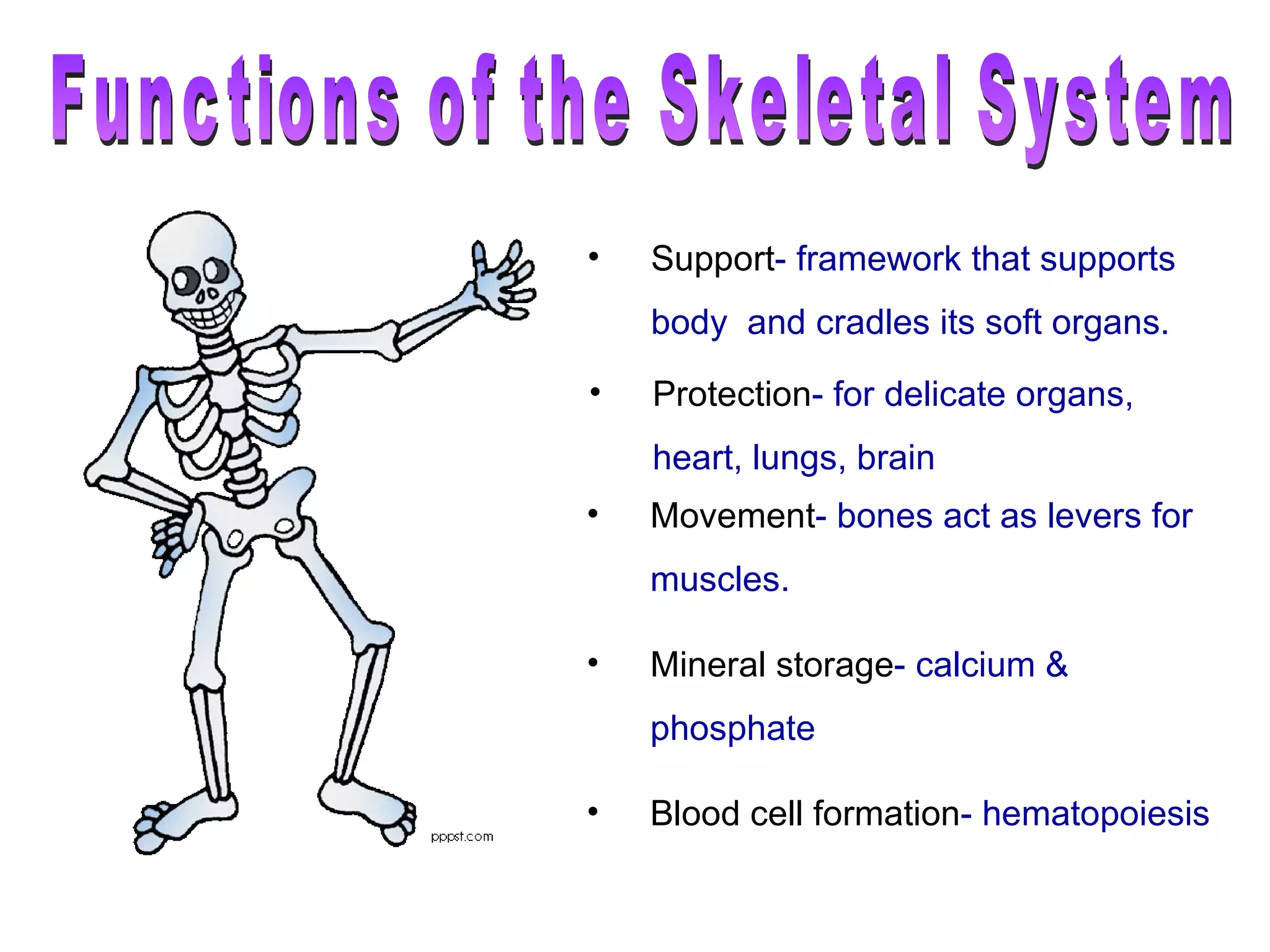
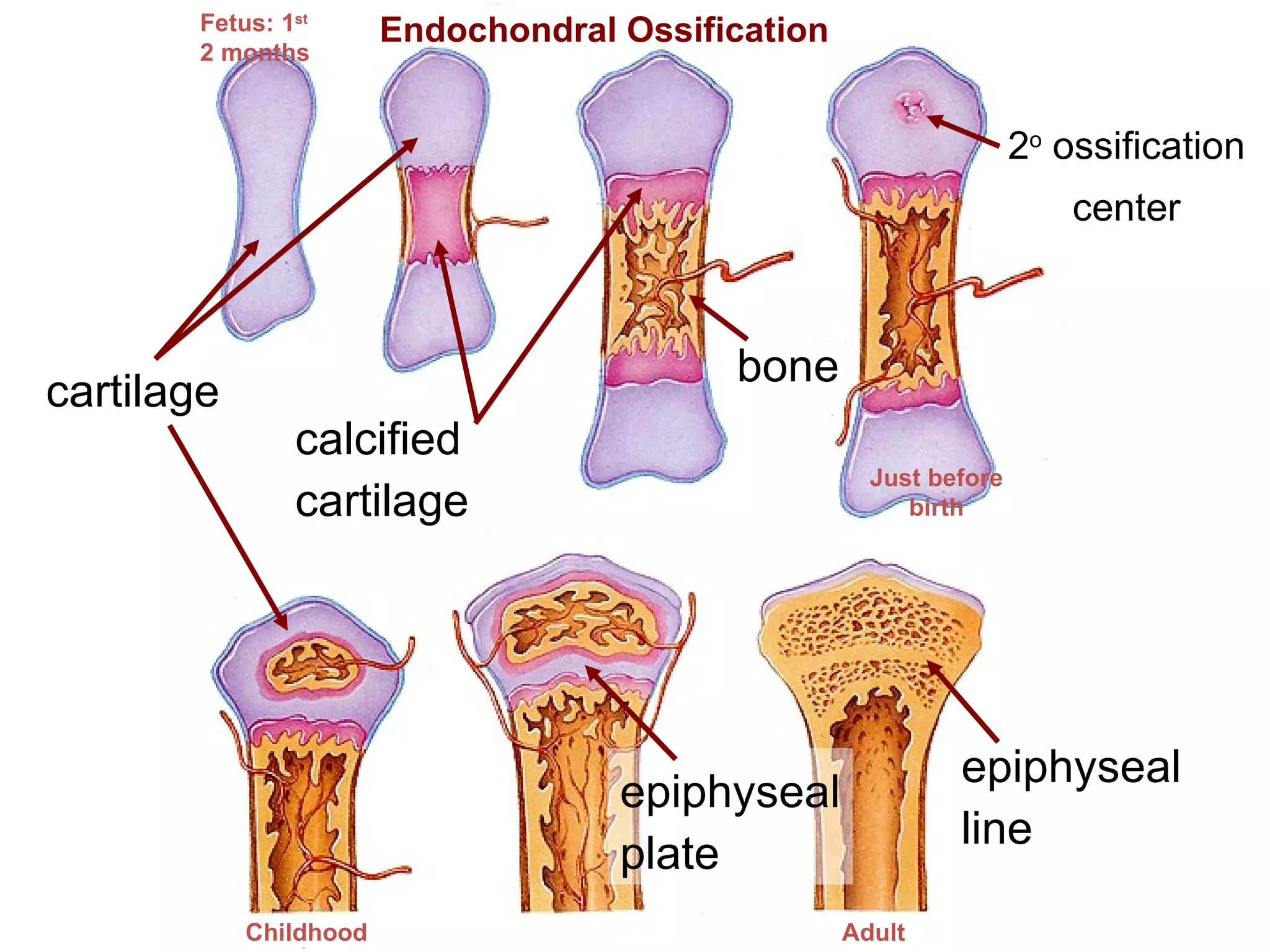
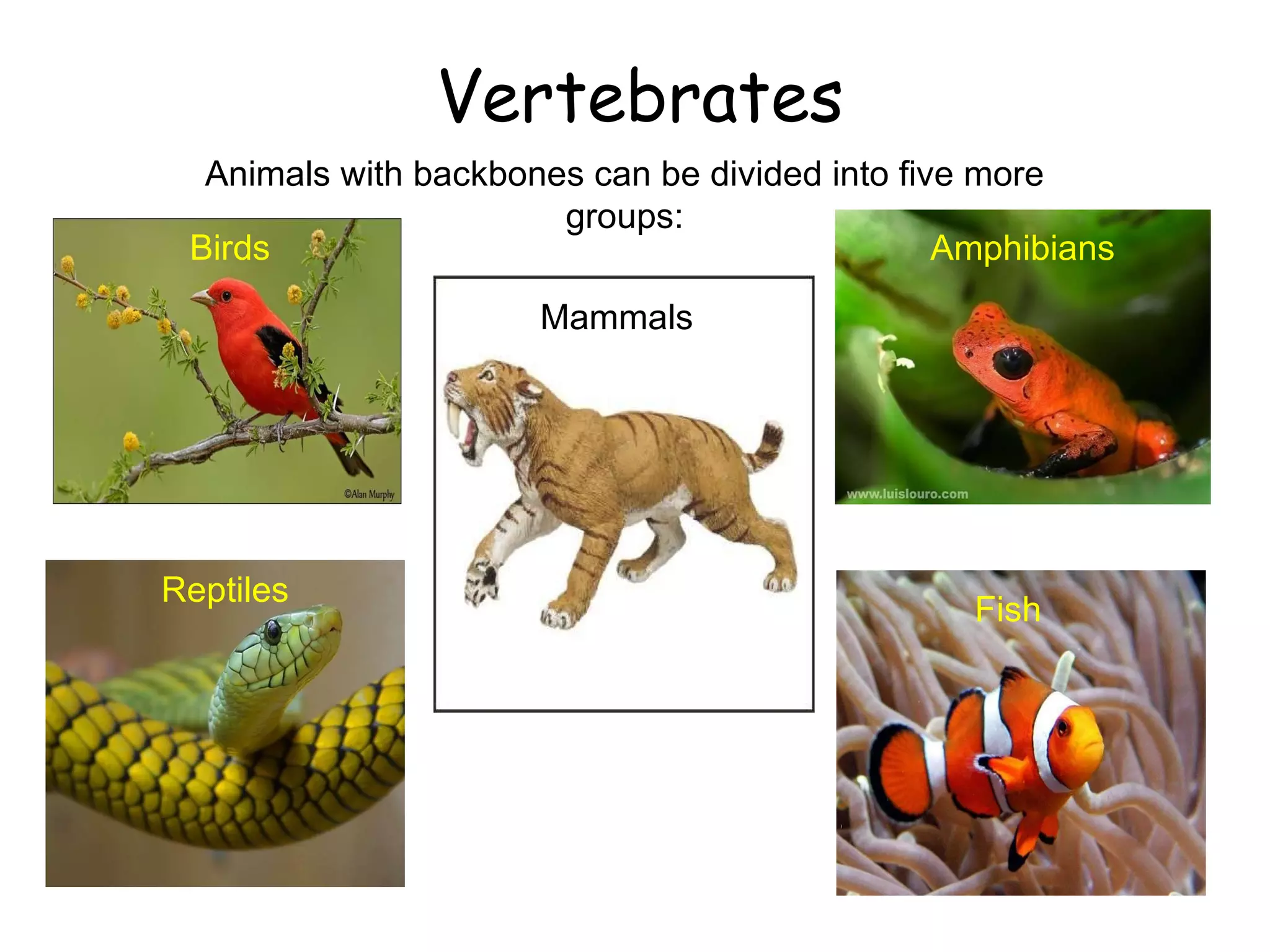
The skeletal system has several important functions:
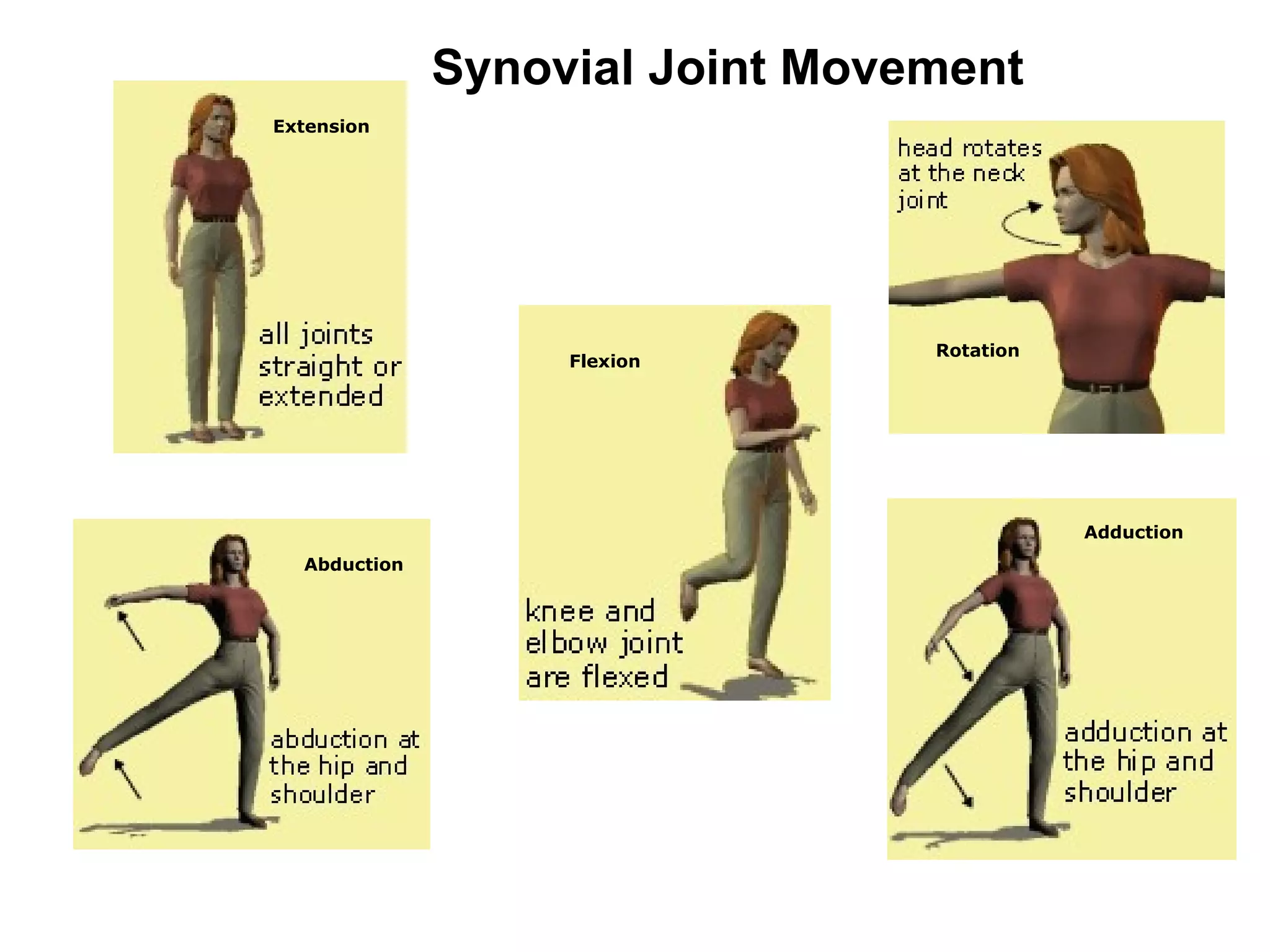
1) It provides structure and support for the body, protects delicate organs, and acts as levers for muscle movement.
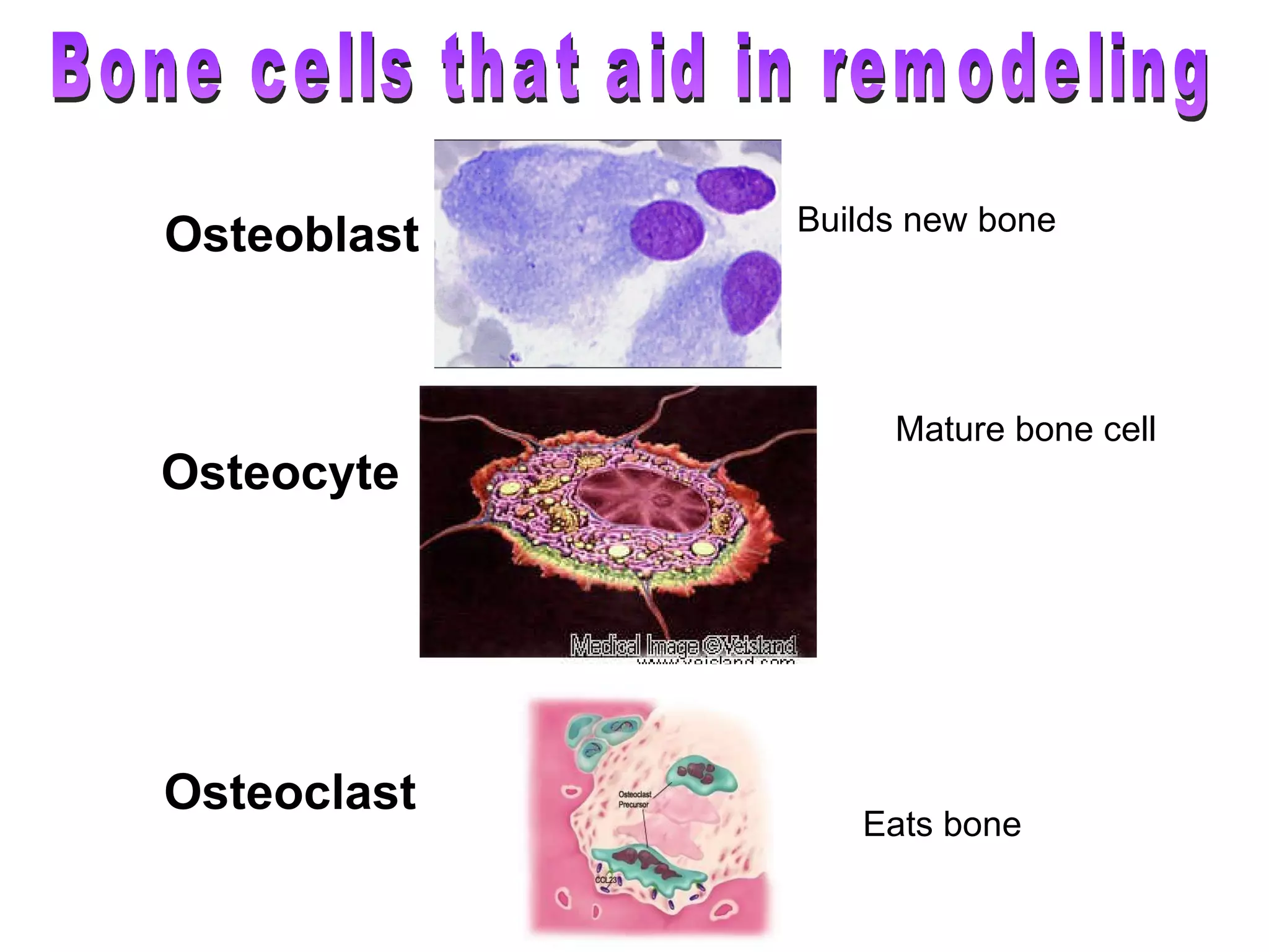
2) Bones also function in mineral storage and blood cell formation.
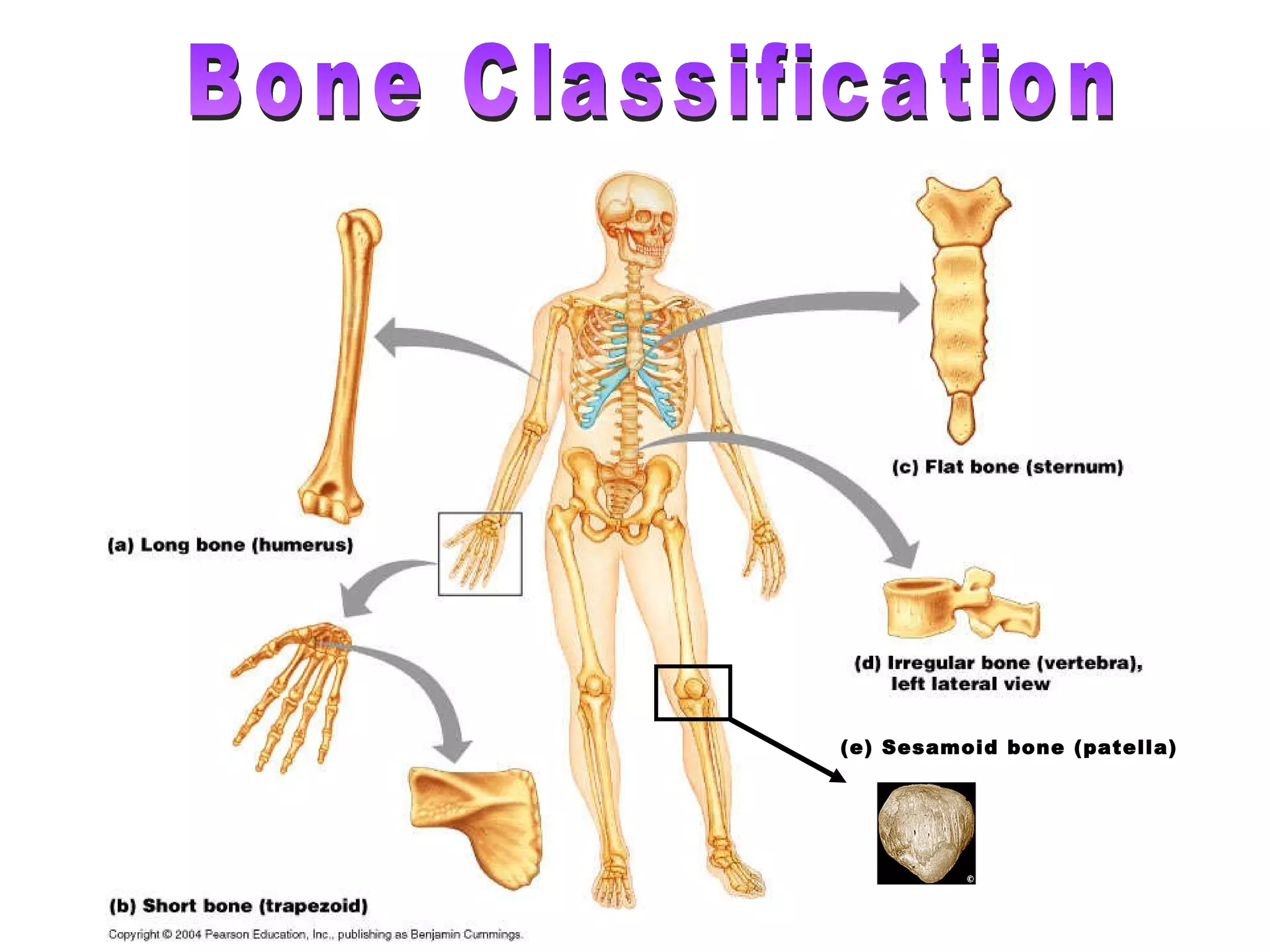
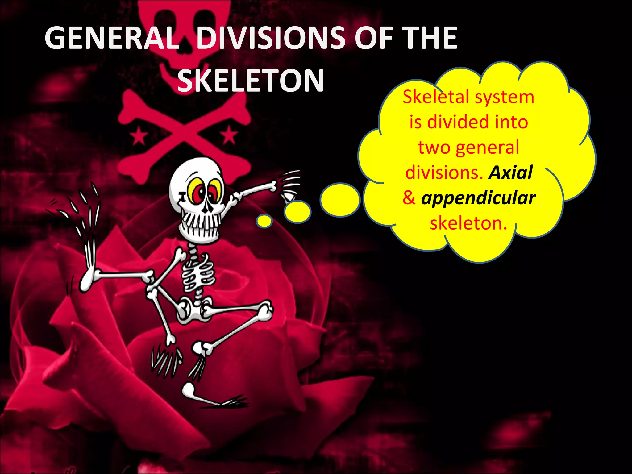
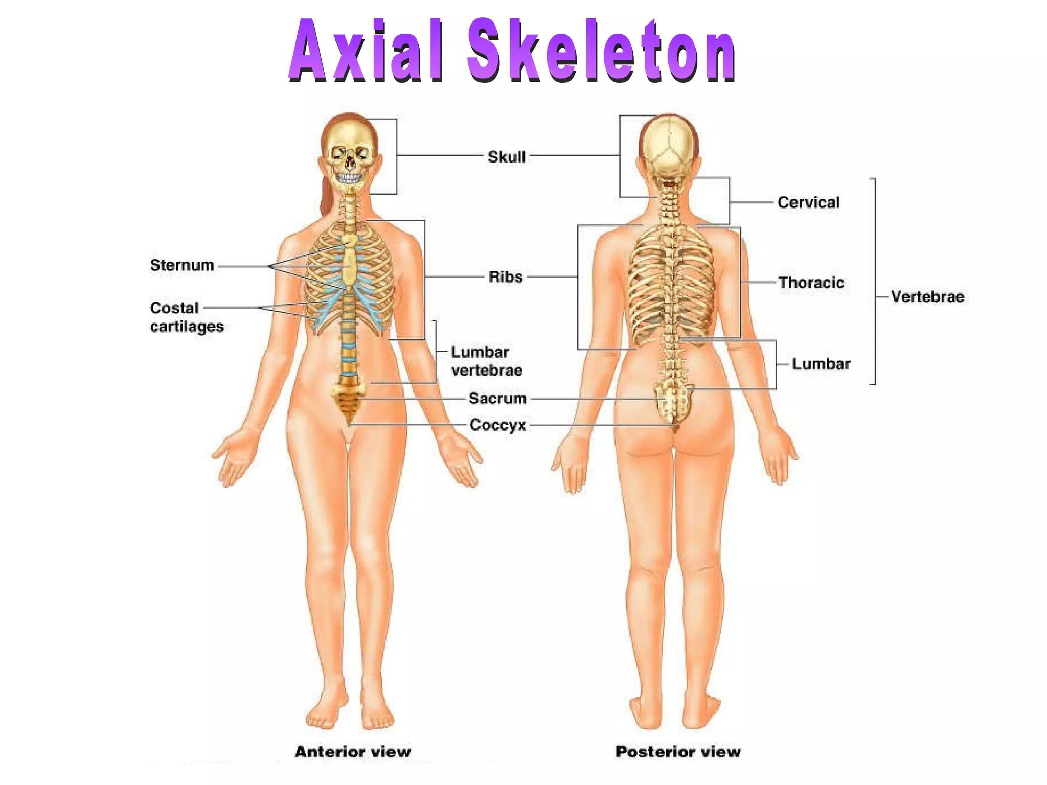
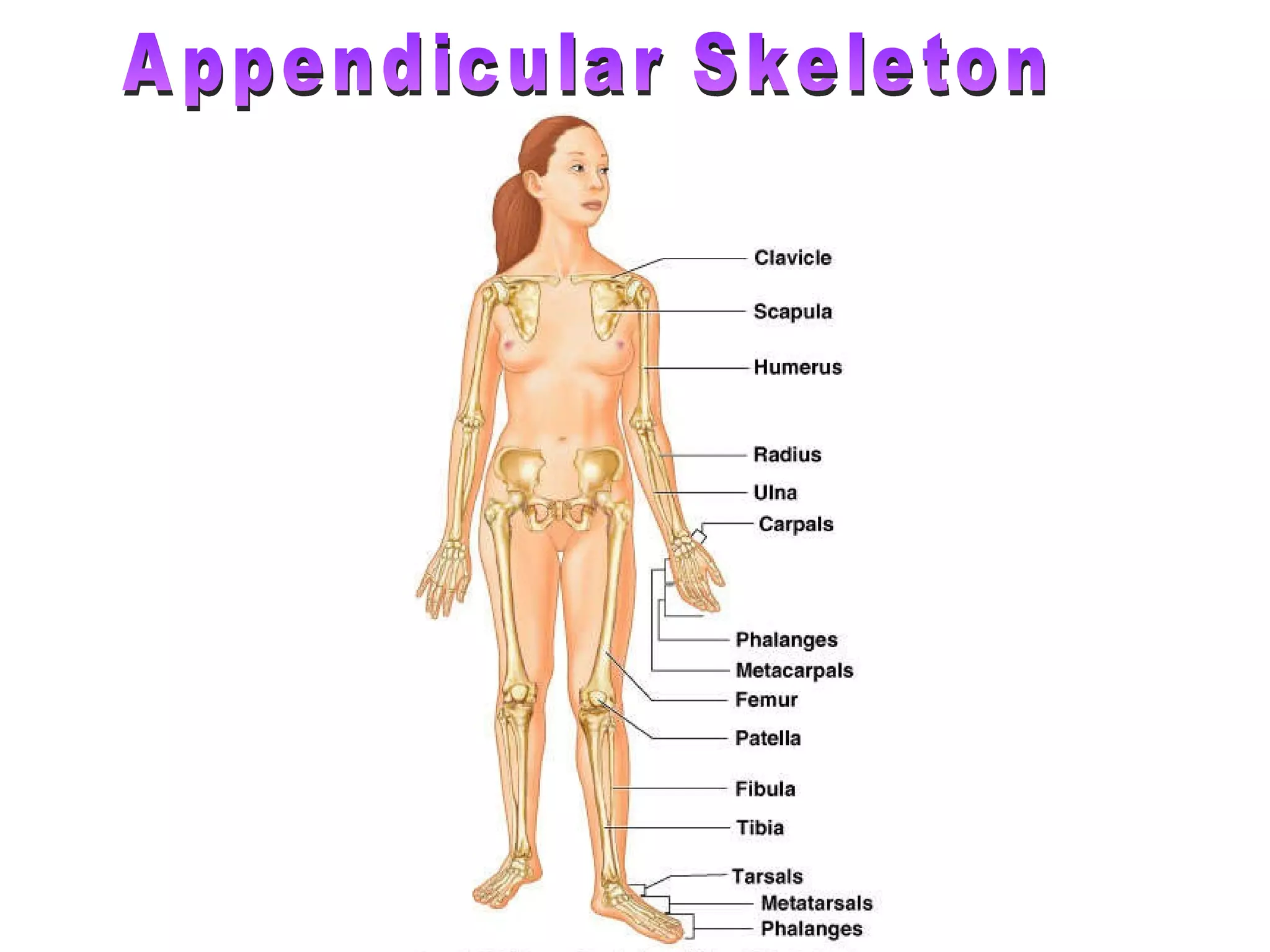
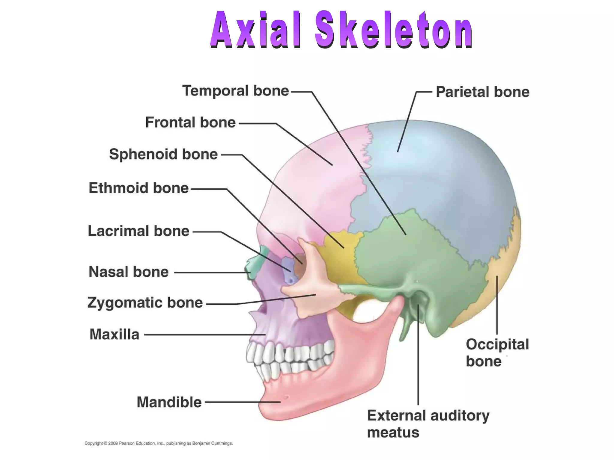
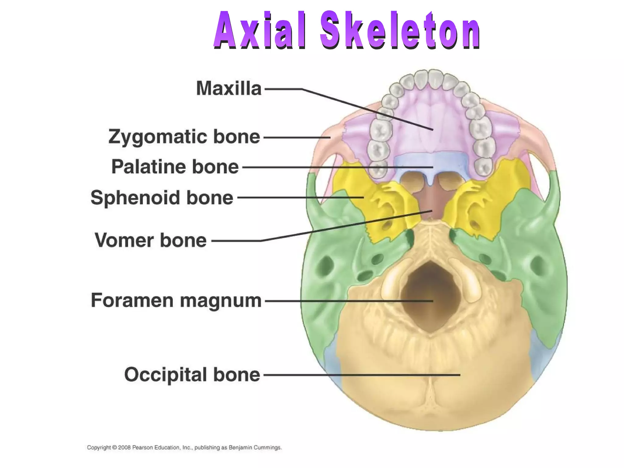
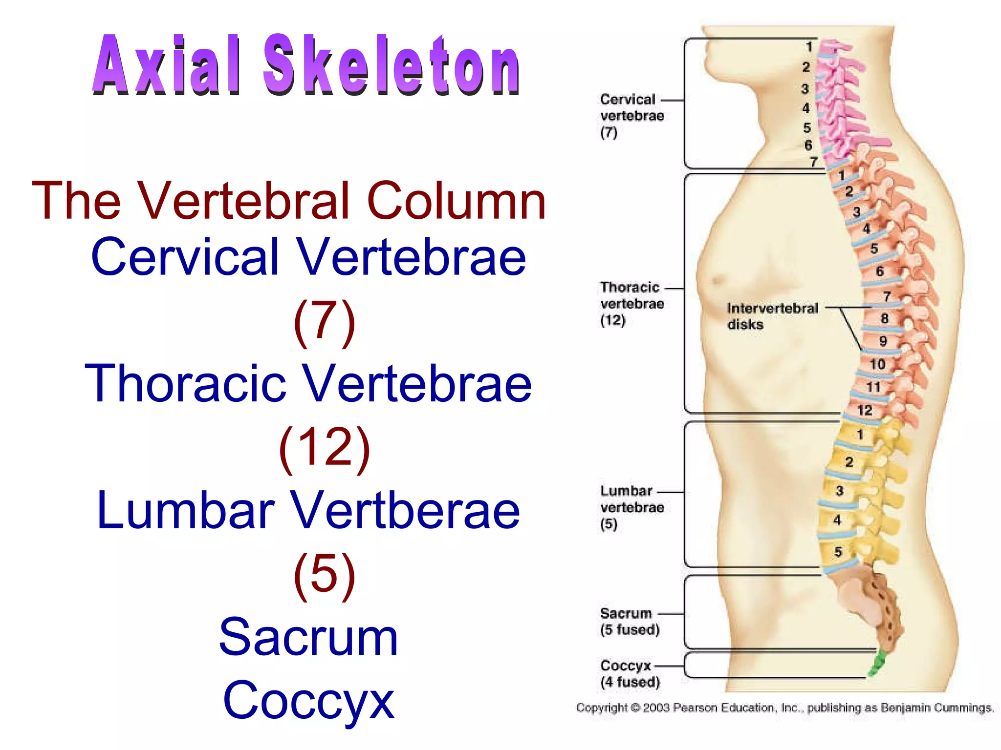
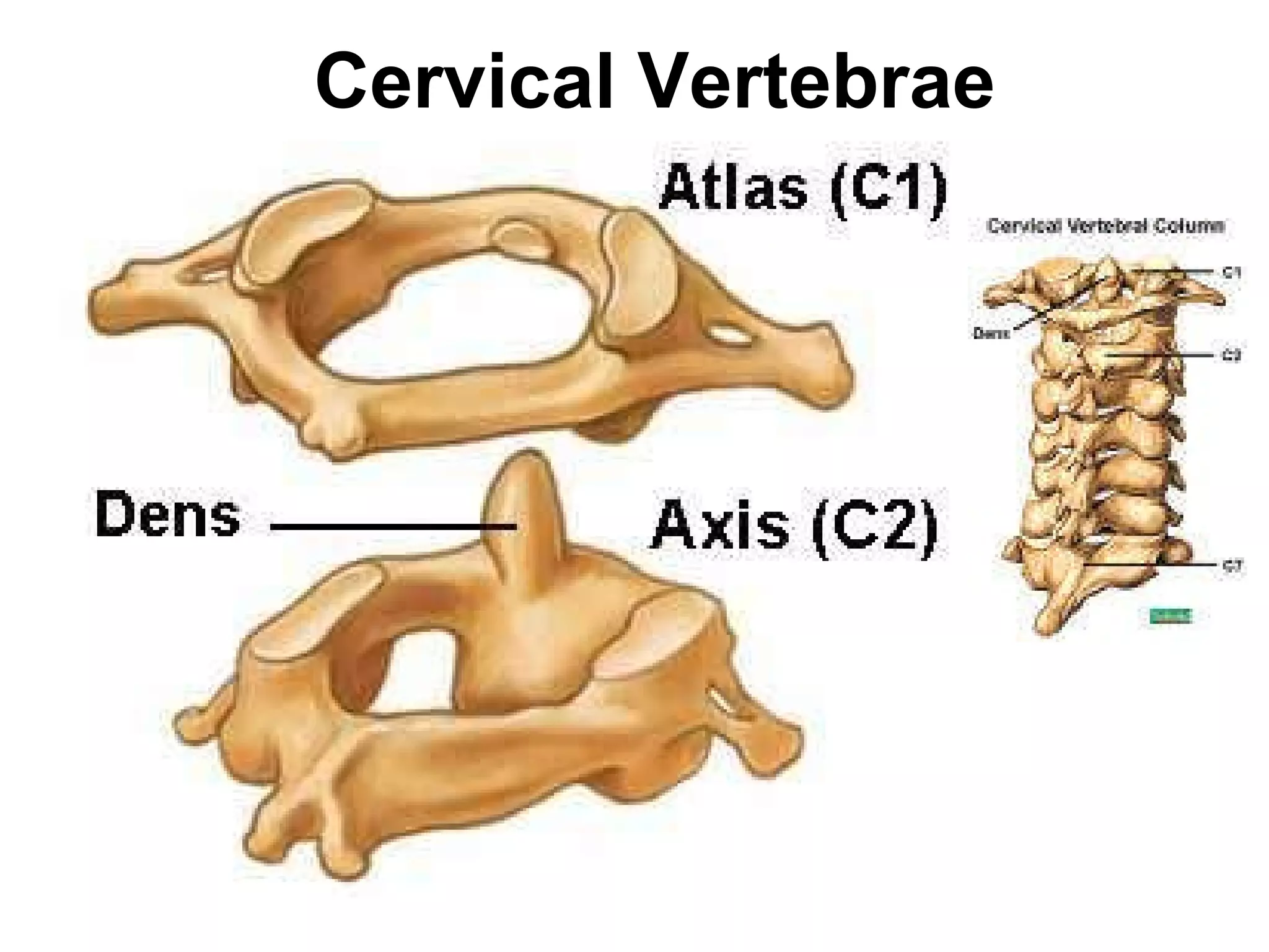
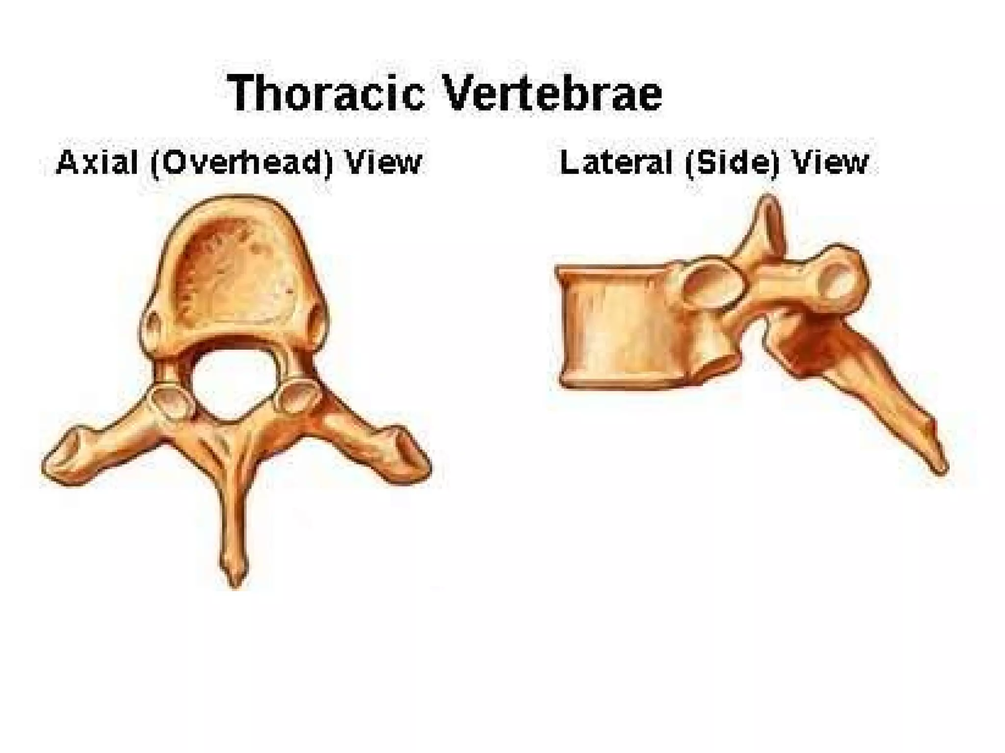
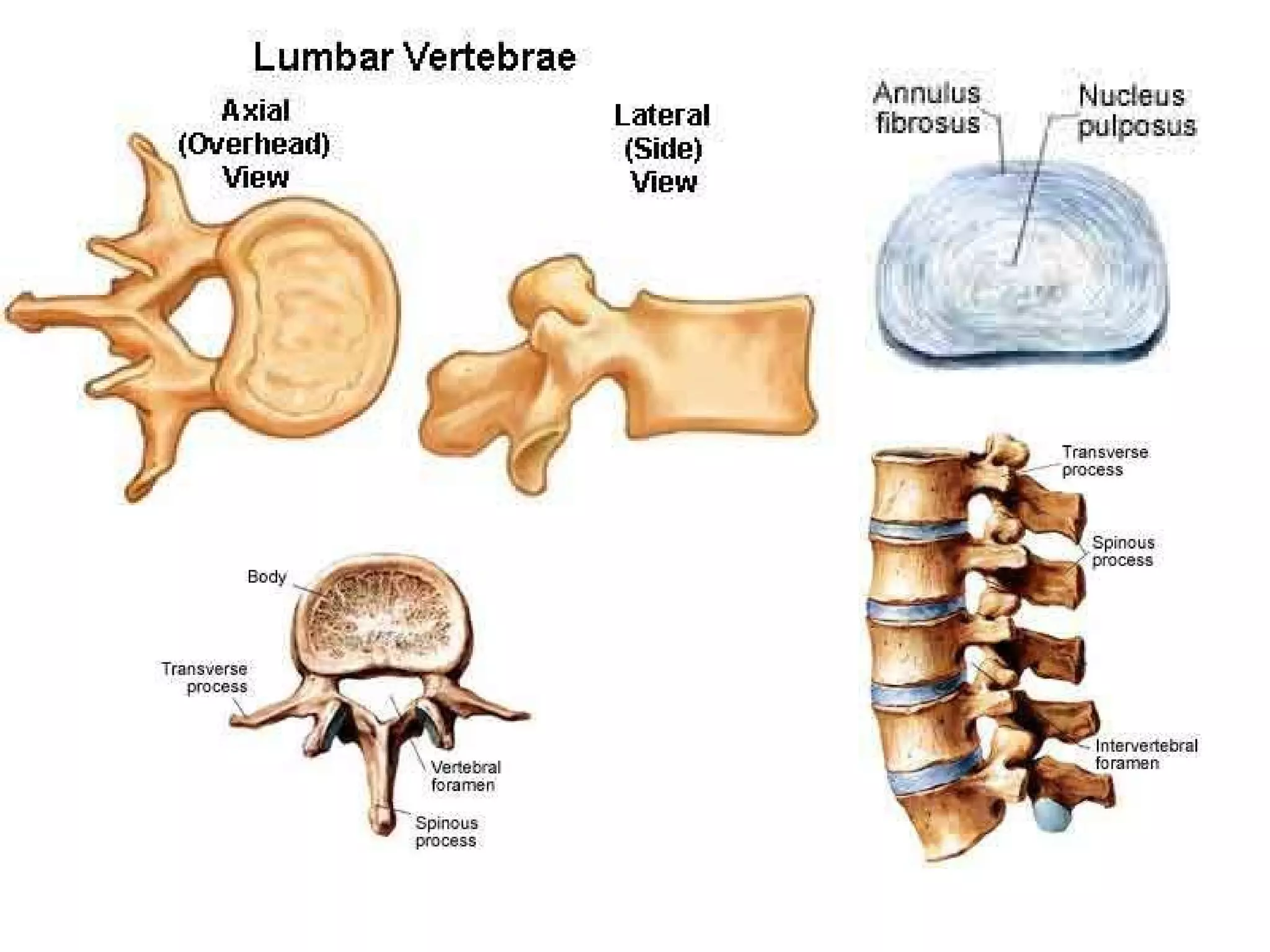
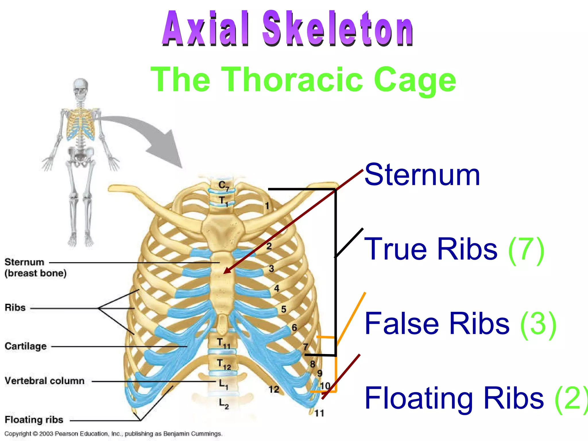
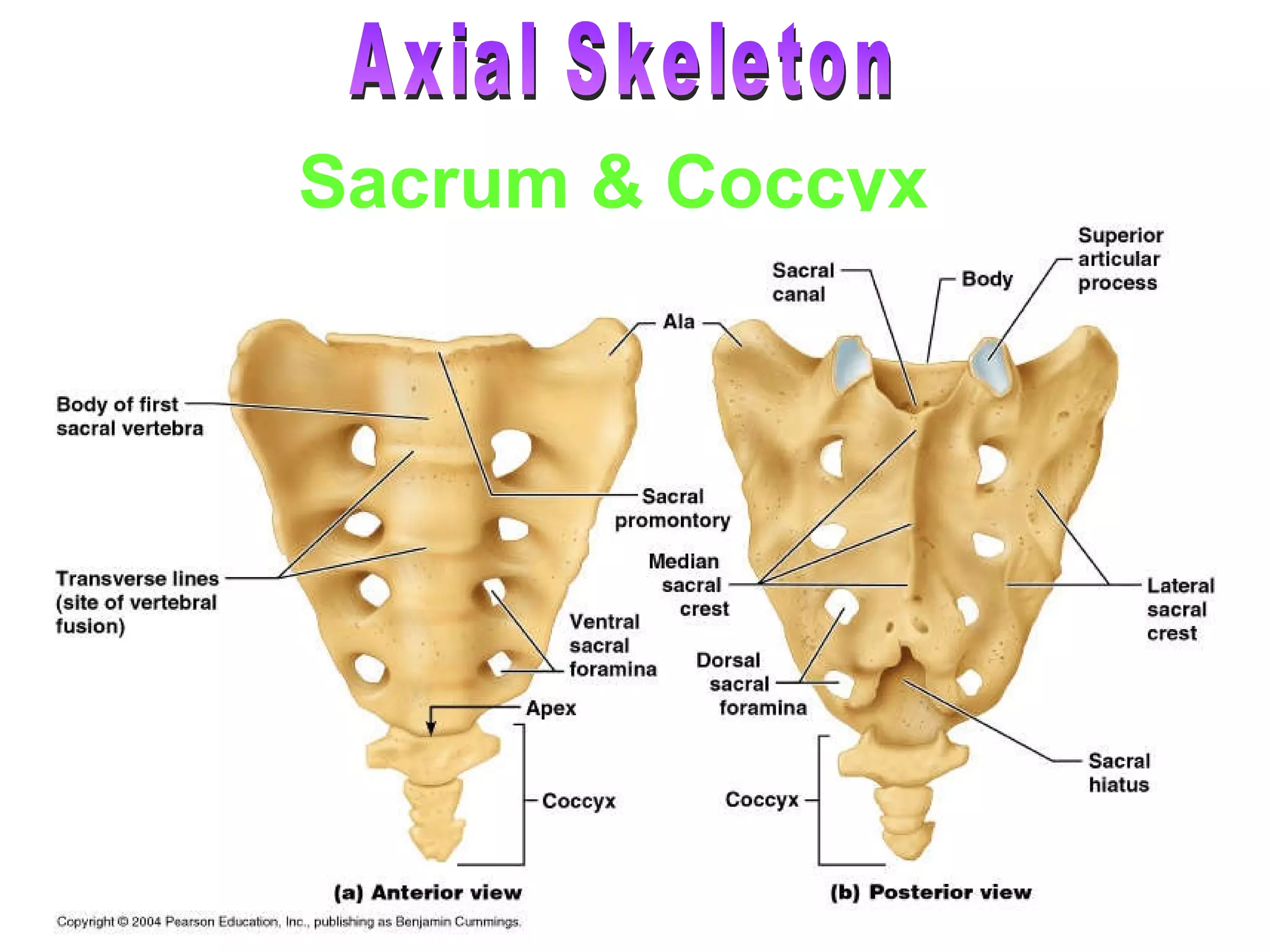
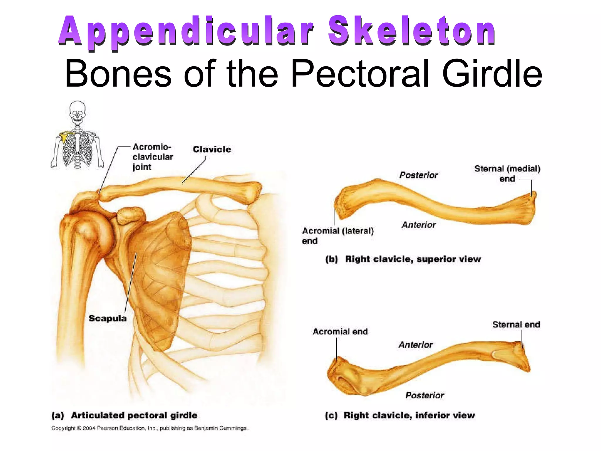
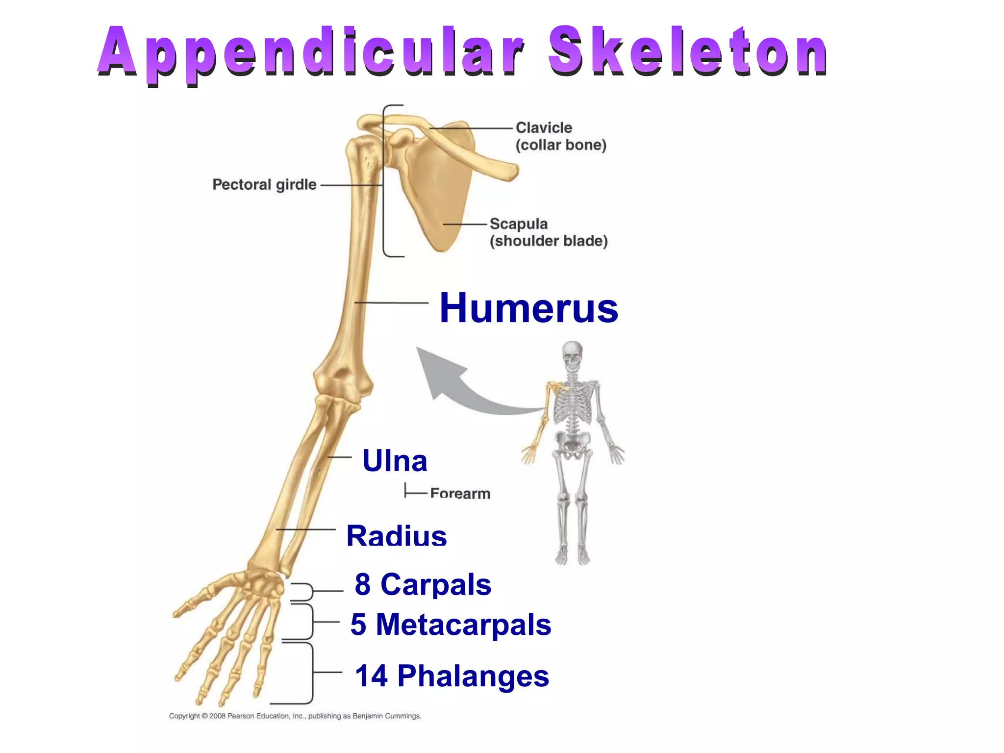
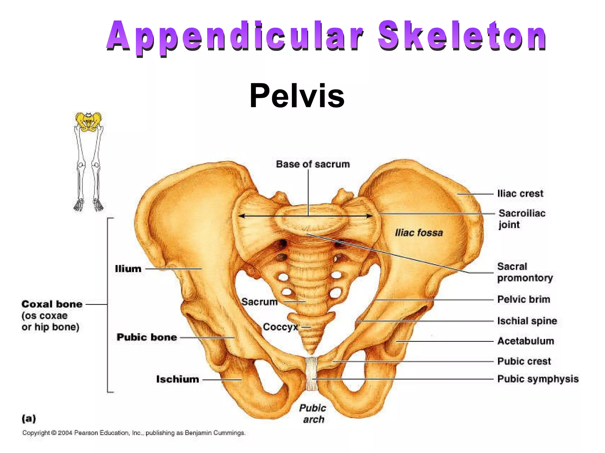
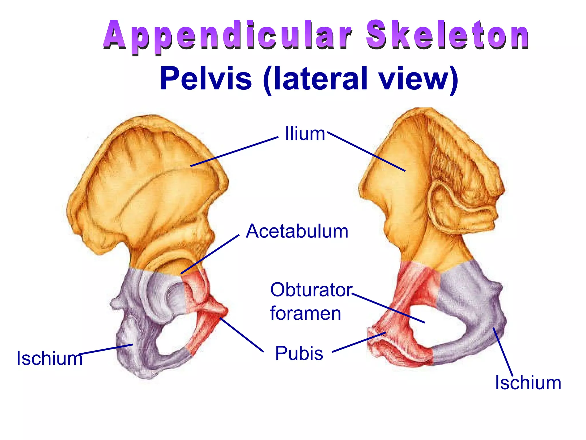
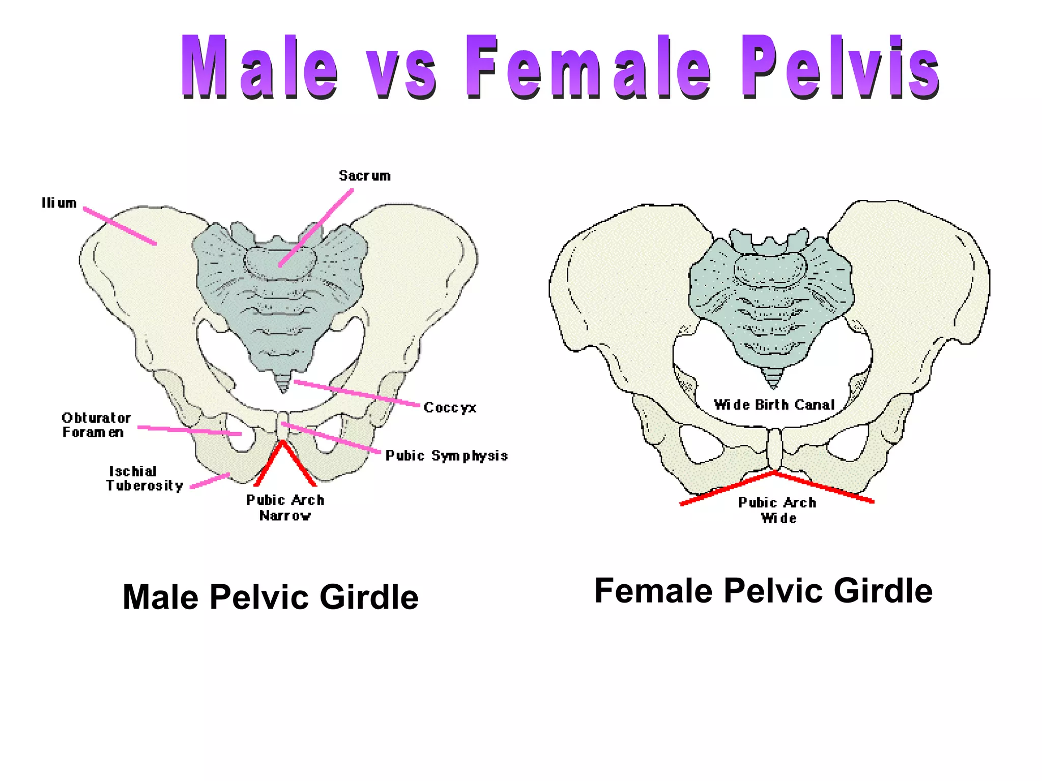
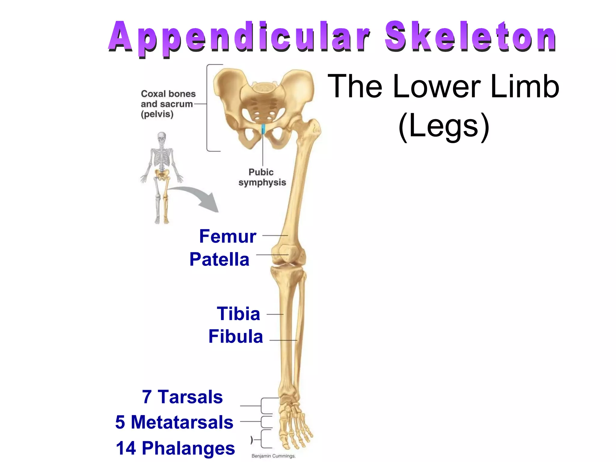
3) The skeletal system is divided into the axial skeleton (which includes bones such as the skull, vertebral column, ribs, and sternum) and appendicular skeleton (which includes the bones of the upper and lower limbs).


![2[1].1 (a) FORM 5](https://cdn.slidesharecdn.com/ss_thumbnails/21-1a-supportlocomotioninhumansanimals-120602235814-phpapp02-thumbnail.jpg?width=640&height=640&fit=bounds)


