SlideShare a Scribd company logo
1 of 40
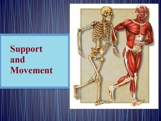
Skeletal Systems
• Skeletons are supportive systems that provide:
1. Rigidity to the body
2. Surfaces for muscle attachment
3. Protection for vulnerable body organs
• Bone of the vertebrate skeleton is only one of several
kinds of supportive and connective tissues serving
various binding and weight-bearing functions
• There are two forms of skeletal systems:
1. Hydrostatic skeletons
2. Rigid Skeletons
Hydrostatic Skeletons
• Not all skeletons are rigid
• Many invertebrate groups use their body fluids as an
internal hydrostatic skeleton
• Muscles in the body wall of the earthworm have no
firm base for attachment but develop muscular force
by contracting against the coelomic fluids
• Earthworms and other annelids are helped by septa
that separate the body into more or less independent
compartments
• An obvious advantage is that if a worm is punctured or
even cut into pieces, each part can still develop
pressure and move
• Worms that lack internal compartments are rendered
helpless if body fluid is lost through a wound
• There are many examples in the animal kingdom of
muscles that not only produce movement but also
provide a unique form of skeletal support
• The elephant’s trunk is an excellent example of a
structure that lacks any obvious form of skeletal
support, yet is capable of bending, twisting, elongating,
and lifting heavy weights
• The elephant’s trunk, tongues of mammals and
reptiles, and tentacles of cephalopod molluscs are
examples of muscular hydrostats
• Like the hydrostatic skeletons of worms, muscular
hydrostats work because they are composed of
incompressible tissues that remain at constant volume
• The remarkably diverse movements of muscular
hydrostats depend on muscles arranged in complex
patterns
Rigid Skeletons
• Rigid skeletons consist of rigid elements, usually
jointed, to which muscles can attach
• Muscles can only contract; to be lengthened they must
be extended by the pull of an antagonistic set of
muscles
• Rigid skeletons provide the anchor points required by
opposing sets of muscles, such as flexors and extensors
• There are two principal types of rigid skeletons:
1. Exoskeleton: Molluscs, Arthropods and many other
invertebrates
2. Endoskeleton: Echinoderms and vertebrates
Invertebrate Exoskeleton
• May be mainly protective
• May also perform a vital role in locomotion
• An exoskeleton may take the form of a shell, a spicule,
or a calcareous, proteinaceous, or chitinous plate
• It may be:
 Rigid, as in molluscs
 Jointed and movable, as in arthropods
• Unlike an endoskeleton, which grows with the animal,
an exoskeleton is often a limiting coat of armor that
must be periodically molted to make way for an
enlarged replacement
 Some invertebrate exoskeletons, such as the shells of
snails and bivalves, grow with the animal
Spicules in Sponges
Chitinous Exoskeleton of Insects
Vertebrate Endoskeleton
• Formed inside the body
• Is composed of bone and cartilage
 Forms of dense connective tissue
• Bone not only supports and protects but is
also the major body reservoir for calcium and
phosphorus
• In amniote vertebrates red blood cells and
certain white blood cells are formed in the
bone marrow
Notochord
• Semirigid supportive axial rod of the
protochordates and all vertebrate larvae and
embryos
 Composed of large, vacuolated cells and is
surrounded by layers of elastic and fibrous
sheaths
 It is a stiffening device, preserving body shape
during locomotion
 Except in the jawless vertebrates (lampreys and
hagfishes), the notochord is surrounded or
replaced by the backbone during embryonic
development
Cartilage
• Major skeletal element of some vertebrates
 The jawless fishes and the elasmobranchs
(sharks, skates, and rays) have purely
cartilaginous skeletons
 Other vertebrates as adults have principally bony
skeletons with some cartilage interspersed
• Cartilage is a soft, pliable, characteristically deep-
lying tissue
 Cartilage is the same wherever it is found
Cartilaginous skeleton of shark
• Hyaline cartilage: The basic form, has a clear,
glassy appearance
 Composed of cartilage cells (chondrocytes)
surrounded by firm complex protein gel
interlaced with a meshwork of collagenous fibers
 Blood vessels are virtually absent (injuries
involving cartilage heal poorly)
 Makes up the articulating surfaces of many bone
joints of most adult vertebrates and the
supporting tracheal, laryngeal, and bronchial
rings
Cartilage similar to hyaline cartilage occurs in some
invertebrates, (A) the radula of gastropod molluscs (B)
lophophore of brachiopods
B
Cartilage of cephalopod molluscs is of a special type with long, branching processes
that resemble the cells of vertebrate bone
Cells of Vertebrate Bone
Bone
• A living tissue that has significant deposits of inorganic calcium salts laid
down in an extracellular matrix
• Its structural organization is such that bone has nearly the tensile strength
of cast iron, yet is only one-third as heavy
• Bone is never formed in vacant space but is always laid down by
replacement in areas occupied by some form of connective tissue
1. Most bone develops from cartilage and is called endochondral (“within
cartilage”) or replacement bone
 Embryonic cartilage is gradually eroded leaving it extensively
honeycombed
 Bone-forming cells then invade these areas and begin depositing calcium
salts around strand like remnants of the cartilage
2. A second type of bone is intramembranous bone
 Develops directly from sheets of embryonic cells
 Dermal bone is a type of intramembranous bone
 In tetrapod vertebrates intramembranous bone is restricted mainly to
bones of the face, cranium and clavicle; the remainder of the skeleton is
endochondral bone
 Whatever the embryonic origin, once fully formed,
endochondral and intramembranous bone look the
same
 Fully formed bone, however, may vary in density
• Cancellous (or spongy) bone consists of an open,
interlacing framework of bony tissue, oriented to give
maximum strength under the normal stresses and
strains that the bone receives
 All bone develops first as cancellous bone
• Some bones, through further deposition of bone salts,
become compact
 Compact bone is dense, appearing solid to the unaided
eye
• Both cancellous and compact bone are found in the
typical long bones of tetrapods
Microscopic Structure of Bone
• Compact bone is composed of a calcified bone matrix arranged in
concentric rings
 Rings contain cavities (lacunae) filled with bone cells (osteocytes),
which are interconnected by many minute passages (canaliculi)
 These passages serve to distribute nutrients throughout the bone
• Entire organization of lacunae and canaliculi is arranged into an
elongated cylinder called an osteon (also called haversian system)
• Bone consists of bundles of osteons cemented together and
interconnected with blood vessels and nerves
• Bone is a living tissue: Blood vessels and nerves throughout
 Nonliving “ground substance” predominates
 As a result of its living state, bone breaks can heal, and bone
diseases can be as painful as any other tissue disease
• Bone growth is a complex restructuring process,
involving both its destruction internally by bone
resorbing cells (osteoclasts) and its deposition
externally by bone building cells (osteoblasts)
• Both processes occur simultaneously so that the
marrow cavity inside grows larger by bone resorption
while new bone is laid down outside by bone
deposition
• Bone growth responds to several hormones:
 Parathyroid hormone from the parathyroid gland
 Stimulates bone resorption
 Calcitonin from the thyroid gland
 Inhibits bone resorption
 These two hormones, together with a derivative of
vitamin D, are responsible for maintaining a constant
level of calcium in the blood
Plan of the Vertebrate Skeleton
• The vertebrate skeleton is composed of two main divisions:
1. Axial skeleton: Skull, vertebral column, sternum, and ribs
2. Appendicular skeleton: Limbs (or fins or wings) and pectoral and
pelvic girdles
• Skeleton has undergone extensive remodeling in the course of
vertebrate evolution
• The move from water to land forced dramatic changes in body form
• With increased cephalization, the further concentration of brain,
sense organs, and food-gathering and respiratory apparatus in the
head, the skull became the most intricate portion of the skeleton
• Some early fishes had as many as 180 skull bones
• Skull bones became greatly reduced in number during evolution of
the tetrapods
 Amphibians and lizards have 50 to 95, and mammals, 35 or fewer,
Humans have 29
Vertebral Column
• Main stiffening axis of the postcranial skeleton
• In fishes it serves much the same function as the notochord
from which it is derived
 Provides points for muscle attachment and prevents
telescoping of the body during muscle contraction
• With evolution of amphibious and terrestrial tetrapods, the
vertebrate body was no longer buoyed by the aquatic
environment
• Vertebral column became structurally adapted to withstand
new regional stresses transmitted to the column by the two
pairs of appendages
• In amniote tetrapods (reptiles, birds, and mammals), the
vertebrae are differentiated into cervical (neck), thoracic
(chest), lumbar (back), sacral (pelvic), and caudal (tail)
vertebrae
• In birds and humans the caudal vertebrae are reduced
in number and size, and the sacral vertebrae are fused
• The number of vertebrae varies among the different
vertebrates:
 Pythons seems to lead the list with more than 400
 In humans there are 33 in a young child, but in adults 5
are fused to form the sacrum and 4 to form the coccyx
 Besides the sacrum and coccyx, humans have 7
cervical, 12 thoracic, and 5 lumbar vertebrae
• Number of cervical vertebrae (7) is constant in nearly
all mammals
 The first two cervical vertebrae, atlas and axis, are
modified to support the skull and permit pivotal
movements
 The atlas bears the globe of the head
 The axis permits the head to turn from side to side
Ribs
• Long or short skeletal structures that articulate medially with
vertebrae and extend into the body wall
• Fishes have a pair of ribs for every vertebra
 They serve as stiffening elements in the connective tissue septa
that separate the muscle segments and thus improve the
effectiveness of muscle contractions
 Many fishes have both dorsal and ventral ribs, and some have
numerous rib-like intermuscular bones as well—all of which
increase the difficulty and reduce the pleasure of eating certain
kinds of fish
• Other vertebrates have a reduced number of ribs, and some, such
as the familiar leopard frog, have no ribs at all
• In mammals the ribs together form the thoracic basket, which
supports the chest wall and prevents collapse of the lungs
• Mammals such as sloths have 24 pairs of ribs, horses posses 18
pairs, primates other than humans have 13 pairs of ribs; humans
have 12 pairs
Intermuscular bones are small, hard spicules of bone
Frog: No ribs at all Humans: 12 pairs of ribs
Appendages
• Most vertebrates, fishes included, have paired appendages
• All fishes except agnathans have thin pectoral and pelvic fins that are
supported by the pectoral and pelvic girdles, respectively
• Tetrapods (except caecilians, snakes, and limbless lizards) have two
pairs of pentadactyl (five toed) limbs, also supported by girdles
Caecilian Legless lizards
• The pentadactyl limb is similar in all tetrapods, alive
and extinct
 Even when highly modified for various modes of life,
the elements are rather easily homologized
• Modifications of the basic pentadactyl limb for life in
different environments involve distal elements much
more frequently than proximal
 It is far more common for bones to be lost or fused
than for new ones to be added
 Horses and their relatives evolved a foot structure for
fleetness by elongation of the third toe
 In effect, a horse stands on its third fingernail (hoof),
much like a ballet dancer standing on the tips of the
toes
• The bird wing is a good example of distal modification
• Bird embryo bears 13 distinct wrist and hand bones
(carpals and metacarpals)
 These are reduced to three digits in the adult
 Most finger bones (phalanges) are lost, leaving four bones
in three digits
 The proximal bones (humerus, radius, and ulna), however,
are only slightly modified in the bird wing
• In nearly all tetrapods the pelvic girdle is firmly attached to
the axial skeleton
• The greatest locomotory forces transmitted to the body
come from the hindlimbs
• The pectoral girdle is much more loosely attached to the
axial skeleton, providing the forelimbs with greater
freedom for manipulative movement
Support and Movement.pptx

More Related Content

Similar to Support and Movement.pptx

1. Unit - 7- Skeleton anatomy by Thiru murugan
1. Unit - 7- Skeleton anatomy by Thiru murugan1. Unit - 7- Skeleton anatomy by Thiru murugan
1. Unit - 7- Skeleton anatomy by Thiru muruganthiru murugan
 
Bonetissuetoskeletalsystemppt 110126162139-phpapp02
Bonetissuetoskeletalsystemppt 110126162139-phpapp02Bonetissuetoskeletalsystemppt 110126162139-phpapp02
Bonetissuetoskeletalsystemppt 110126162139-phpapp02IICCAA
 
Unit 16 support and movement
Unit 16 support and movementUnit 16 support and movement
Unit 16 support and movementIshtiaqAhmad114
 
Basic Structures in human body
Basic Structures in human bodyBasic Structures in human body
Basic Structures in human bodyssuseraf61fb
 
Lecture 1 (introduction to bone)
Lecture 1 (introduction to bone)Lecture 1 (introduction to bone)
Lecture 1 (introduction to bone)Ayub Abdi
 
ANATOMY OF THE HUMAN SKELETON POWERPOINT
ANATOMY OF THE HUMAN SKELETON POWERPOINTANATOMY OF THE HUMAN SKELETON POWERPOINT
ANATOMY OF THE HUMAN SKELETON POWERPOINTmhixgoodie
 
peer tutorial for body systems and anatomy
peer tutorial for body systems and anatomypeer tutorial for body systems and anatomy
peer tutorial for body systems and anatomysondos59
 
Ls2 afet unit 6 support systems in animals
Ls2 afet unit 6 support systems in animalsLs2 afet unit 6 support systems in animals
Ls2 afet unit 6 support systems in animalsgoogle
 
Human skeleton system
Human skeleton systemHuman skeleton system
Human skeleton systemAsma Amir
 
Presentation on bones
Presentation on bonesPresentation on bones
Presentation on bonesSam Shaikh
 
Introduction to osteology
Introduction to osteologyIntroduction to osteology
Introduction to osteologyAfTaab AfRaz
 
Introduction to osteology
Introduction to osteologyIntroduction to osteology
Introduction to osteologyAfTaab AfRaz
 
support systems in animals
 support systems in animals support systems in animals
support systems in animalsThabang Mokoena
 

Similar to Support and Movement.pptx (20)

Skeletal system
Skeletal systemSkeletal system
Skeletal system
 
1. Unit - 7- Skeleton anatomy by Thiru murugan
1. Unit - 7- Skeleton anatomy by Thiru murugan1. Unit - 7- Skeleton anatomy by Thiru murugan
1. Unit - 7- Skeleton anatomy by Thiru murugan
 
Skeletal System
Skeletal SystemSkeletal System
Skeletal System
 
Bonetissuetoskeletalsystemppt 110126162139-phpapp02
Bonetissuetoskeletalsystemppt 110126162139-phpapp02Bonetissuetoskeletalsystemppt 110126162139-phpapp02
Bonetissuetoskeletalsystemppt 110126162139-phpapp02
 
LP 1-cartilage ppt.pptx
LP 1-cartilage ppt.pptxLP 1-cartilage ppt.pptx
LP 1-cartilage ppt.pptx
 
The skeletomuscular.ppsx
The skeletomuscular.ppsxThe skeletomuscular.ppsx
The skeletomuscular.ppsx
 
Unit 16 support and movement
Unit 16 support and movementUnit 16 support and movement
Unit 16 support and movement
 
Basic Structures in human body
Basic Structures in human bodyBasic Structures in human body
Basic Structures in human body
 
Lecture 1 (introduction to bone)
Lecture 1 (introduction to bone)Lecture 1 (introduction to bone)
Lecture 1 (introduction to bone)
 
Bones anatomy
Bones anatomyBones anatomy
Bones anatomy
 
Skeletons grade 10
Skeletons grade 10Skeletons grade 10
Skeletons grade 10
 
ANATOMY OF THE HUMAN SKELETON POWERPOINT
ANATOMY OF THE HUMAN SKELETON POWERPOINTANATOMY OF THE HUMAN SKELETON POWERPOINT
ANATOMY OF THE HUMAN SKELETON POWERPOINT
 
peer tutorial for body systems and anatomy
peer tutorial for body systems and anatomypeer tutorial for body systems and anatomy
peer tutorial for body systems and anatomy
 
Ls2 afet unit 6 support systems in animals
Ls2 afet unit 6 support systems in animalsLs2 afet unit 6 support systems in animals
Ls2 afet unit 6 support systems in animals
 
Human skeleton system
Human skeleton systemHuman skeleton system
Human skeleton system
 
Presentation on bones
Presentation on bonesPresentation on bones
Presentation on bones
 
Introduction to osteology
Introduction to osteologyIntroduction to osteology
Introduction to osteology
 
Introduction to osteology
Introduction to osteologyIntroduction to osteology
Introduction to osteology
 
support systems in animals
 support systems in animals support systems in animals
support systems in animals
 
Human Skeletal system
Human Skeletal systemHuman Skeletal system
Human Skeletal system
 

More from adesiredWish

Animal Movement.pptx
Animal Movement.pptxAnimal Movement.pptx
Animal Movement.pptxadesiredWish
 
Communication-I.pptx
Communication-I.pptxCommunication-I.pptx
Communication-I.pptxadesiredWish
 
Movement in Mammals.pptx
Movement in Mammals.pptxMovement in Mammals.pptx
Movement in Mammals.pptxadesiredWish
 
Defence and Protection in Mammals.pptx
Defence and Protection in Mammals.pptxDefence and Protection in Mammals.pptx
Defence and Protection in Mammals.pptxadesiredWish
 
Origin and Evolution of Mammals.pptx
Origin and Evolution of Mammals.pptxOrigin and Evolution of Mammals.pptx
Origin and Evolution of Mammals.pptxadesiredWish
 
1. Protection, Support, and Movement.pptx
1. Protection, Support, and Movement.pptx1. Protection, Support, and Movement.pptx
1. Protection, Support, and Movement.pptxadesiredWish
 

More from adesiredWish (6)

Animal Movement.pptx
Animal Movement.pptxAnimal Movement.pptx
Animal Movement.pptx
 
Communication-I.pptx
Communication-I.pptxCommunication-I.pptx
Communication-I.pptx
 
Movement in Mammals.pptx
Movement in Mammals.pptxMovement in Mammals.pptx
Movement in Mammals.pptx
 
Defence and Protection in Mammals.pptx
Defence and Protection in Mammals.pptxDefence and Protection in Mammals.pptx
Defence and Protection in Mammals.pptx
 
Origin and Evolution of Mammals.pptx
Origin and Evolution of Mammals.pptxOrigin and Evolution of Mammals.pptx
Origin and Evolution of Mammals.pptx
 
1. Protection, Support, and Movement.pptx
1. Protection, Support, and Movement.pptx1. Protection, Support, and Movement.pptx
1. Protection, Support, and Movement.pptx
 

Recently uploaded

STERILITY TESTING OF PHARMACEUTICALS ppt by DR.C.P.PRINCE
STERILITY TESTING OF PHARMACEUTICALS ppt by DR.C.P.PRINCESTERILITY TESTING OF PHARMACEUTICALS ppt by DR.C.P.PRINCE
STERILITY TESTING OF PHARMACEUTICALS ppt by DR.C.P.PRINCEPRINCE C P
 
GFP in rDNA Technology (Biotechnology).pptx
GFP in rDNA Technology (Biotechnology).pptxGFP in rDNA Technology (Biotechnology).pptx
GFP in rDNA Technology (Biotechnology).pptxAleenaTreesaSaji
 
Pests of cotton_Borer_Pests_Binomics_Dr.UPR.pdf
Pests of cotton_Borer_Pests_Binomics_Dr.UPR.pdfPests of cotton_Borer_Pests_Binomics_Dr.UPR.pdf
Pests of cotton_Borer_Pests_Binomics_Dr.UPR.pdfPirithiRaju
 
Botany krishna series 2nd semester Only Mcq type questions
Botany krishna series 2nd semester Only Mcq type questionsBotany krishna series 2nd semester Only Mcq type questions
Botany krishna series 2nd semester Only Mcq type questionsSumit Kumar yadav
 
PossibleEoarcheanRecordsoftheGeomagneticFieldPreservedintheIsuaSupracrustalBe...
PossibleEoarcheanRecordsoftheGeomagneticFieldPreservedintheIsuaSupracrustalBe...PossibleEoarcheanRecordsoftheGeomagneticFieldPreservedintheIsuaSupracrustalBe...
PossibleEoarcheanRecordsoftheGeomagneticFieldPreservedintheIsuaSupracrustalBe...Sérgio Sacani
 
Chemistry 4th semester series (krishna).pdf
Chemistry 4th semester series (krishna).pdfChemistry 4th semester series (krishna).pdf
Chemistry 4th semester series (krishna).pdfSumit Kumar yadav
 
DIFFERENCE IN BACK CROSS AND TEST CROSS
DIFFERENCE IN  BACK CROSS AND TEST CROSSDIFFERENCE IN  BACK CROSS AND TEST CROSS
DIFFERENCE IN BACK CROSS AND TEST CROSSLeenakshiTyagi
 
Recombinant DNA technology (Immunological screening)
Recombinant DNA technology (Immunological screening)Recombinant DNA technology (Immunological screening)
Recombinant DNA technology (Immunological screening)PraveenaKalaiselvan1
 
Nightside clouds and disequilibrium chemistry on the hot Jupiter WASP-43b
Nightside clouds and disequilibrium chemistry on the hot Jupiter WASP-43bNightside clouds and disequilibrium chemistry on the hot Jupiter WASP-43b
Nightside clouds and disequilibrium chemistry on the hot Jupiter WASP-43bSérgio Sacani
 
Animal Communication- Auditory and Visual.pptx
Animal Communication- Auditory and Visual.pptxAnimal Communication- Auditory and Visual.pptx
Animal Communication- Auditory and Visual.pptxUmerFayaz5
 
Botany 4th semester file By Sumit Kumar yadav.pdf
Botany 4th semester file By Sumit Kumar yadav.pdfBotany 4th semester file By Sumit Kumar yadav.pdf
Botany 4th semester file By Sumit Kumar yadav.pdfSumit Kumar yadav
 
Raman spectroscopy.pptx M Pharm, M Sc, Advanced Spectral Analysis
Raman spectroscopy.pptx M Pharm, M Sc, Advanced Spectral AnalysisRaman spectroscopy.pptx M Pharm, M Sc, Advanced Spectral Analysis
Raman spectroscopy.pptx M Pharm, M Sc, Advanced Spectral AnalysisDiwakar Mishra
 
Chromatin Structure | EUCHROMATIN | HETEROCHROMATIN
Chromatin Structure | EUCHROMATIN | HETEROCHROMATINChromatin Structure | EUCHROMATIN | HETEROCHROMATIN
Chromatin Structure | EUCHROMATIN | HETEROCHROMATINsankalpkumarsahoo174
 
Stunning ➥8448380779▻ Call Girls In Panchshil Enclave Delhi NCR
Stunning ➥8448380779▻ Call Girls In Panchshil Enclave Delhi NCRStunning ➥8448380779▻ Call Girls In Panchshil Enclave Delhi NCR
Stunning ➥8448380779▻ Call Girls In Panchshil Enclave Delhi NCRDelhi Call girls
 
SOLUBLE PATTERN RECOGNITION RECEPTORS.pptx
SOLUBLE PATTERN RECOGNITION RECEPTORS.pptxSOLUBLE PATTERN RECOGNITION RECEPTORS.pptx
SOLUBLE PATTERN RECOGNITION RECEPTORS.pptxkessiyaTpeter
 
Biological Classification BioHack (3).pdf
Biological Classification BioHack (3).pdfBiological Classification BioHack (3).pdf
Biological Classification BioHack (3).pdfmuntazimhurra
 
VIRUSES structure and classification ppt by Dr.Prince C P
VIRUSES structure and classification ppt by Dr.Prince C PVIRUSES structure and classification ppt by Dr.Prince C P
VIRUSES structure and classification ppt by Dr.Prince C PPRINCE C P
 
GBSN - Microbiology (Unit 1)
GBSN - Microbiology (Unit 1)GBSN - Microbiology (Unit 1)
GBSN - Microbiology (Unit 1)Areesha Ahmad
 
Pests of cotton_Sucking_Pests_Dr.UPR.pdf
Pests of cotton_Sucking_Pests_Dr.UPR.pdfPests of cotton_Sucking_Pests_Dr.UPR.pdf
Pests of cotton_Sucking_Pests_Dr.UPR.pdfPirithiRaju
 
Zoology 4th semester series (krishna).pdf
Zoology 4th semester series (krishna).pdfZoology 4th semester series (krishna).pdf
Zoology 4th semester series (krishna).pdfSumit Kumar yadav
 

Recently uploaded (20)

STERILITY TESTING OF PHARMACEUTICALS ppt by DR.C.P.PRINCE
STERILITY TESTING OF PHARMACEUTICALS ppt by DR.C.P.PRINCESTERILITY TESTING OF PHARMACEUTICALS ppt by DR.C.P.PRINCE
STERILITY TESTING OF PHARMACEUTICALS ppt by DR.C.P.PRINCE
 
GFP in rDNA Technology (Biotechnology).pptx
GFP in rDNA Technology (Biotechnology).pptxGFP in rDNA Technology (Biotechnology).pptx
GFP in rDNA Technology (Biotechnology).pptx
 
Pests of cotton_Borer_Pests_Binomics_Dr.UPR.pdf
Pests of cotton_Borer_Pests_Binomics_Dr.UPR.pdfPests of cotton_Borer_Pests_Binomics_Dr.UPR.pdf
Pests of cotton_Borer_Pests_Binomics_Dr.UPR.pdf
 
Botany krishna series 2nd semester Only Mcq type questions
Botany krishna series 2nd semester Only Mcq type questionsBotany krishna series 2nd semester Only Mcq type questions
Botany krishna series 2nd semester Only Mcq type questions
 
PossibleEoarcheanRecordsoftheGeomagneticFieldPreservedintheIsuaSupracrustalBe...
PossibleEoarcheanRecordsoftheGeomagneticFieldPreservedintheIsuaSupracrustalBe...PossibleEoarcheanRecordsoftheGeomagneticFieldPreservedintheIsuaSupracrustalBe...
PossibleEoarcheanRecordsoftheGeomagneticFieldPreservedintheIsuaSupracrustalBe...
 
Chemistry 4th semester series (krishna).pdf
Chemistry 4th semester series (krishna).pdfChemistry 4th semester series (krishna).pdf
Chemistry 4th semester series (krishna).pdf
 
DIFFERENCE IN BACK CROSS AND TEST CROSS
DIFFERENCE IN  BACK CROSS AND TEST CROSSDIFFERENCE IN  BACK CROSS AND TEST CROSS
DIFFERENCE IN BACK CROSS AND TEST CROSS
 
Recombinant DNA technology (Immunological screening)
Recombinant DNA technology (Immunological screening)Recombinant DNA technology (Immunological screening)
Recombinant DNA technology (Immunological screening)
 
Nightside clouds and disequilibrium chemistry on the hot Jupiter WASP-43b
Nightside clouds and disequilibrium chemistry on the hot Jupiter WASP-43bNightside clouds and disequilibrium chemistry on the hot Jupiter WASP-43b
Nightside clouds and disequilibrium chemistry on the hot Jupiter WASP-43b
 
Animal Communication- Auditory and Visual.pptx
Animal Communication- Auditory and Visual.pptxAnimal Communication- Auditory and Visual.pptx
Animal Communication- Auditory and Visual.pptx
 
Botany 4th semester file By Sumit Kumar yadav.pdf
Botany 4th semester file By Sumit Kumar yadav.pdfBotany 4th semester file By Sumit Kumar yadav.pdf
Botany 4th semester file By Sumit Kumar yadav.pdf
 
Raman spectroscopy.pptx M Pharm, M Sc, Advanced Spectral Analysis
Raman spectroscopy.pptx M Pharm, M Sc, Advanced Spectral AnalysisRaman spectroscopy.pptx M Pharm, M Sc, Advanced Spectral Analysis
Raman spectroscopy.pptx M Pharm, M Sc, Advanced Spectral Analysis
 
Chromatin Structure | EUCHROMATIN | HETEROCHROMATIN
Chromatin Structure | EUCHROMATIN | HETEROCHROMATINChromatin Structure | EUCHROMATIN | HETEROCHROMATIN
Chromatin Structure | EUCHROMATIN | HETEROCHROMATIN
 
Stunning ➥8448380779▻ Call Girls In Panchshil Enclave Delhi NCR
Stunning ➥8448380779▻ Call Girls In Panchshil Enclave Delhi NCRStunning ➥8448380779▻ Call Girls In Panchshil Enclave Delhi NCR
Stunning ➥8448380779▻ Call Girls In Panchshil Enclave Delhi NCR
 
SOLUBLE PATTERN RECOGNITION RECEPTORS.pptx
SOLUBLE PATTERN RECOGNITION RECEPTORS.pptxSOLUBLE PATTERN RECOGNITION RECEPTORS.pptx
SOLUBLE PATTERN RECOGNITION RECEPTORS.pptx
 
Biological Classification BioHack (3).pdf
Biological Classification BioHack (3).pdfBiological Classification BioHack (3).pdf
Biological Classification BioHack (3).pdf
 
VIRUSES structure and classification ppt by Dr.Prince C P
VIRUSES structure and classification ppt by Dr.Prince C PVIRUSES structure and classification ppt by Dr.Prince C P
VIRUSES structure and classification ppt by Dr.Prince C P
 
GBSN - Microbiology (Unit 1)
GBSN - Microbiology (Unit 1)GBSN - Microbiology (Unit 1)
GBSN - Microbiology (Unit 1)
 
Pests of cotton_Sucking_Pests_Dr.UPR.pdf
Pests of cotton_Sucking_Pests_Dr.UPR.pdfPests of cotton_Sucking_Pests_Dr.UPR.pdf
Pests of cotton_Sucking_Pests_Dr.UPR.pdf
 
Zoology 4th semester series (krishna).pdf
Zoology 4th semester series (krishna).pdfZoology 4th semester series (krishna).pdf
Zoology 4th semester series (krishna).pdf
 

Support and Movement.pptx

  • 1.
  • 2. Skeletal Systems • Skeletons are supportive systems that provide: 1. Rigidity to the body 2. Surfaces for muscle attachment 3. Protection for vulnerable body organs • Bone of the vertebrate skeleton is only one of several kinds of supportive and connective tissues serving various binding and weight-bearing functions • There are two forms of skeletal systems: 1. Hydrostatic skeletons 2. Rigid Skeletons
  • 3. Hydrostatic Skeletons • Not all skeletons are rigid • Many invertebrate groups use their body fluids as an internal hydrostatic skeleton • Muscles in the body wall of the earthworm have no firm base for attachment but develop muscular force by contracting against the coelomic fluids • Earthworms and other annelids are helped by septa that separate the body into more or less independent compartments • An obvious advantage is that if a worm is punctured or even cut into pieces, each part can still develop pressure and move • Worms that lack internal compartments are rendered helpless if body fluid is lost through a wound
  • 4.
  • 5. • There are many examples in the animal kingdom of muscles that not only produce movement but also provide a unique form of skeletal support • The elephant’s trunk is an excellent example of a structure that lacks any obvious form of skeletal support, yet is capable of bending, twisting, elongating, and lifting heavy weights • The elephant’s trunk, tongues of mammals and reptiles, and tentacles of cephalopod molluscs are examples of muscular hydrostats • Like the hydrostatic skeletons of worms, muscular hydrostats work because they are composed of incompressible tissues that remain at constant volume • The remarkably diverse movements of muscular hydrostats depend on muscles arranged in complex patterns
  • 6.
  • 7. Rigid Skeletons • Rigid skeletons consist of rigid elements, usually jointed, to which muscles can attach • Muscles can only contract; to be lengthened they must be extended by the pull of an antagonistic set of muscles • Rigid skeletons provide the anchor points required by opposing sets of muscles, such as flexors and extensors • There are two principal types of rigid skeletons: 1. Exoskeleton: Molluscs, Arthropods and many other invertebrates 2. Endoskeleton: Echinoderms and vertebrates
  • 8.
  • 9. Invertebrate Exoskeleton • May be mainly protective • May also perform a vital role in locomotion • An exoskeleton may take the form of a shell, a spicule, or a calcareous, proteinaceous, or chitinous plate • It may be:  Rigid, as in molluscs  Jointed and movable, as in arthropods • Unlike an endoskeleton, which grows with the animal, an exoskeleton is often a limiting coat of armor that must be periodically molted to make way for an enlarged replacement  Some invertebrate exoskeletons, such as the shells of snails and bivalves, grow with the animal
  • 10. Spicules in Sponges Chitinous Exoskeleton of Insects
  • 11. Vertebrate Endoskeleton • Formed inside the body • Is composed of bone and cartilage  Forms of dense connective tissue • Bone not only supports and protects but is also the major body reservoir for calcium and phosphorus • In amniote vertebrates red blood cells and certain white blood cells are formed in the bone marrow
  • 12. Notochord • Semirigid supportive axial rod of the protochordates and all vertebrate larvae and embryos  Composed of large, vacuolated cells and is surrounded by layers of elastic and fibrous sheaths  It is a stiffening device, preserving body shape during locomotion  Except in the jawless vertebrates (lampreys and hagfishes), the notochord is surrounded or replaced by the backbone during embryonic development
  • 13.
  • 14. Cartilage • Major skeletal element of some vertebrates  The jawless fishes and the elasmobranchs (sharks, skates, and rays) have purely cartilaginous skeletons  Other vertebrates as adults have principally bony skeletons with some cartilage interspersed • Cartilage is a soft, pliable, characteristically deep- lying tissue  Cartilage is the same wherever it is found
  • 16. • Hyaline cartilage: The basic form, has a clear, glassy appearance  Composed of cartilage cells (chondrocytes) surrounded by firm complex protein gel interlaced with a meshwork of collagenous fibers  Blood vessels are virtually absent (injuries involving cartilage heal poorly)  Makes up the articulating surfaces of many bone joints of most adult vertebrates and the supporting tracheal, laryngeal, and bronchial rings
  • 17.
  • 18. Cartilage similar to hyaline cartilage occurs in some invertebrates, (A) the radula of gastropod molluscs (B) lophophore of brachiopods B
  • 19. Cartilage of cephalopod molluscs is of a special type with long, branching processes that resemble the cells of vertebrate bone Cells of Vertebrate Bone
  • 20. Bone • A living tissue that has significant deposits of inorganic calcium salts laid down in an extracellular matrix • Its structural organization is such that bone has nearly the tensile strength of cast iron, yet is only one-third as heavy • Bone is never formed in vacant space but is always laid down by replacement in areas occupied by some form of connective tissue 1. Most bone develops from cartilage and is called endochondral (“within cartilage”) or replacement bone  Embryonic cartilage is gradually eroded leaving it extensively honeycombed  Bone-forming cells then invade these areas and begin depositing calcium salts around strand like remnants of the cartilage 2. A second type of bone is intramembranous bone  Develops directly from sheets of embryonic cells  Dermal bone is a type of intramembranous bone  In tetrapod vertebrates intramembranous bone is restricted mainly to bones of the face, cranium and clavicle; the remainder of the skeleton is endochondral bone
  • 21.  Whatever the embryonic origin, once fully formed, endochondral and intramembranous bone look the same  Fully formed bone, however, may vary in density • Cancellous (or spongy) bone consists of an open, interlacing framework of bony tissue, oriented to give maximum strength under the normal stresses and strains that the bone receives  All bone develops first as cancellous bone • Some bones, through further deposition of bone salts, become compact  Compact bone is dense, appearing solid to the unaided eye • Both cancellous and compact bone are found in the typical long bones of tetrapods
  • 22. Microscopic Structure of Bone • Compact bone is composed of a calcified bone matrix arranged in concentric rings  Rings contain cavities (lacunae) filled with bone cells (osteocytes), which are interconnected by many minute passages (canaliculi)  These passages serve to distribute nutrients throughout the bone • Entire organization of lacunae and canaliculi is arranged into an elongated cylinder called an osteon (also called haversian system) • Bone consists of bundles of osteons cemented together and interconnected with blood vessels and nerves • Bone is a living tissue: Blood vessels and nerves throughout  Nonliving “ground substance” predominates  As a result of its living state, bone breaks can heal, and bone diseases can be as painful as any other tissue disease
  • 23.
  • 24. • Bone growth is a complex restructuring process, involving both its destruction internally by bone resorbing cells (osteoclasts) and its deposition externally by bone building cells (osteoblasts) • Both processes occur simultaneously so that the marrow cavity inside grows larger by bone resorption while new bone is laid down outside by bone deposition • Bone growth responds to several hormones:  Parathyroid hormone from the parathyroid gland  Stimulates bone resorption  Calcitonin from the thyroid gland  Inhibits bone resorption  These two hormones, together with a derivative of vitamin D, are responsible for maintaining a constant level of calcium in the blood
  • 25.
  • 26. Plan of the Vertebrate Skeleton • The vertebrate skeleton is composed of two main divisions: 1. Axial skeleton: Skull, vertebral column, sternum, and ribs 2. Appendicular skeleton: Limbs (or fins or wings) and pectoral and pelvic girdles • Skeleton has undergone extensive remodeling in the course of vertebrate evolution • The move from water to land forced dramatic changes in body form • With increased cephalization, the further concentration of brain, sense organs, and food-gathering and respiratory apparatus in the head, the skull became the most intricate portion of the skeleton • Some early fishes had as many as 180 skull bones • Skull bones became greatly reduced in number during evolution of the tetrapods  Amphibians and lizards have 50 to 95, and mammals, 35 or fewer, Humans have 29
  • 27.
  • 28. Vertebral Column • Main stiffening axis of the postcranial skeleton • In fishes it serves much the same function as the notochord from which it is derived  Provides points for muscle attachment and prevents telescoping of the body during muscle contraction • With evolution of amphibious and terrestrial tetrapods, the vertebrate body was no longer buoyed by the aquatic environment • Vertebral column became structurally adapted to withstand new regional stresses transmitted to the column by the two pairs of appendages • In amniote tetrapods (reptiles, birds, and mammals), the vertebrae are differentiated into cervical (neck), thoracic (chest), lumbar (back), sacral (pelvic), and caudal (tail) vertebrae
  • 29.
  • 30. • In birds and humans the caudal vertebrae are reduced in number and size, and the sacral vertebrae are fused • The number of vertebrae varies among the different vertebrates:  Pythons seems to lead the list with more than 400  In humans there are 33 in a young child, but in adults 5 are fused to form the sacrum and 4 to form the coccyx  Besides the sacrum and coccyx, humans have 7 cervical, 12 thoracic, and 5 lumbar vertebrae • Number of cervical vertebrae (7) is constant in nearly all mammals  The first two cervical vertebrae, atlas and axis, are modified to support the skull and permit pivotal movements  The atlas bears the globe of the head  The axis permits the head to turn from side to side
  • 31.
  • 32. Ribs • Long or short skeletal structures that articulate medially with vertebrae and extend into the body wall • Fishes have a pair of ribs for every vertebra  They serve as stiffening elements in the connective tissue septa that separate the muscle segments and thus improve the effectiveness of muscle contractions  Many fishes have both dorsal and ventral ribs, and some have numerous rib-like intermuscular bones as well—all of which increase the difficulty and reduce the pleasure of eating certain kinds of fish • Other vertebrates have a reduced number of ribs, and some, such as the familiar leopard frog, have no ribs at all • In mammals the ribs together form the thoracic basket, which supports the chest wall and prevents collapse of the lungs • Mammals such as sloths have 24 pairs of ribs, horses posses 18 pairs, primates other than humans have 13 pairs of ribs; humans have 12 pairs
  • 33.
  • 34. Intermuscular bones are small, hard spicules of bone
  • 35. Frog: No ribs at all Humans: 12 pairs of ribs
  • 36. Appendages • Most vertebrates, fishes included, have paired appendages • All fishes except agnathans have thin pectoral and pelvic fins that are supported by the pectoral and pelvic girdles, respectively • Tetrapods (except caecilians, snakes, and limbless lizards) have two pairs of pentadactyl (five toed) limbs, also supported by girdles Caecilian Legless lizards
  • 37. • The pentadactyl limb is similar in all tetrapods, alive and extinct  Even when highly modified for various modes of life, the elements are rather easily homologized • Modifications of the basic pentadactyl limb for life in different environments involve distal elements much more frequently than proximal  It is far more common for bones to be lost or fused than for new ones to be added  Horses and their relatives evolved a foot structure for fleetness by elongation of the third toe  In effect, a horse stands on its third fingernail (hoof), much like a ballet dancer standing on the tips of the toes
  • 38.
  • 39. • The bird wing is a good example of distal modification • Bird embryo bears 13 distinct wrist and hand bones (carpals and metacarpals)  These are reduced to three digits in the adult  Most finger bones (phalanges) are lost, leaving four bones in three digits  The proximal bones (humerus, radius, and ulna), however, are only slightly modified in the bird wing • In nearly all tetrapods the pelvic girdle is firmly attached to the axial skeleton • The greatest locomotory forces transmitted to the body come from the hindlimbs • The pectoral girdle is much more loosely attached to the axial skeleton, providing the forelimbs with greater freedom for manipulative movement

Editor's Notes

  1. Articulating: form a joint
  2. Auricle: outer ear
  3. Intricate: Complex Skull bones reduced through loss of some bones and fusion of others
  4. Telescoping: Compress buoyed: not sinking
  5. Agnatha: Jawless fishes