The document provides information on the human skeletal system including:
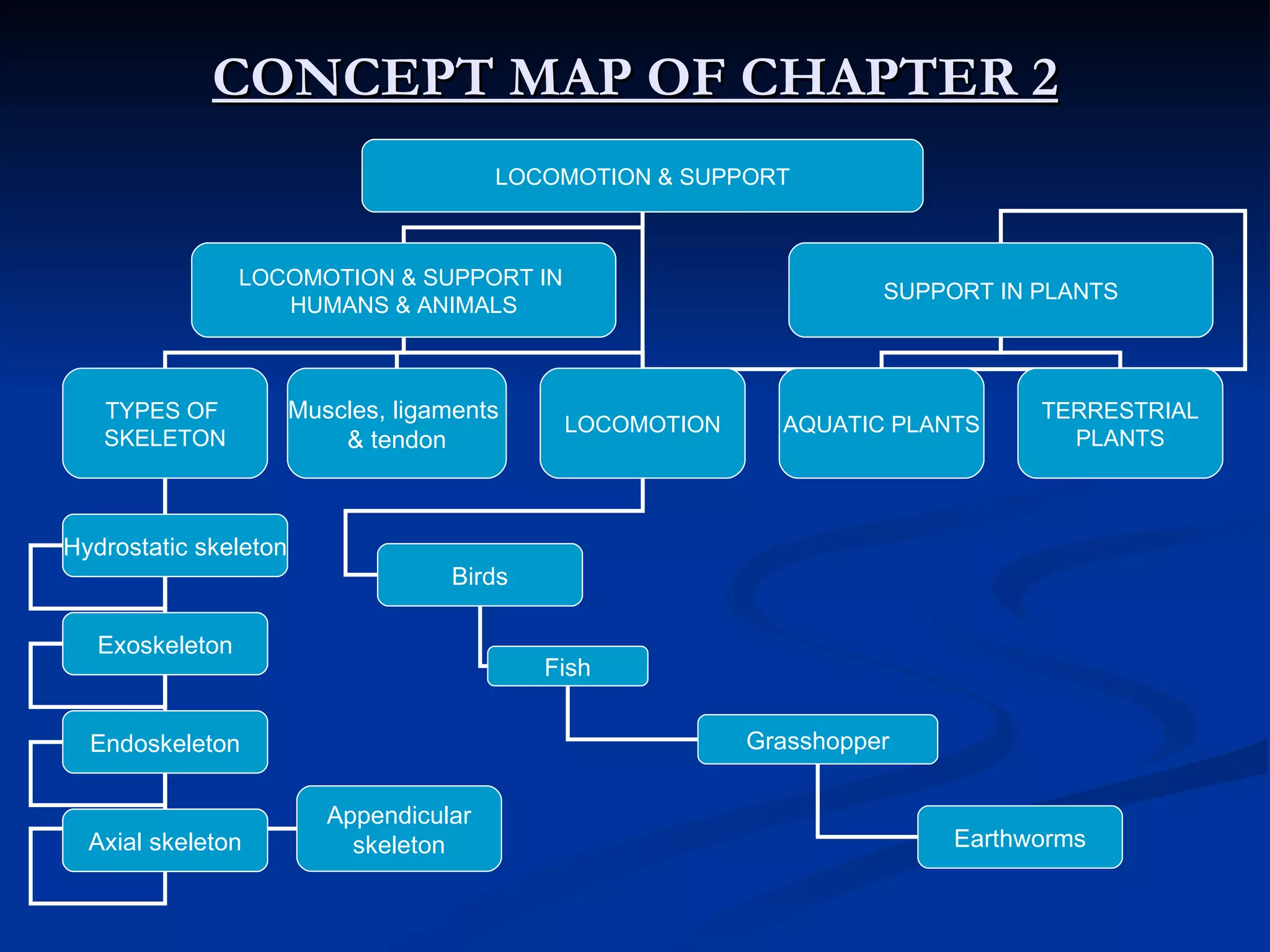
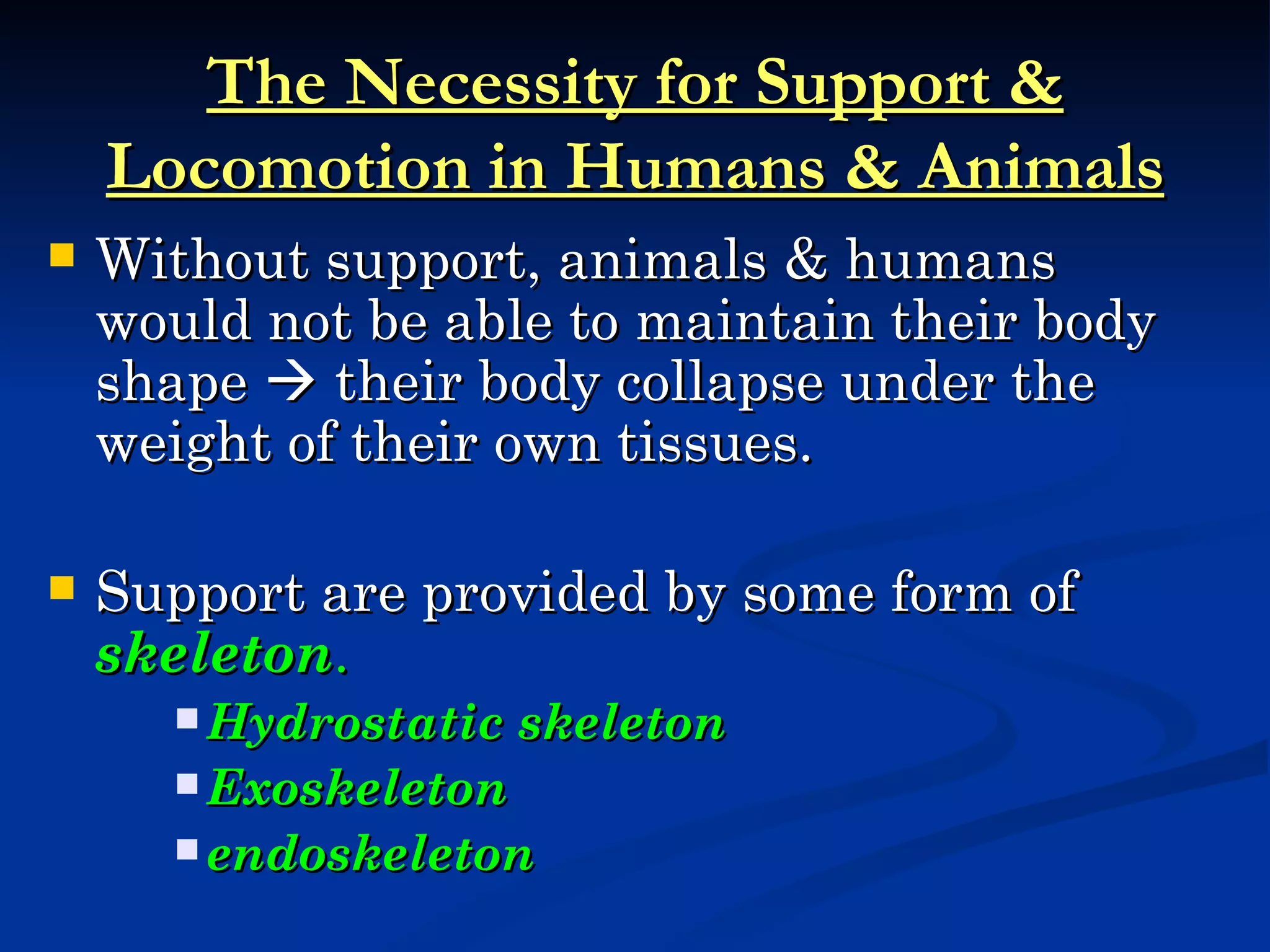
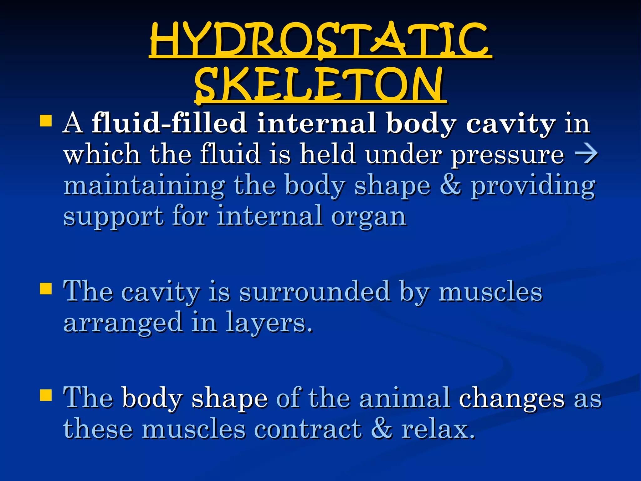
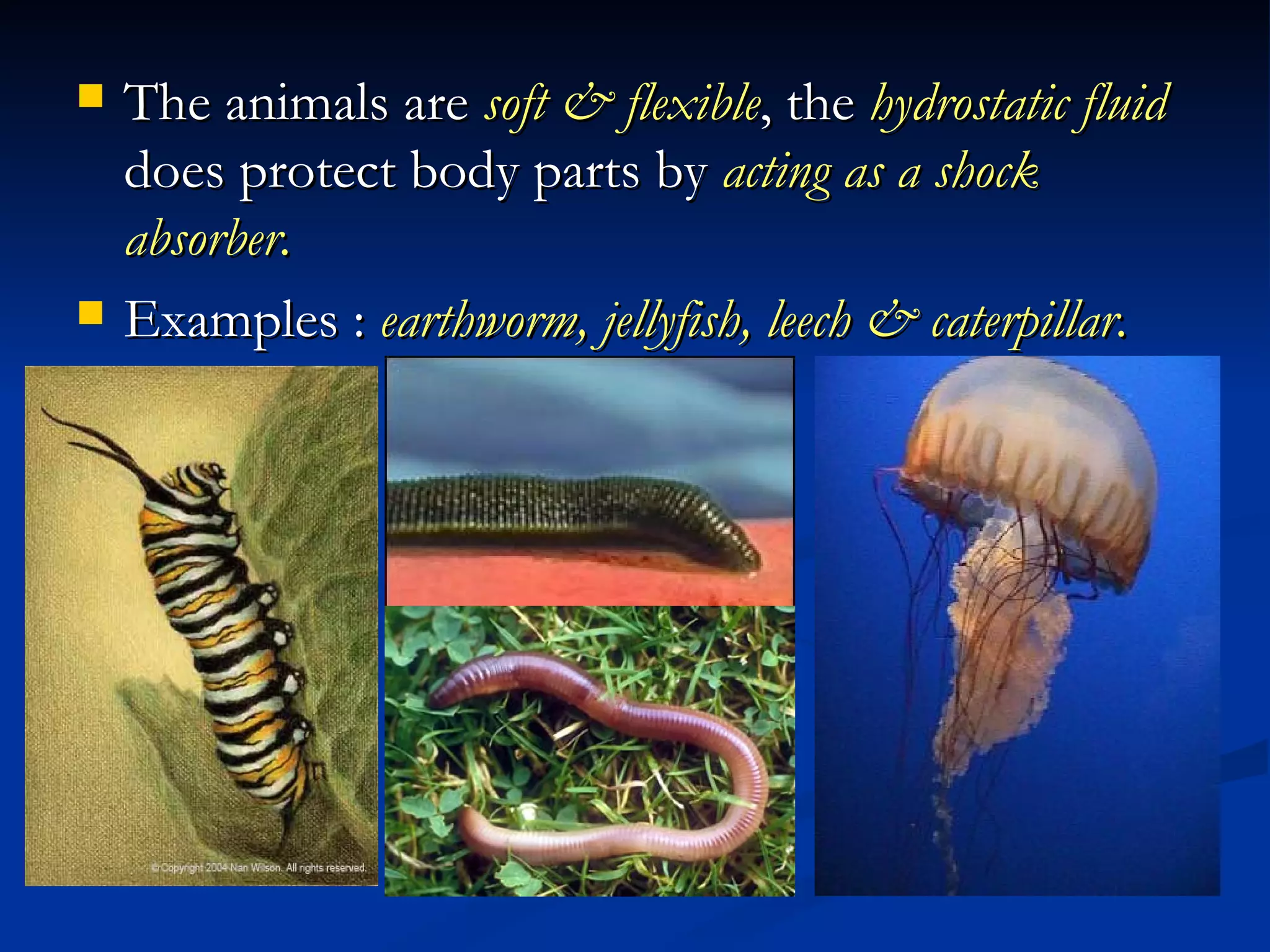
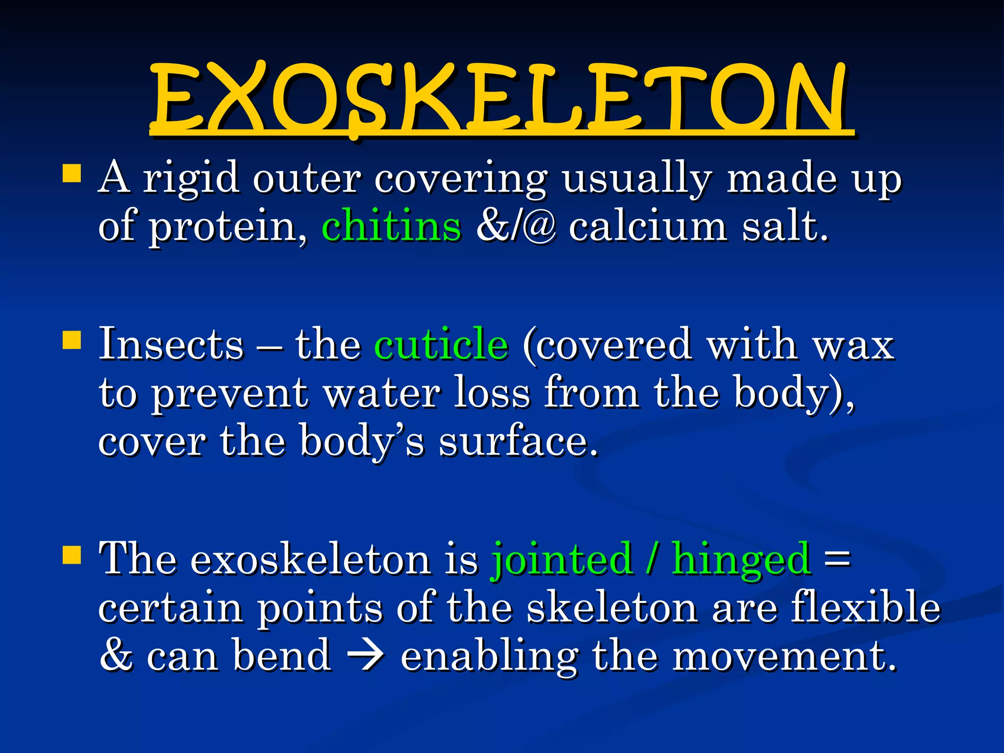
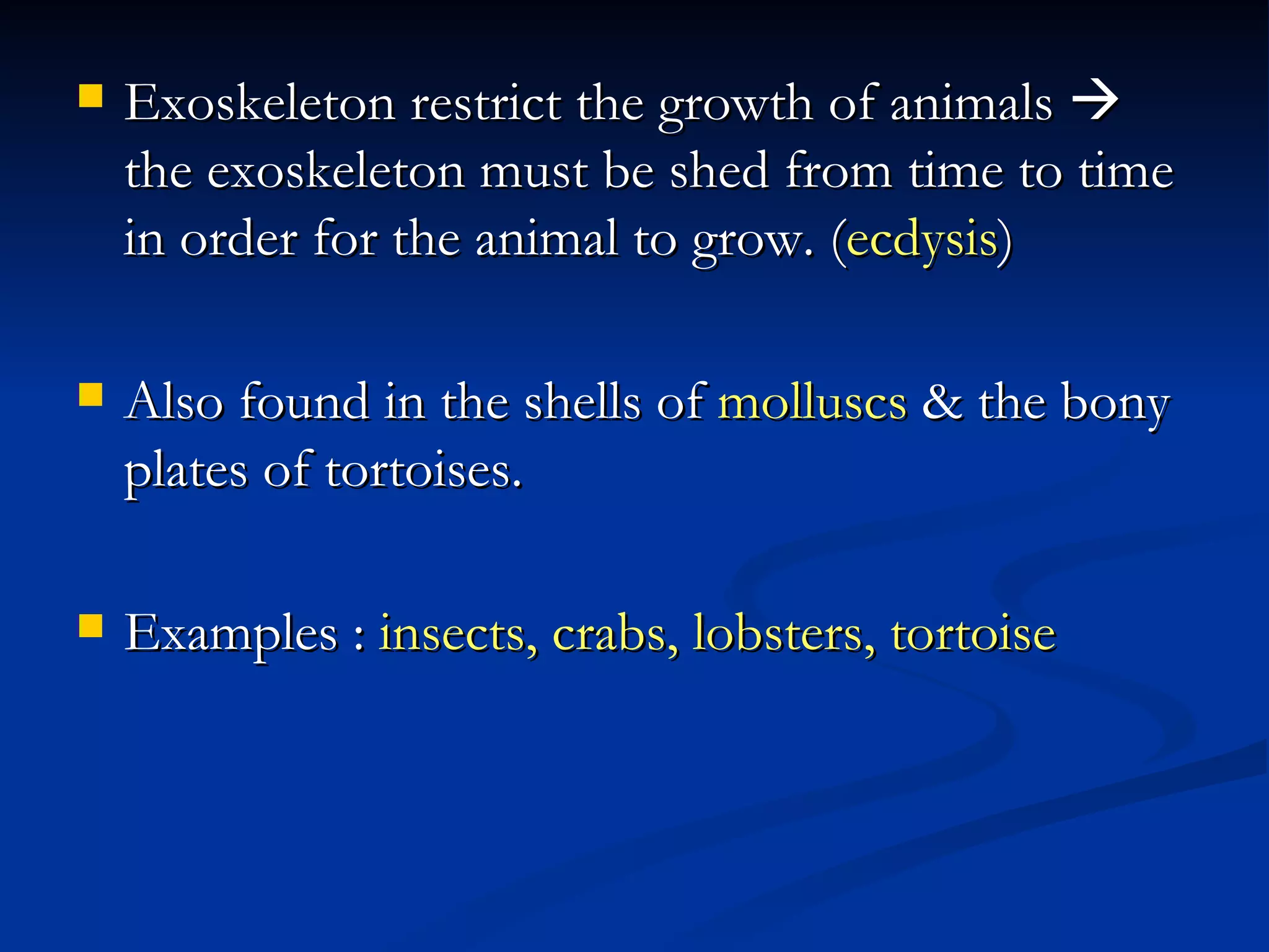
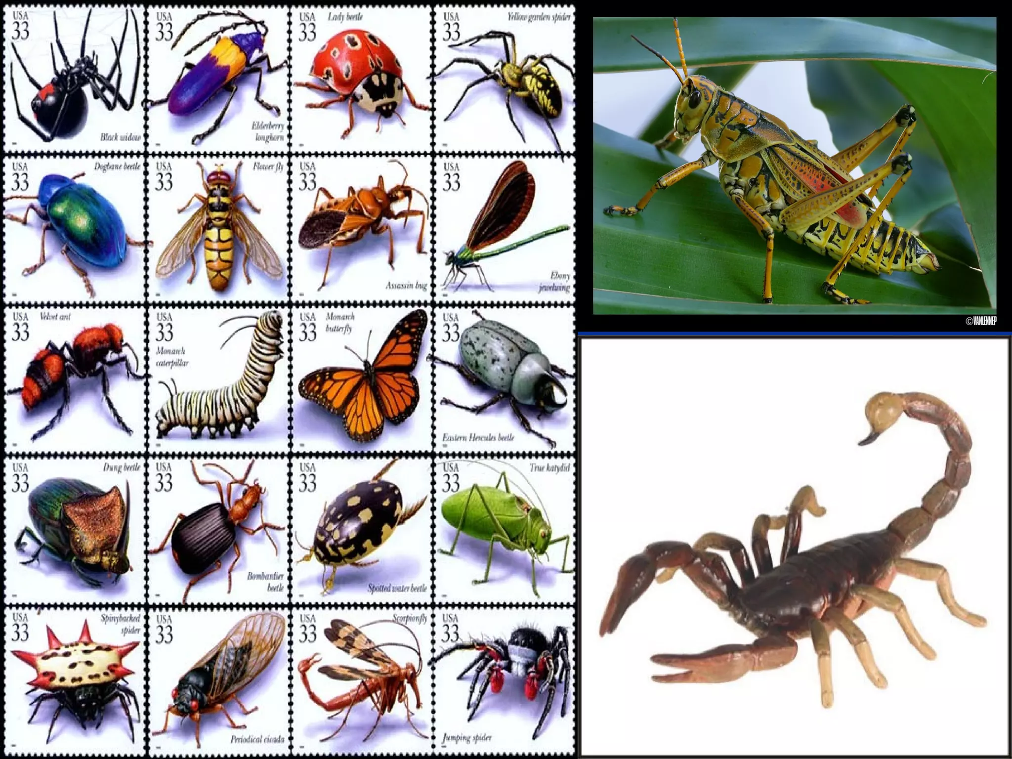
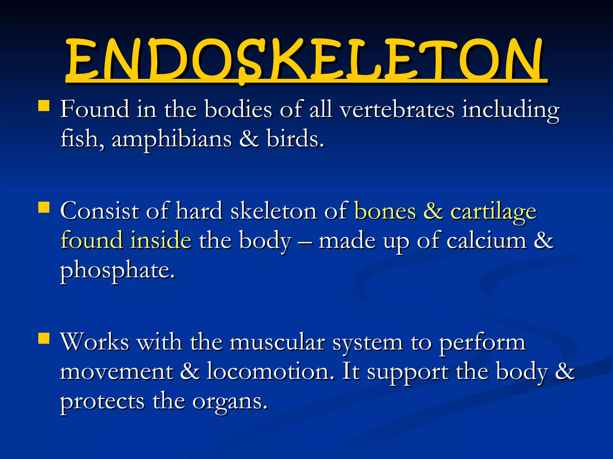
- It describes the main types of skeletons in humans and animals - endoskeleton, exoskeleton, and hydrostatic skeleton.
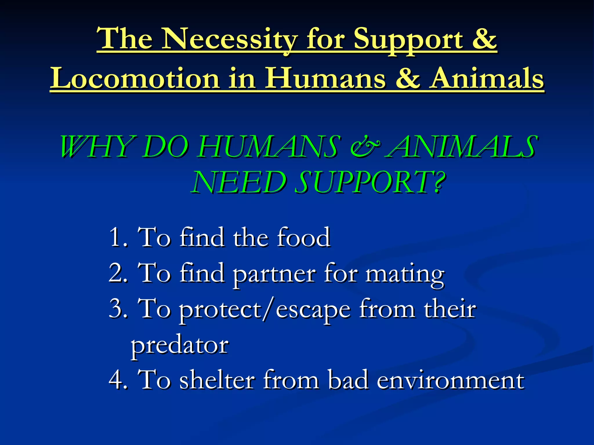
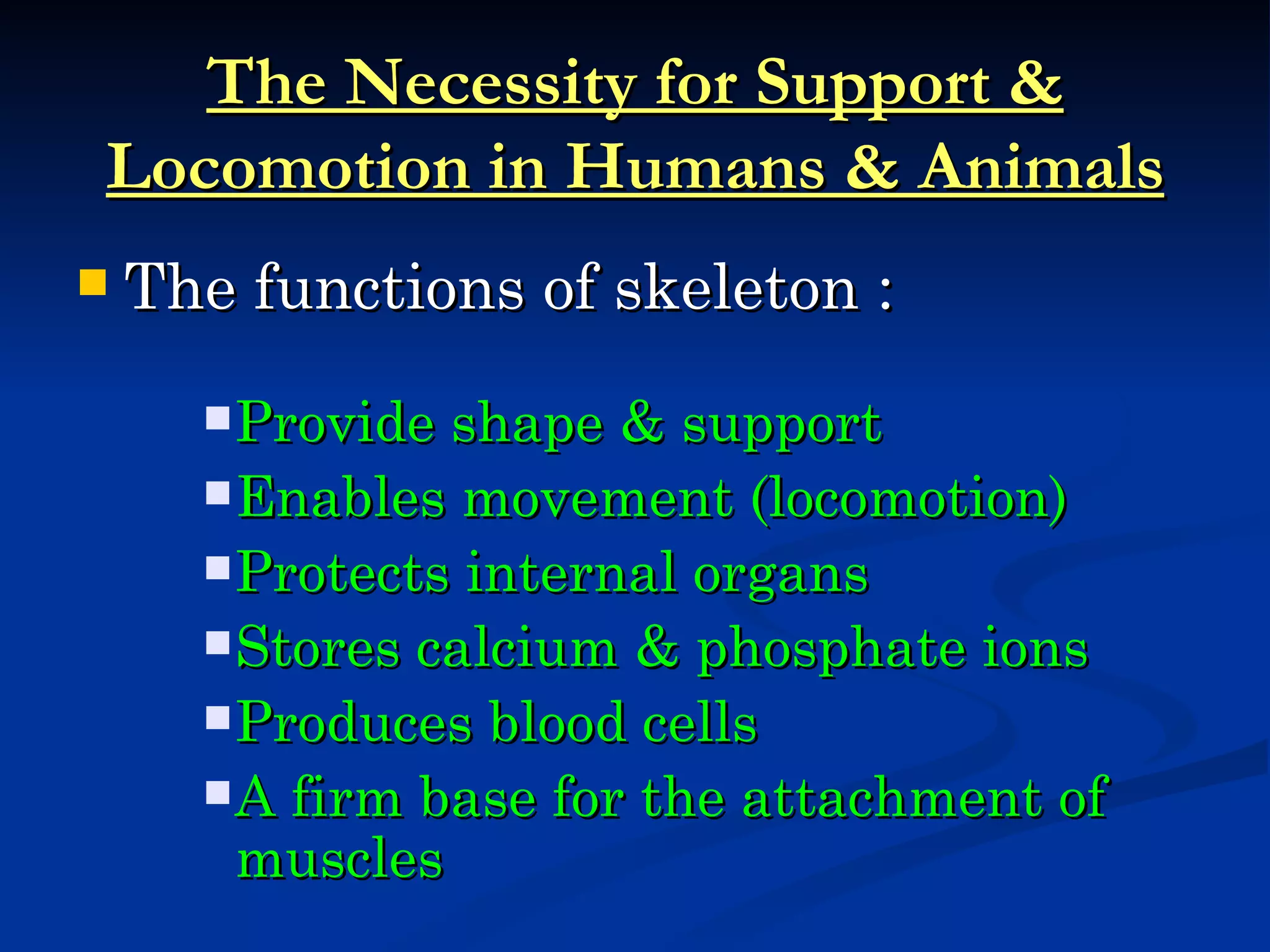
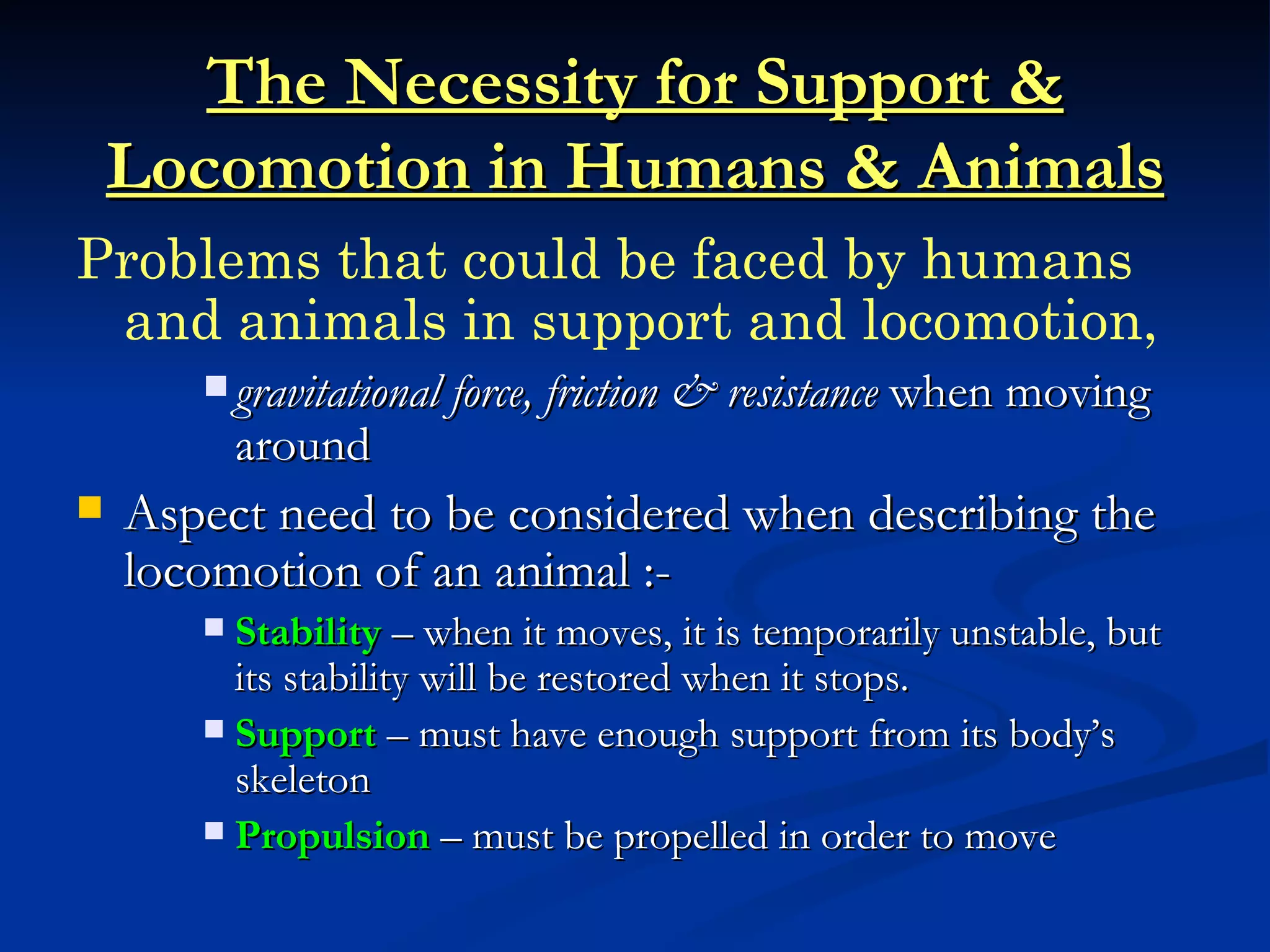
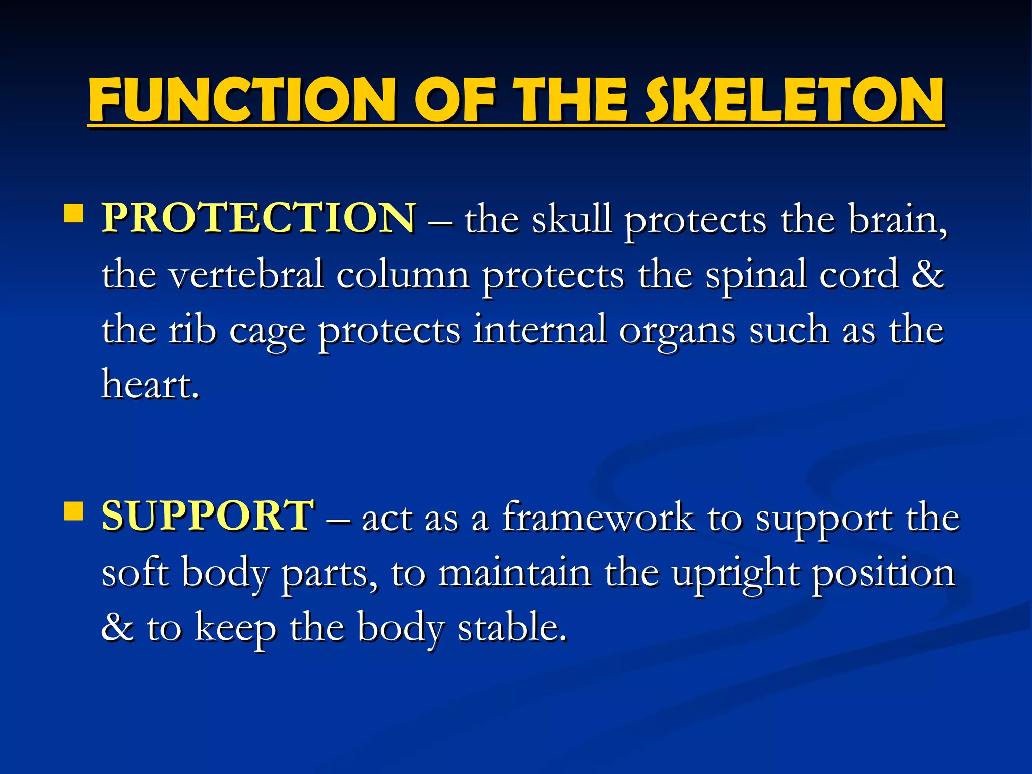
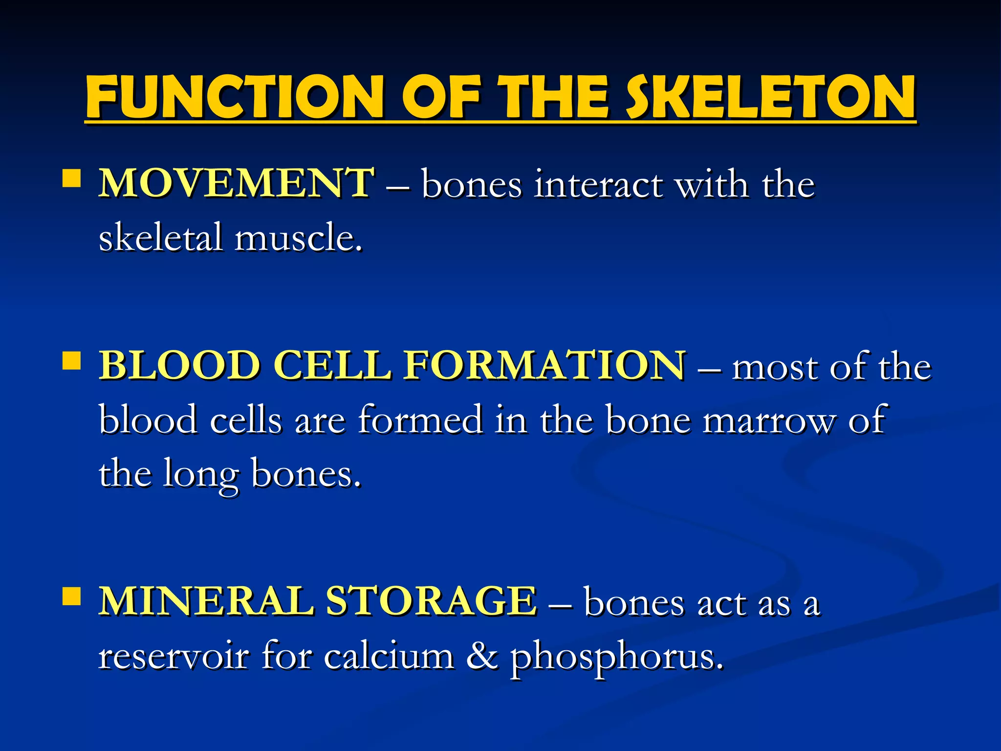
- It explains the functions of the skeletal system which include protection, support, movement, blood cell formation, and mineral storage.
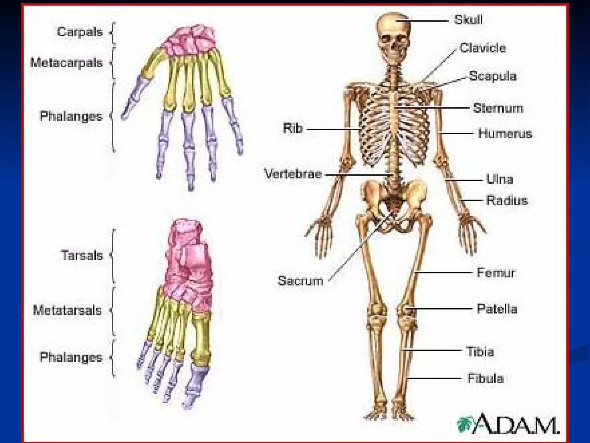
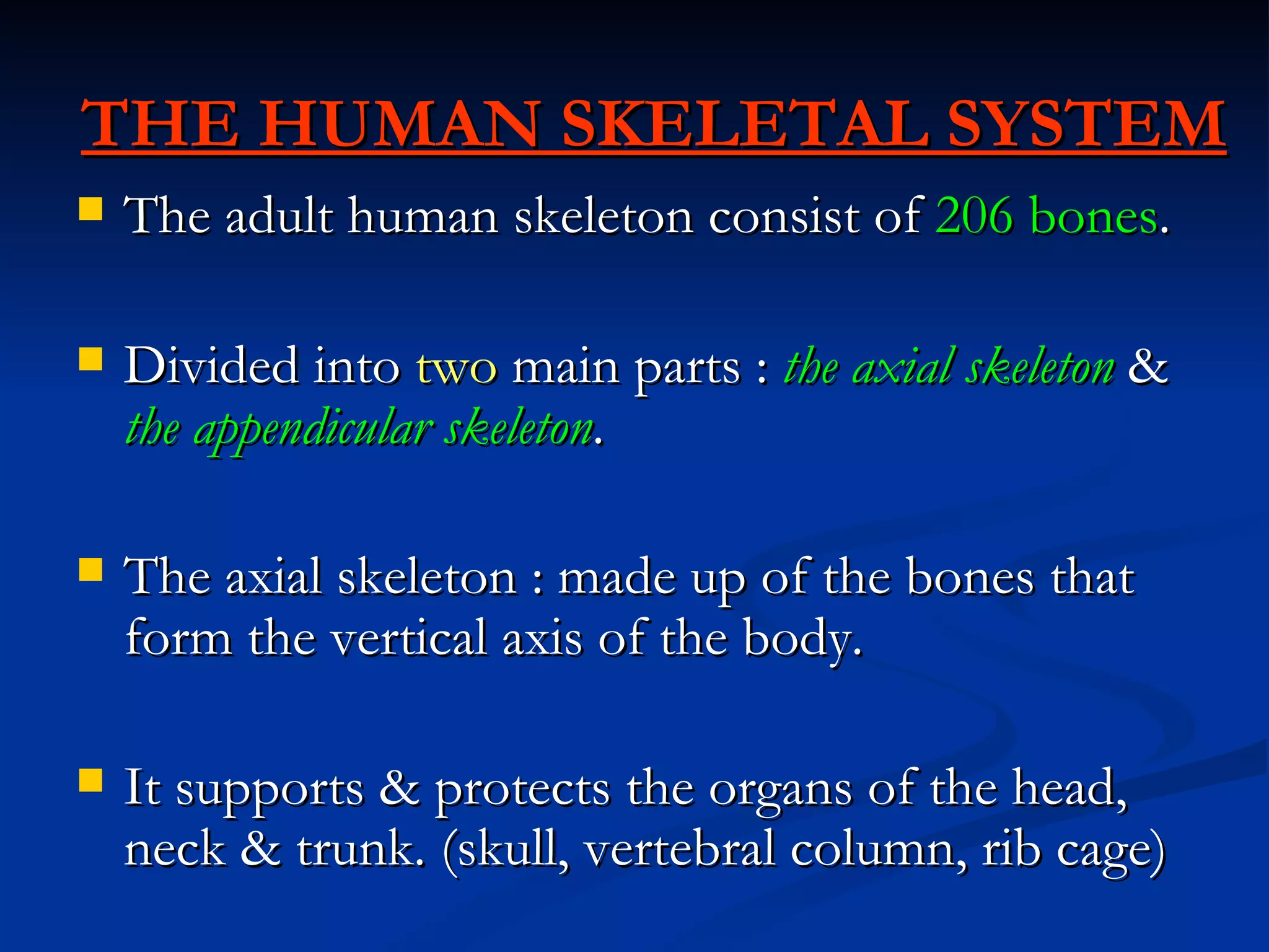
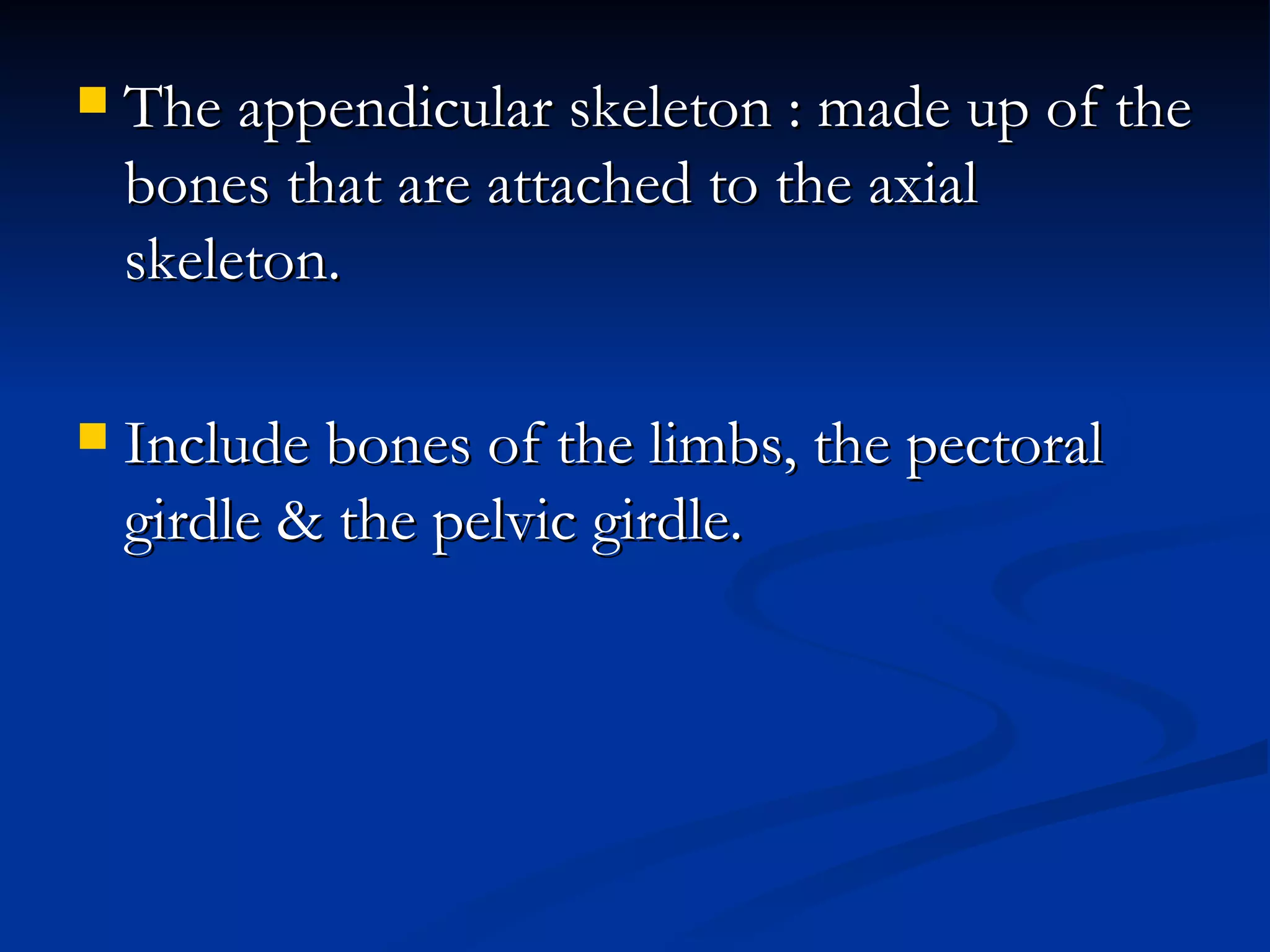
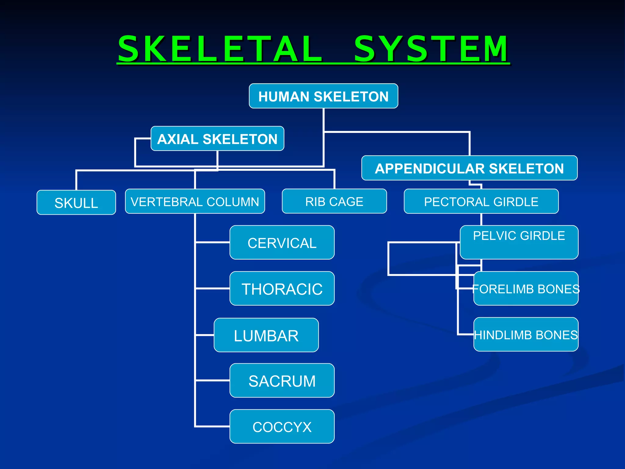
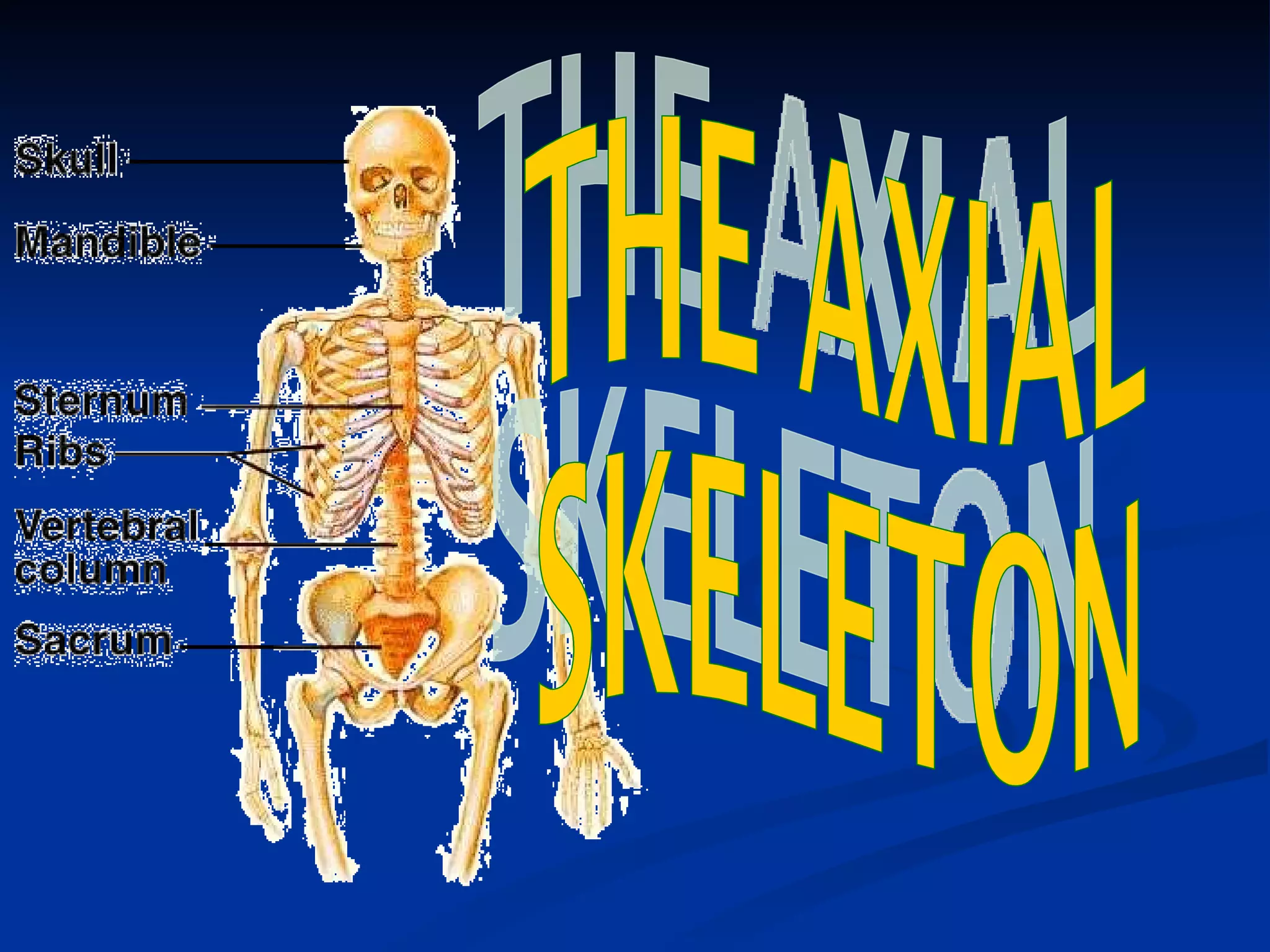
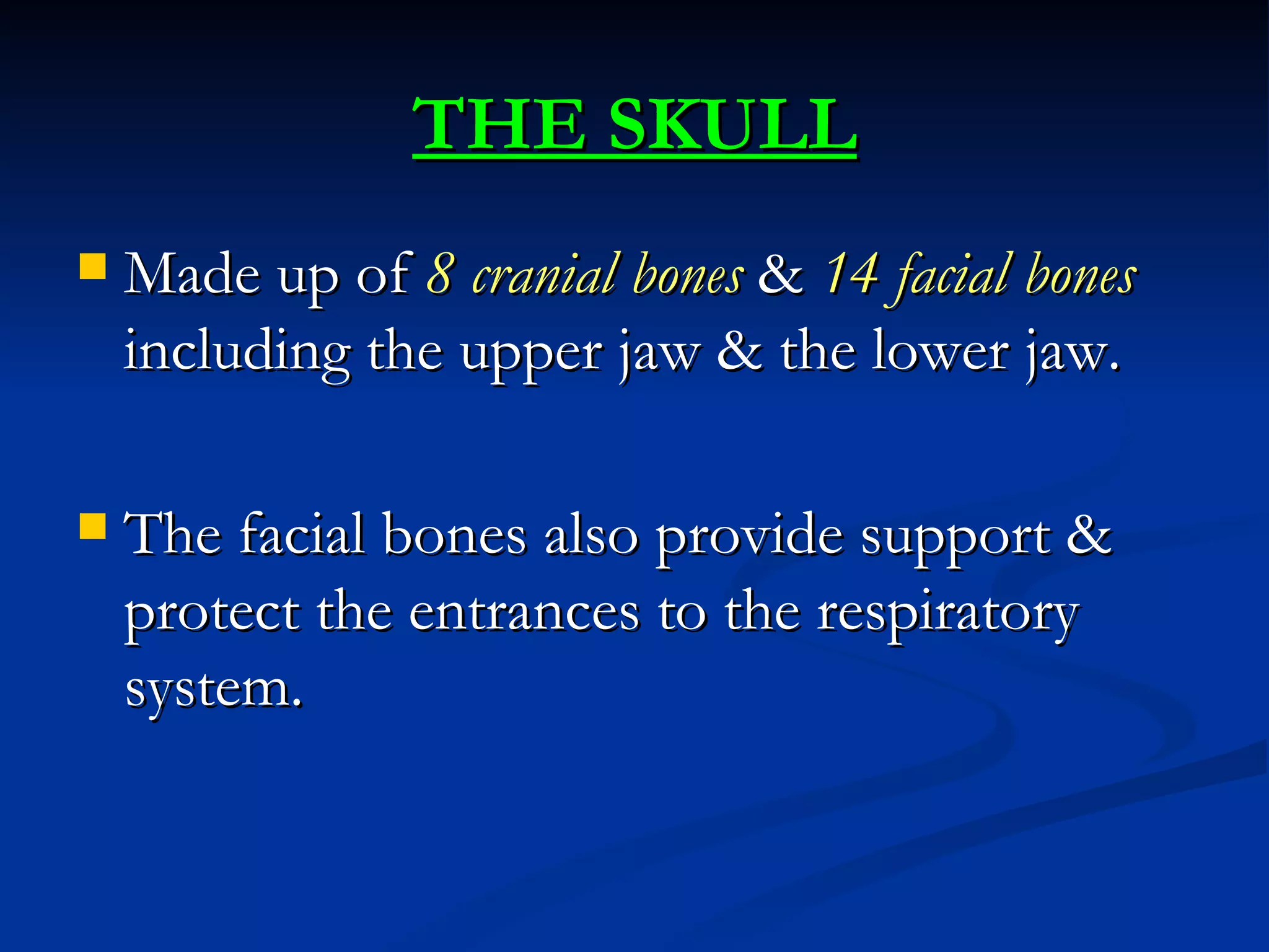
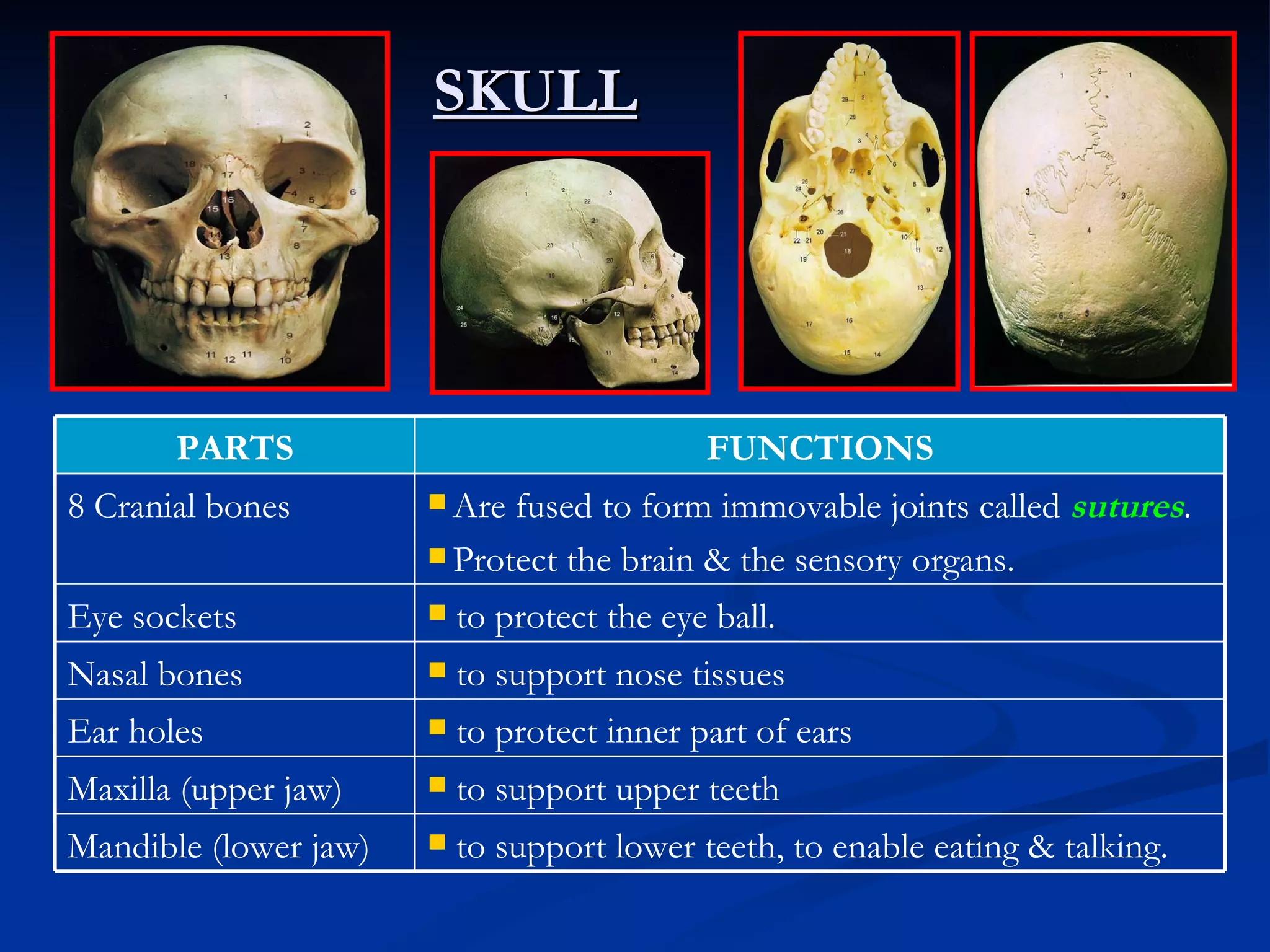
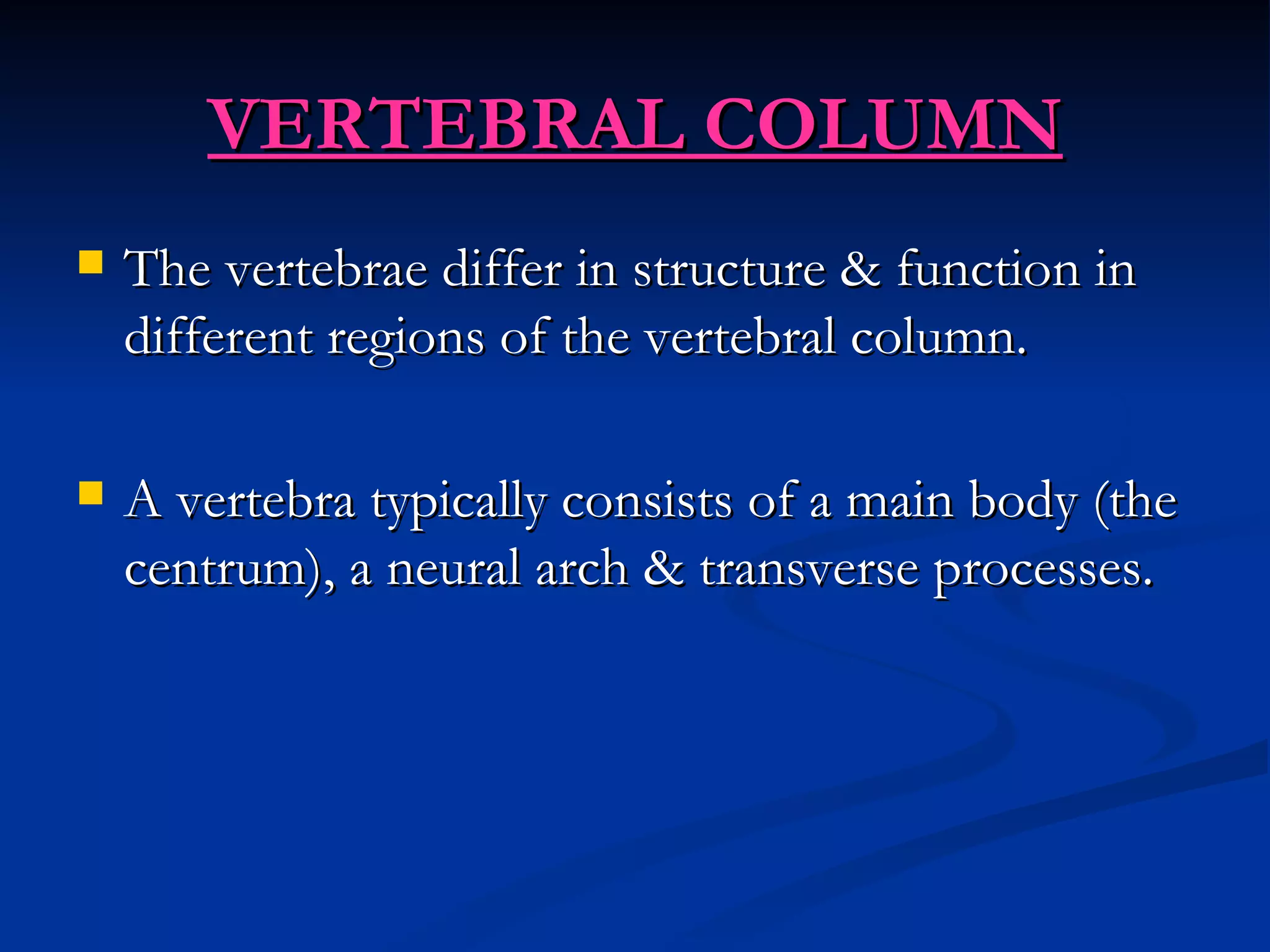
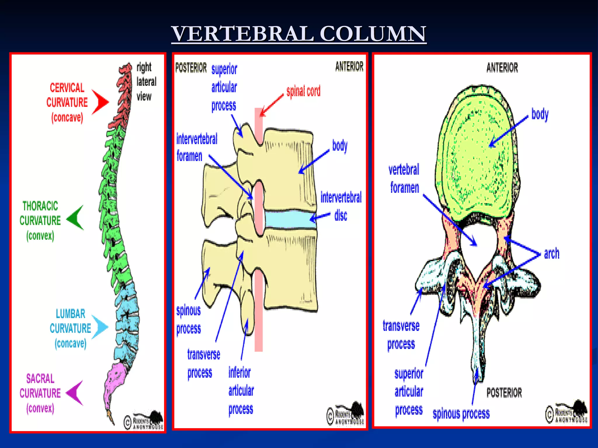
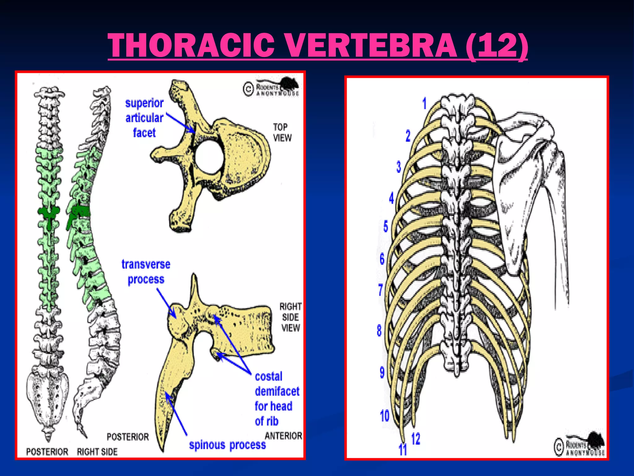
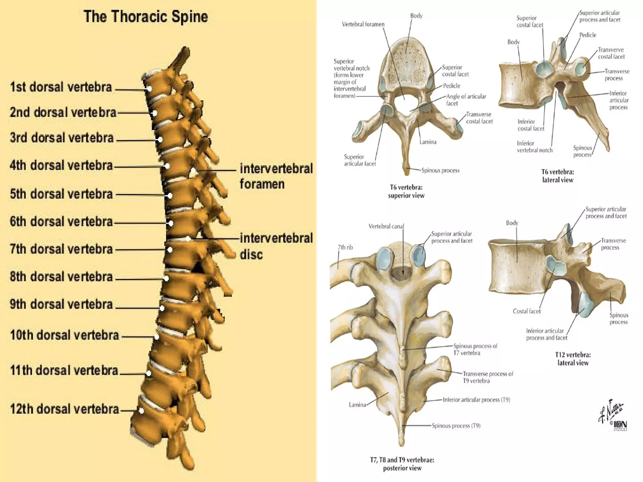
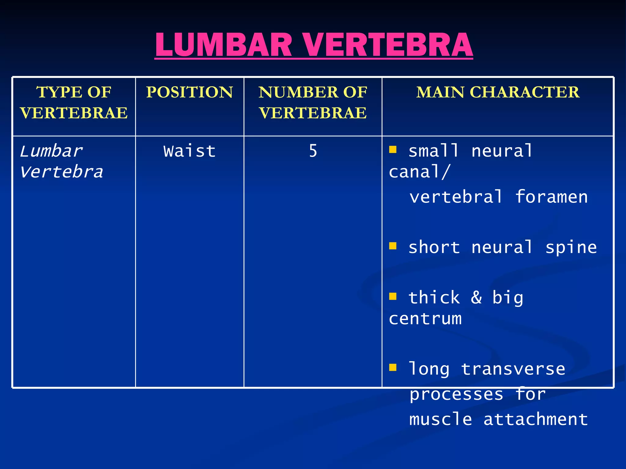
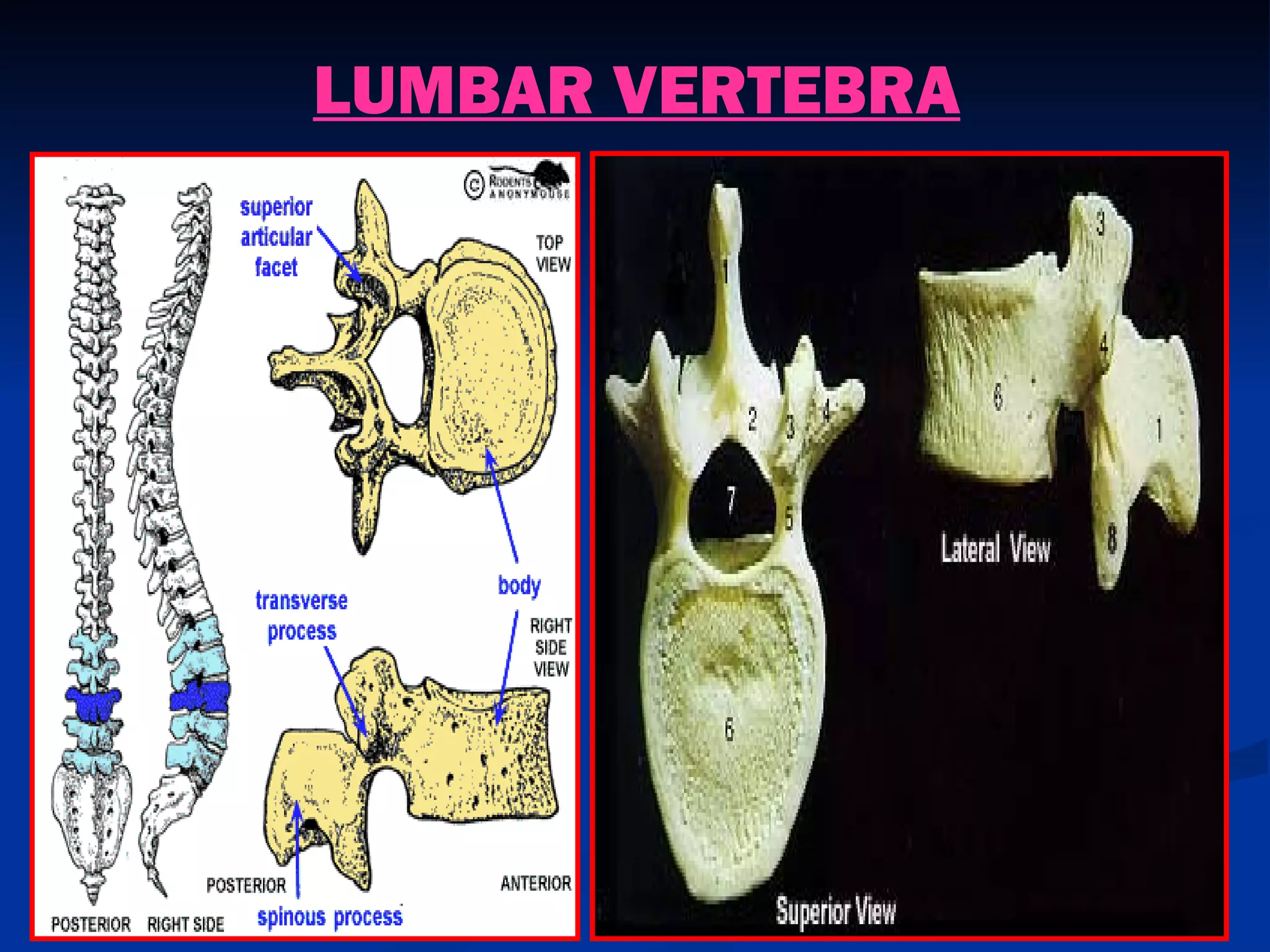
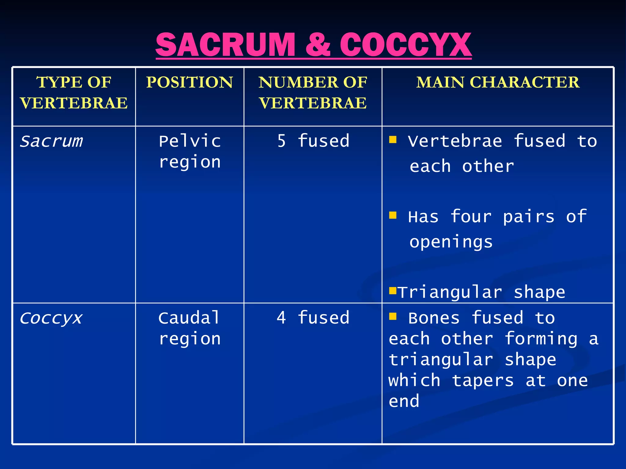
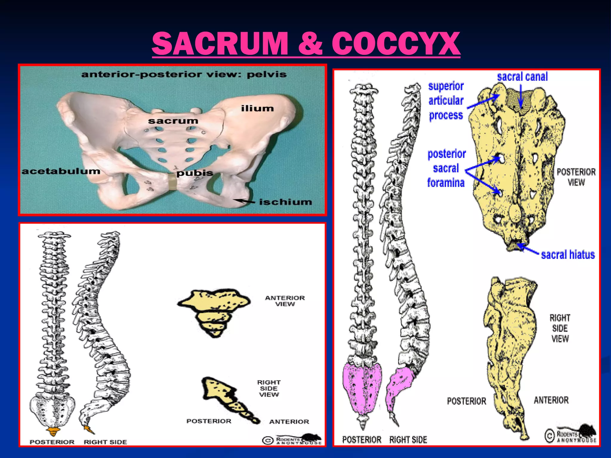
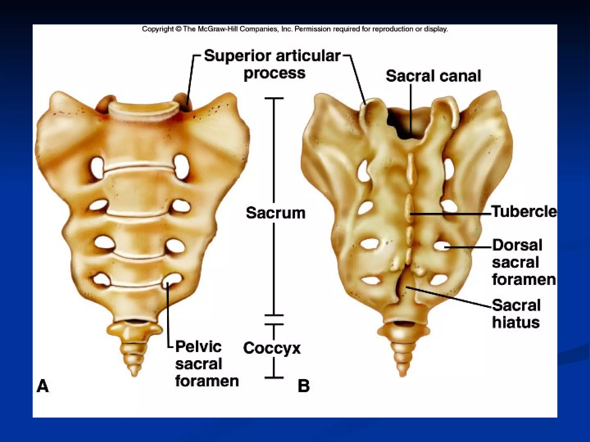
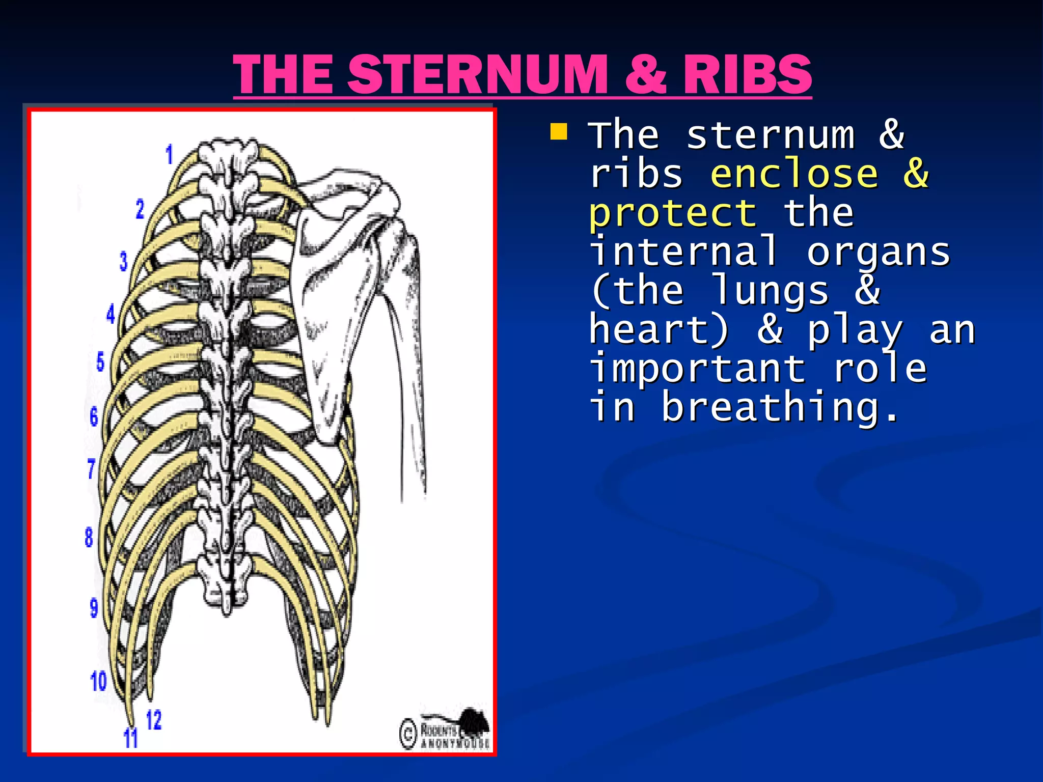
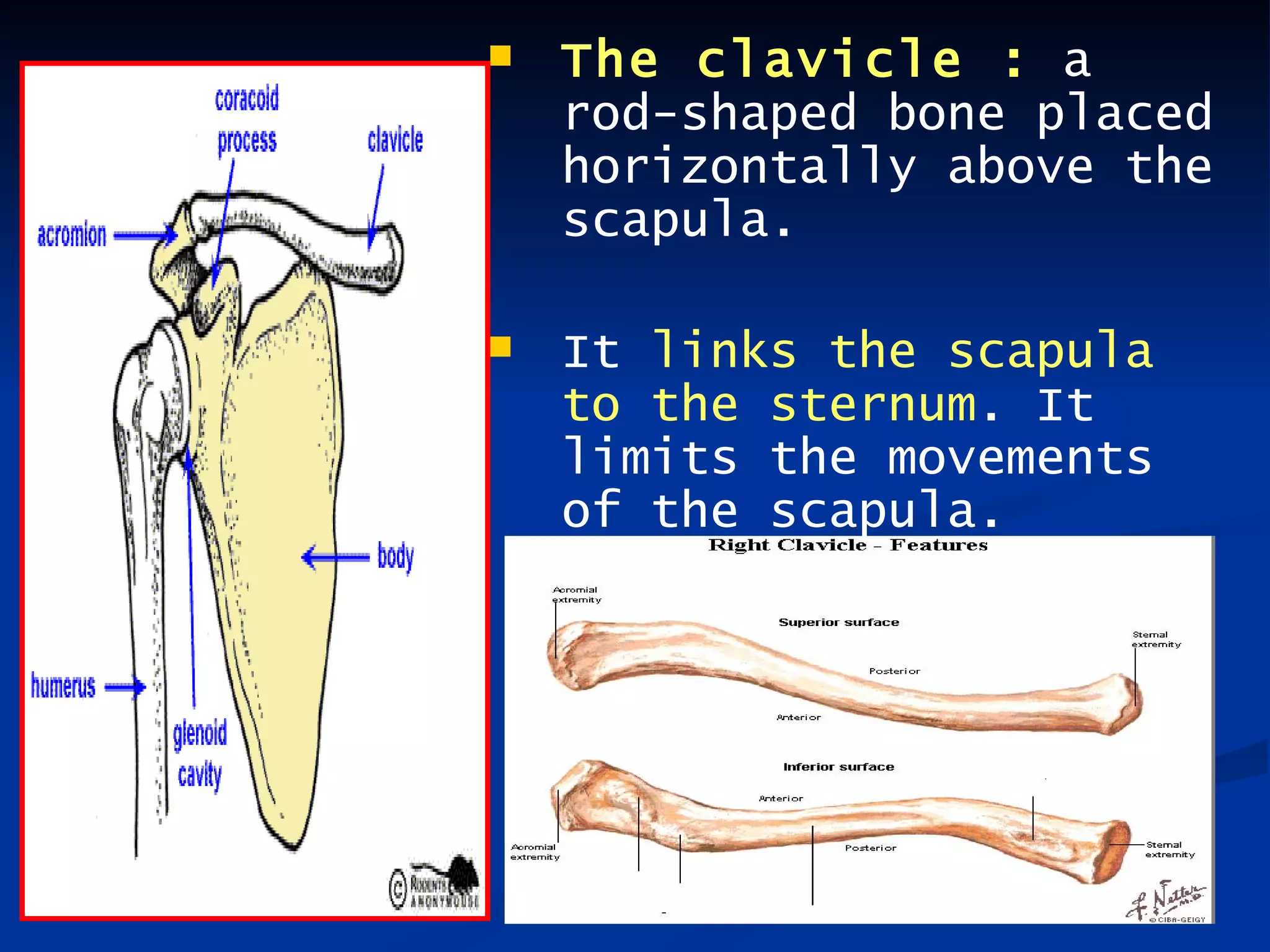
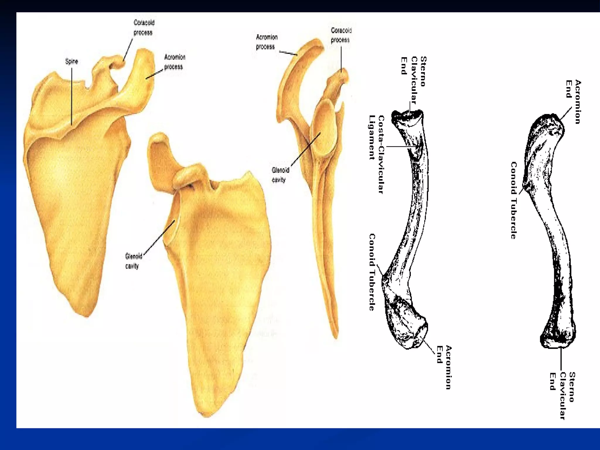
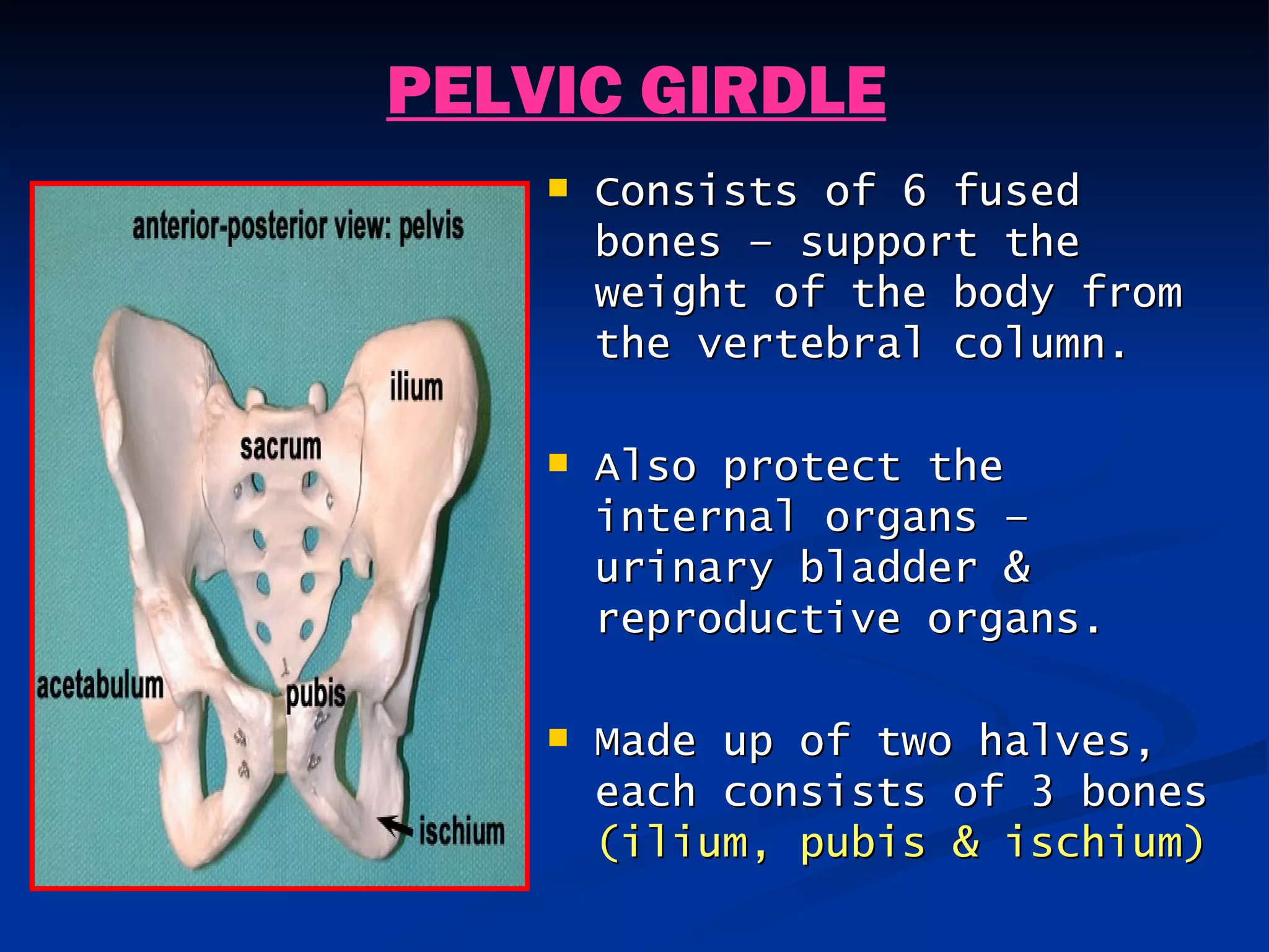
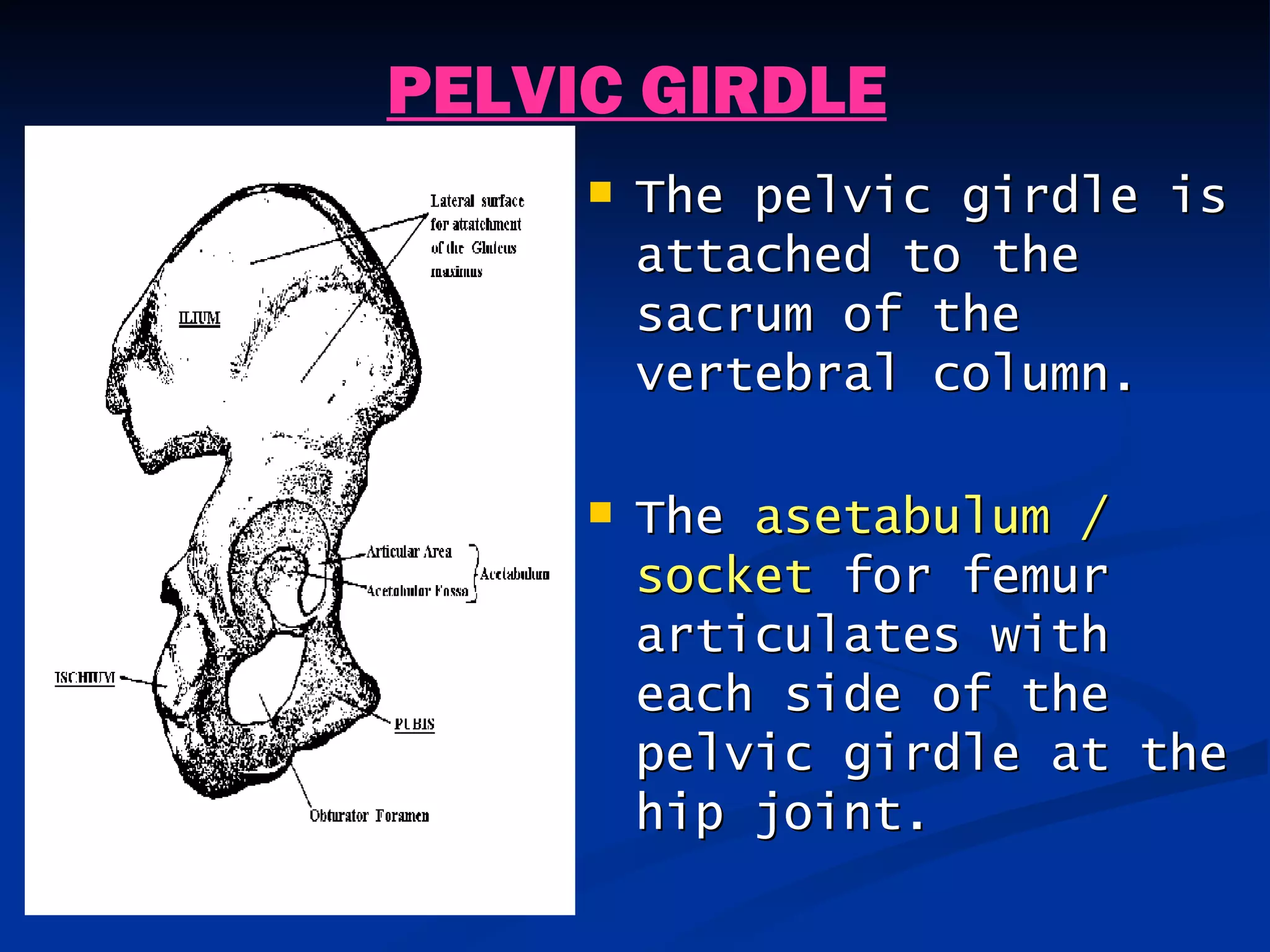
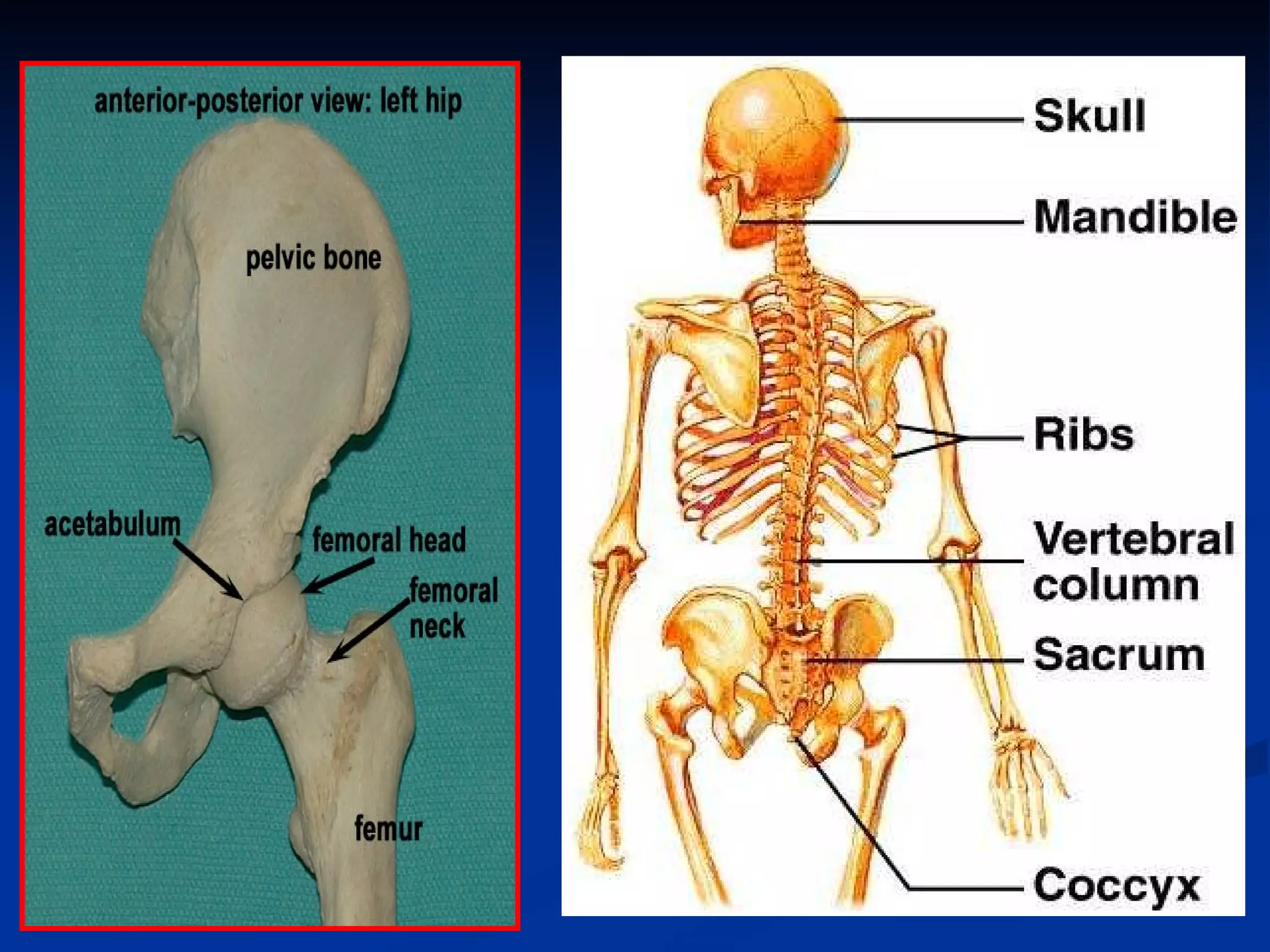
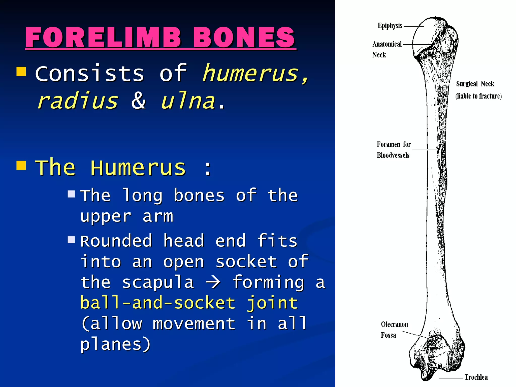
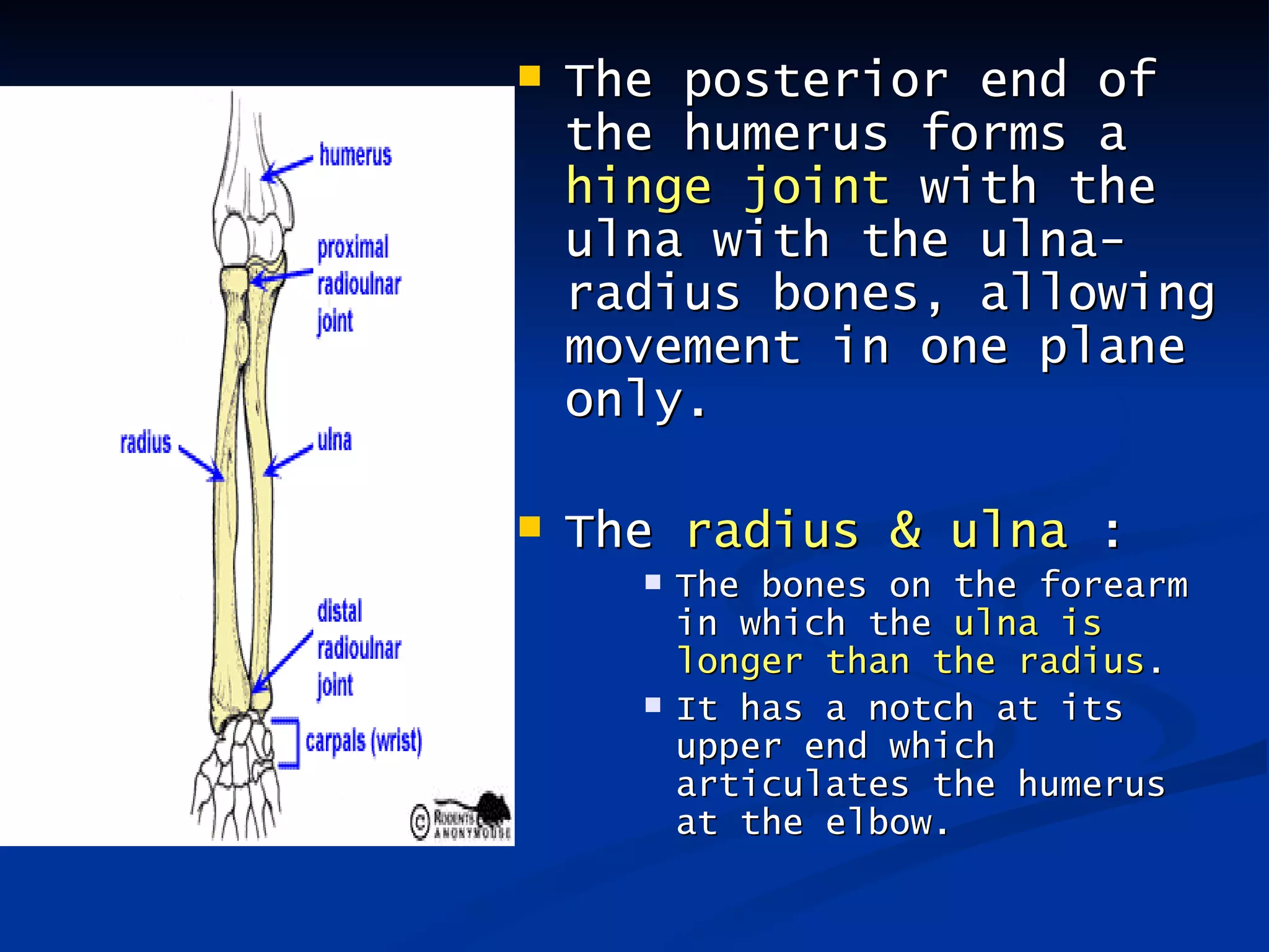
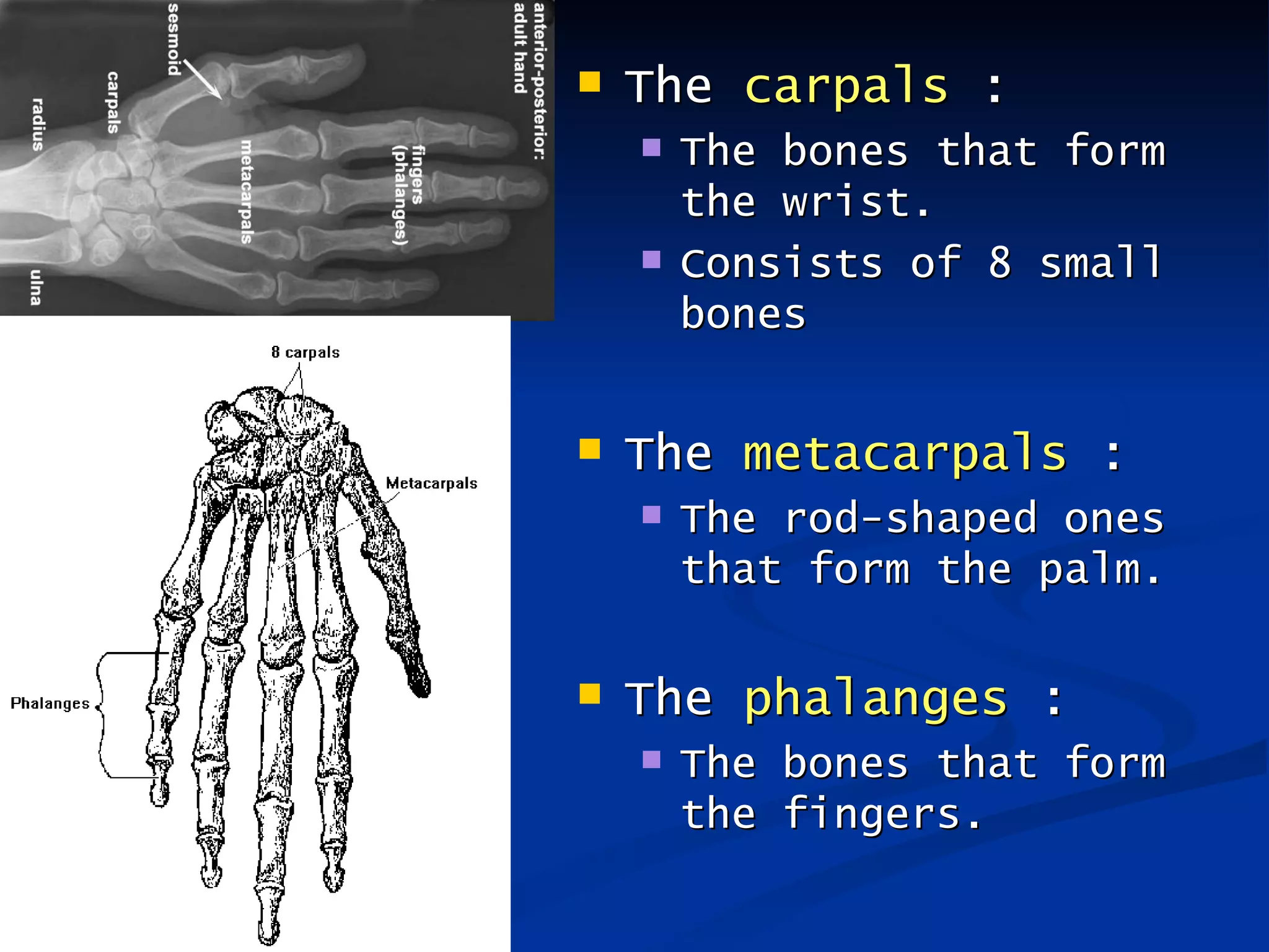
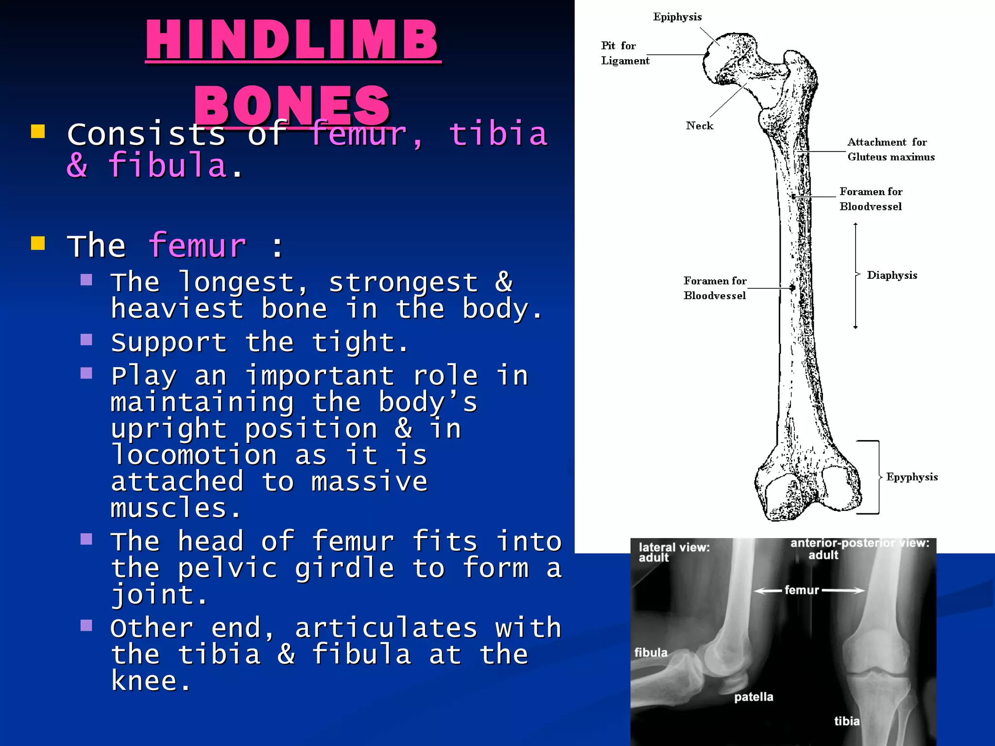
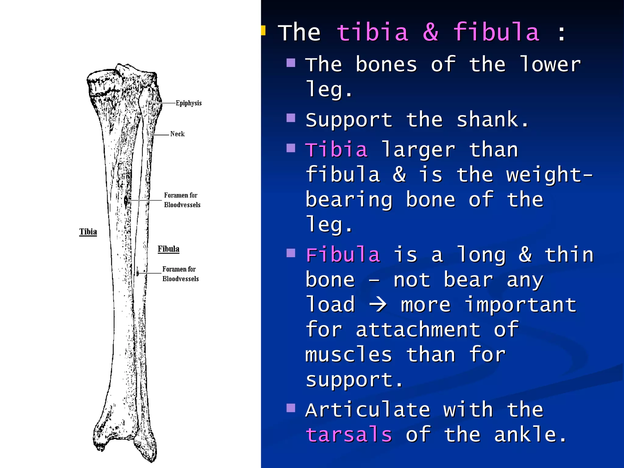
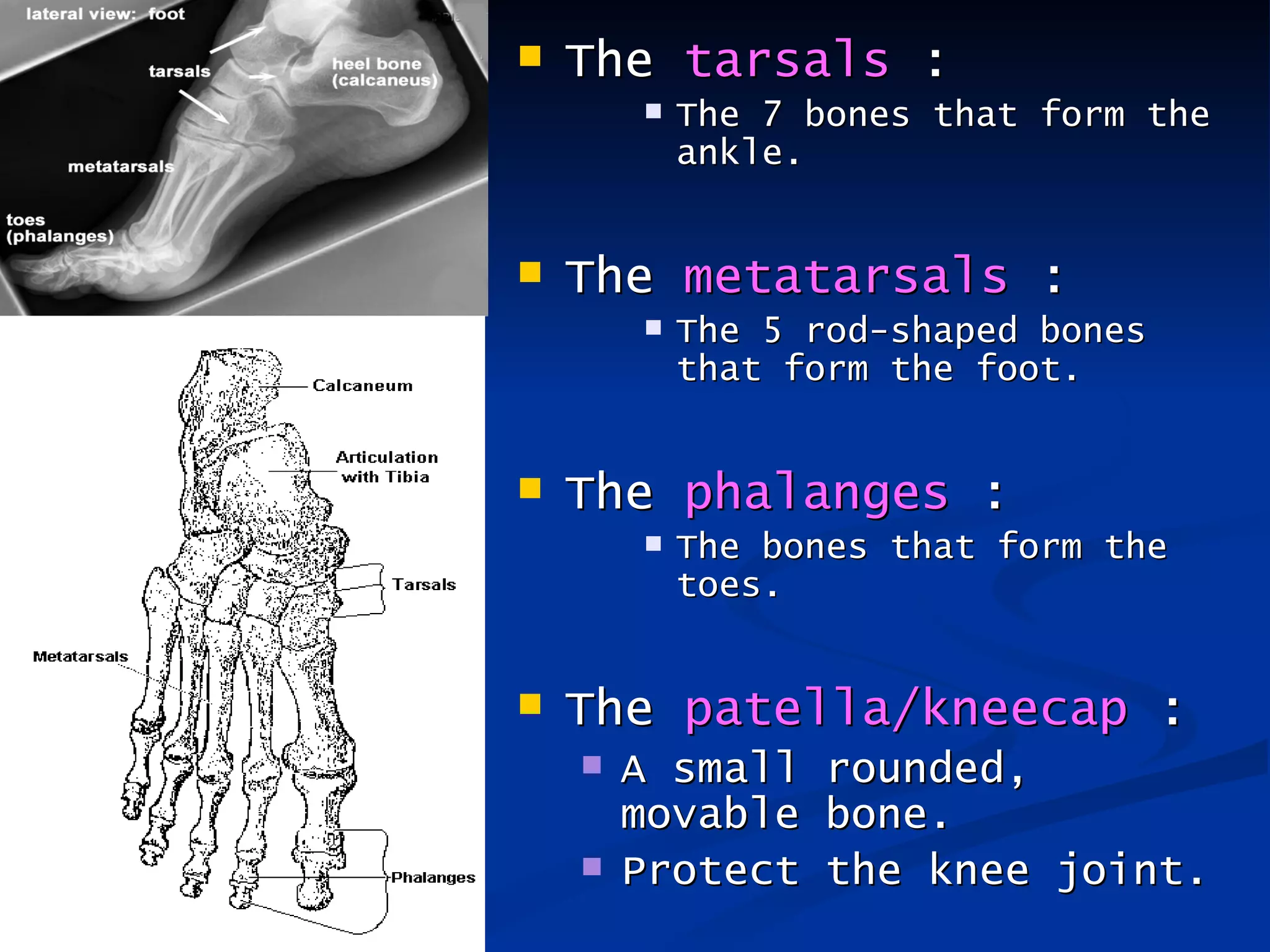
- It outlines the main bones that make up the axial skeleton (skull, vertebral column, rib cage) and appendicular skeleton (pectoral girdle, pelvic girdle, forelimb bones, hindlimb bones).


![Endocrine System, Nervous System And Homeostatic Control[1]](https://cdn.slidesharecdn.com/ss_thumbnails/endocrinesystemnervoussystemandhomeostaticcontrol1-1222215609200686-9-thumbnail.jpg?width=640&height=640&fit=bounds)


![2[1].1 (c) FORM 5](https://cdn.slidesharecdn.com/ss_thumbnails/21-1c-themechanisimoflocomotioninanimals-120602235904-phpapp01-thumbnail.jpg?width=640&height=640&fit=bounds)
![2[1].1 (b) FORM 5](https://cdn.slidesharecdn.com/ss_thumbnails/21-1b-roleofmusclesligamentstendonsin-120602235830-phpapp02-thumbnail.jpg?width=640&height=640&fit=bounds)
![Subtopic 3 1[1] FORM 5](https://cdn.slidesharecdn.com/ss_thumbnails/subtopic3-11-120603001001-phpapp01-thumbnail.jpg?width=640&height=640&fit=bounds)
![Subtopic 3[1].2 FORM 5](https://cdn.slidesharecdn.com/ss_thumbnails/subtopic31-2-120603000911-phpapp01-thumbnail.jpg?width=640&height=640&fit=bounds)
![4[1].5 FORM 5](https://cdn.slidesharecdn.com/ss_thumbnails/41-5-floweringplant-120603003443-phpapp01-thumbnail.jpg?width=640&height=640&fit=bounds)
![Sub 2[1].3 FORM 5](https://cdn.slidesharecdn.com/ss_thumbnails/sub21-3supportinplants-120602235702-phpapp02-thumbnail.jpg?width=640&height=640&fit=bounds)
![4[1].1 FORM 5](https://cdn.slidesharecdn.com/ss_thumbnails/41-1theformationofgametes-120603003019-phpapp01-thumbnail.jpg?width=640&height=640&fit=bounds)
![4[1].6 FORM 5](https://cdn.slidesharecdn.com/ss_thumbnails/41-6-growthinmulticellularorganisms-120603003459-phpapp02-thumbnail.jpg?width=640&height=640&fit=bounds)
![5[1].3 form 5](https://cdn.slidesharecdn.com/ss_thumbnails/51-3geneschromosomes-120603004012-phpapp02-thumbnail.jpg?width=640&height=640&fit=bounds)
![4[1].7 FORM 5](https://cdn.slidesharecdn.com/ss_thumbnails/41-7-thegrowthcurve-120603002724-phpapp02-thumbnail.jpg?width=640&height=640&fit=bounds)

![5[1].1 form 5](https://cdn.slidesharecdn.com/ss_thumbnails/51-1theconceptofinheritance-120603003907-phpapp02-thumbnail.jpg?width=640&height=640&fit=bounds)
![4[1].3 FORM 5](https://cdn.slidesharecdn.com/ss_thumbnails/41-3earlydevelopmentofzygote-120603003204-phpapp02-thumbnail.jpg?width=640&height=640&fit=bounds)
![Sub 2[1].2 FORM5](https://cdn.slidesharecdn.com/ss_thumbnails/sub21-2healthymusculoskeletalsystem-120602235534-phpapp02-thumbnail.jpg?width=640&height=640&fit=bounds)
![6[1].1 form 5](https://cdn.slidesharecdn.com/ss_thumbnails/61-1-variationinorganisms-120603005250-phpapp01-thumbnail.jpg?width=640&height=640&fit=bounds)
![4[1].2 FORM 5](https://cdn.slidesharecdn.com/ss_thumbnails/41-2-menstrualcycle-120603003033-phpapp01-thumbnail.jpg?width=640&height=640&fit=bounds)
![4[1].4 FORM 5](https://cdn.slidesharecdn.com/ss_thumbnails/41-4-thecontributionofst-120603003308-phpapp01-thumbnail.jpg?width=640&height=640&fit=bounds)
![3[1].3 FORM 5](https://cdn.slidesharecdn.com/ss_thumbnails/31-3-appreciate-120603000912-phpapp02-thumbnail.jpg?width=640&height=640&fit=bounds)
![6[1].2 form 5](https://cdn.slidesharecdn.com/ss_thumbnails/61-2-causesofvariation-120603003705-phpapp02-thumbnail.jpg?width=640&height=640&fit=bounds)
![6[1].3 form 5](https://cdn.slidesharecdn.com/ss_thumbnails/61-3-respectfultowardsanother-120603003718-phpapp02-thumbnail.jpg?width=640&height=640&fit=bounds)


