
Recommended
More Related Content
What's hot
What's hot (20)
Similar to Skeleton System Explained in 40 Characters
Similar to Skeleton System Explained in 40 Characters (20)
More from bhartisharma175
More from bhartisharma175 (16)
Recently uploaded
Recently uploaded (20)
Skeleton System Explained in 40 Characters
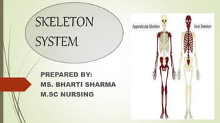
- 1. SKELETON SYSTEM PREPARED BY: MS. BHARTI SHARMA M.SC NURSING
- 2. SKELETON The skeleton is the bony framework of the body. It forms the cavities & fossae ( depression or hollow) that protect some structures from the joints & give attachment to the muscles. The skeleton is described in two parts: Axial (80) and Appendicular (126) skeleton. The human skeleton consists of 206 bones, most of which are paired with one member of each pair on the right and left sides of the body. The infants have more than 206 bones because some of their bones ( sacrum & coccyx) fuse later in life.
- 3. The Axial Skeleton: It consists of : skull, vertebral column, sternum and bones of ribs.
- 4. A. SKULL: Consist of two parts: Cranium and face. Formed by number of flat and irregular bones that provide a bony protection for the brain. It has a base on which brain rest upon and vault that surrounds and cover it. The only movable bone is the mandible / lower jaw. Lining of inner surface of skull is called as periosteum that forms the outer layer of dura mater. In the mature skull the joints ( sutures) between the bones are immovable (fibrous). The bones have numerous perforations ( foramina fissures) through which nerves, blood and lymph vessels pass.
- 6. The bones of the cranium are:- 1 frontal bone 2 parietal bones 2 temporal bones 1 occipital bone 1 sphenoid bone 1 ethmoid bone
- 7. SUTURES: The sutures are the immovable joints between the bones of the skull. They are- coronal suture, frontal sutures, sagittal suture, lambdoidal suture and squamous suture.
- 8. FONTANELLE:
- 9. FACE The skeleton of the face is formed by 13 bones, in addition to the frontal bone. They are- 2 Zygomatic (cheek) bone 1 Maxilla 2 Nasal bones 2 Lacrimal bones 1 Vomer 2 Palatine bones 2 Inferior conchae 1 Mandible
- 10. HYOID BONE: This is an isolated horseshoe- shaped bone lying in the soft tissues of the neck just above the larynx and below the mandible. It does not articulate with any other bone, but is attached to the styloid process of the temporal bone by ligaments. It support the larynx and gives attachment to the base of the tongue.
- 11. SINUSES: Sinuses containing air are present in the sphenoid, ethmoid, maxillary and frontal bones. They all communicate with the nasal cavity and are lined with ciliated mucous membrane. Their functions are to give resonance to the voice and to reduce the weight of the skull, making it easier to carry.
- 12. FUNCTIONS OF THE SKULL: The various parts of the skull have specific and different functions: The cranium protects the delicate tissues of the brain. The bony eye sockets provide the eyes with some protection against injury and helps in eye movement. The temporal bone protects the delicate structures of the ear. Some bones of the face and the base of the skull give resonance to the voice. The maxilla and mandible provide alveolar ridges in which the teeth are embedded. The mandible is the only movable bone of the skull and help in chewing food.
- 13. B. VERTEBRAL COLUMN: There are 26 bones in the vertebral column. 24 separate vertebrae extend downwards from the occipital bone of the skull; then there is the sacrum, formed from five fused vertebrae, and lastly the coccyx, or tail, which is formed from between three to five small fused vertebrae. The vertebral column is divided into different regions. The first seven form the cervical region; the next twelve vertebrae are the thoracic spine, and next five the lumbar spine, the lowest vertebrae of which articulates with the sacrum. Each vertebra is identified by the first letter of its region in the spine, followed by a number indicating its position.
- 15. CHARACTERISTICS OF A TYPICAL VERTEBRA: THE BODY : The body of each vertebra is situated anteriorly. The size varies with the site. They are smallest in the cervical region and become larger towards the lumbar region. THE VERTEBRAL (NEURAL) ARCH: This encloses a large vertebral foramen. It is the area behind the body, and forms the posterior and lateral walls of the vertebral foramen. The lateral walls are formed from plates of bone called pedicles, and the posterior walls are formed from laminae. Projecting from the regions where the pedicle meets the lamina is a lateral prominence called a transverse process, and where the two laminae meet at the back is a process called the spinous process.
- 20. FEATURES OF THE VERTEBRAL COLUMN: INTERVERTEBRAL DISCS: The bodies of adjacent vertebrae are separated by intervertebral discs, consisting of an outer rim of fibrocartilage and a central core of soft gelatinous material. They are thinnest in the cervical region and become progressively thicker towards the lumbar region, as spinal loading increases. The posterior longitudinal ligament in the vertebral canal helps to keep them in place. They have a shock- absorbing function and the cartilaginous joints they form contribute to the flexibility of the vertebral column as a whole.
- 21. INTERVERTEBRAL FORAMINA: When two adjacent vertebrae are viewed from the side, a foramen formed by a gap between the vertebral pedicles can be seen. Throughout the length of the column there is an intervertebral foramen on each side between every pair of vertebrae, through which spinal nerves, blood vessels and lymph vessels pass.
- 22. CURVES OF THE VERTEBRAL COLUMN: When viewed from the side, the vertebral column presents four curves: two primary and two secondary. The fetus in the uterus lies curled up so that the head and the knees are more or less touching. This position shows the primary curvature. The secondary cervical curve develops when the child can hold up his head ( after about 3 months) and the secondary curve develops when he stands upright ( after 12 to 15 months). The thoracic and sacral primary curves are retained.
- 23. FUNCTION OF THE VERTEBRAL COLUMN: Collectively the vertebral foramina form the vertebral canal, which provides a strong bony protection for the delicate spinal cord lying within it. The pedicles of adjacent vertebrae from intervertebral foramina, one on each side, providing access to the spinal cord for spinal nerves, blood vessels and lymph vessels. The numerous individual bones enable a certain amount of movement. It supports the skull. The intervertebral discs act as shock absorbers, protecting the brain. It forms the axis of the trunk, giving attachment to the ribs, shoulder girdle and upper limbs, and the pelvic girdle and lower limbs.
- 24. THE RIB CAGE
- 25. THORACIC / RIB CAGE: The thorax is formed by sternum anteriorly, twelve pairs of ribs forming the lateral bony cages, and the twelve thoracic vertebrae. STERNUM ( BREAST BONE) : This flat bone can be felt just under the skin in the middle of the front of the chest. The manubrium is the uppermost section and articulates with the clavicles at the sternoclavicular joints and with the first pairs of ribs. The body or middle portion gives attachment to the ribs. The xiphoid process is the tip of the bone. It gives attachment to the diaphragm, muscles of the anterior abdominal wall and the linea alba.
- 26. RIBS: The 12 pairs of ribs from the lateral walls of the thoracic cage. They are elongated curved bones that articulate posteriorly with the vertebral column. Anteriorly, the first 7 pairs of ribs articulate directly with the sternum and are known as the true ribs. The next 3 pairs articulate only indirectly and they are called false rib. In both cases, costal cartilages, attach the ribs to the sternum. The lowest 2 pairs of ribs, referred to as floating ribs, do not join the sternum at all, their anterior tips being free. The first rib is firmly fixed to the sternum and to the first thoracic vertebra, and does not move during inspiration. Because it is a fixed point, when the intercostal muscles contract, they pull the entire rib cage upwards towards the first rib.
- 29. THE APPENDICULAR SKELETON: The appendicular skeleton consists of the shoulder girdle with the upper limbs and the pelvic girdle with the lower limbs.
- 30. SHOULDER GIRDLE AND UPPER LIMB: The upper limbs forms a joint with the trunk via the shoulder (pectoral) girdle. SHOULDER GIRDLE: The shoulder girdle consists of two scapulae and two clavicles. CLAVICLE (COLLAR BONE): The clavicle is an S- shaped long bone. It articulates with the manubrium of the sternum at the sternoclavicular joint and forms the acromioclavicular joint with the acromion process of the scapula. It provide the only bony link between the upper limb and the axial skeleton.
- 31. SCAPULA ( SHOULDER BLADE): The scapula is a flat triangular-shaped bone, lying on the posterior chest wall superficial to the ribs and separated from them by muscles. At the lateral angle is a shallow articular surface, the glenoid cavity, which, with the head of the humerus, forms the shoulder joint. The prominent overhang, which can be felt through the skin as the highest point of the shoulder, is called the acromion process.
- 34. THE UPPER LIMBS: HUMERUS: This is the bone of the upper arm. The head sits with the glenoid cavity of the scapula, forming the shoulder joint. Distal to the head are two roughened projections of bone, the greater and lesser tubercles, and between them there is a deep groove. The distal end of the bone presents two surfaces that articulates with the radius and ulna to form the elbow joint.
- 35. ULNA AND RADIUS: These are the two bones of the forearm. The ulna is longer than and medial to the radius and when the arm is in the anatomical position. They articulate with the humerus at the elbow joint, the carpal bones at the wrist joint and with each other at the proximal and distal radioulnar joints.
- 36. CARPAL (WRIST ) BONE There are eight carpal bones arranged in two rows of four. From outside inwards they are: Proximal row: scaphoid, lunate, triquetral, pisiform Diatal row: trapezium, trapezoid, capitate, hamate. These bones are closely fitted together and held in position by ligaments that allow a limited amount of movement between them.
- 38. METACARPAL BONES: These five bones form the palm of the hand. They are numbered from the thumb side inwards. The proximal ends articulate with the carpal bones and the distal ends with the phalanges. PHALANGES: There are 14 phalanges, three in each finger and two in the thumb. They articulate with the metacarpal bones and with each other, by hinge joints.
- 39. PELVIC GIRDLE AND LOWER LIMB: The lower limbs forms a joint with the trunk at the pelvic girdle. THE PELVIC GIRDLE: The pelvic girdle is formed from two innominate (hip) bones. The pelvis is the term given to the basin-shaped structure formed by the pelvic girdle and its associated sacrum. The pelvis is formed by the hip bones, the sacrum and the coccyx. It is divided into upper and lower parts by the brim of the pelvis, consisting of the promontory of the sacrum and the iliopectineal lines of the innominate bones.
- 40. INNOMINATE (HIP) BONE: Each hip bone consists of three fused bones: the ilium, ischium and pubis. The ilium is the upper flattened part of the bone and it presents the iliac crest, the anterior curve of which is called anterior superior iliac supine. It forms sacroiliac joint with sacrum. The pubis is the anterior part of the bone and it articulates with the pubis of the other hip bone at the symphysis pubis. The ischium is the inferior and posterior part. The rough inferior projections of the ischia, the ischial tuberosities, bear the weight of the body when seated. The union of the three parts takes place in the acetabulum.
- 41. THE LOWER LIMB FEMUR (THIGH BONE): The femur is the longest and heaviest bone of the body. The head is almost spherical and fits into the acetabulum of the hip bone to form the hip joint. The posterior surface of the lower third forms a flat triangular area called popliteal surface. The distal extremity has two articular condyles, which, with the tibia and patella, form the knee joint. The function of the femur is to transmit the body weight of the body through the bones below the knee to the foot.
- 43. PATELLA ( KNEE CAP): Roughly triangular-shaped sesamoid bone associated with the knee joint.
- 44. TIBIA(SHIN BONE): The tibia is the medial of the two bones of the lower leg. The proximal extremity is broad and flat and presents two condyles for articulation with the femur at the knee joint. The distal extremity of the tibia forms the ankle joint with the talus and the fibula. FIBULA: The fibula is the long slender lateral bone in the leg. The head of the upper extremity articulates with the lateral condyle of the tibia, forming the proximal tibiofibular joint, and the lower extremity articulates with the tibia.It helps to stabilize the ankle joint.
- 45. TARSAL (ANKLE) BONES: The seven bones forming the posterior part of the foot are the talus, calcaneus, navicular, cuboid and three cuneiform bones. The talus articulates with the tibia and fibula at the ankle joint. The calcaneous forms the heel of the foot. The other bones articulate with each other and with metatarsal bones. METATARSAL (BONE OF THE FOOT): These are five bones, numbered from inside out, which form the greater part of the dorsum of the foot. At their proximal they articulate with the tarsal bones and at the distal end with the phalanges.
- 46. PHALANGES ( TOE BONES): There are 14 phalanges arranged in a similar manner to those in the fingers, i.e. two in the great toe (hallux) and three in each of the toe.
- 47. THANKS