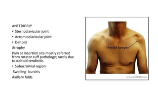
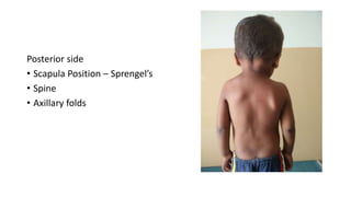
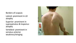
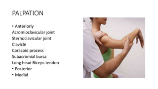
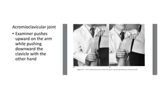
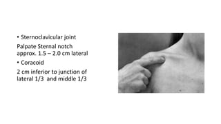
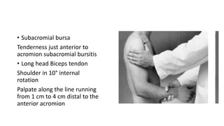
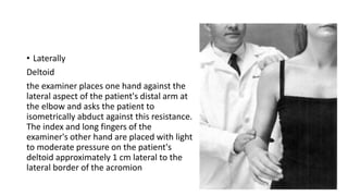
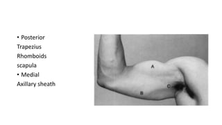
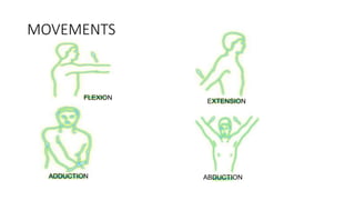
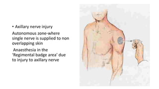
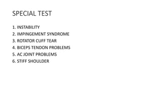
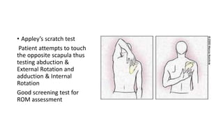
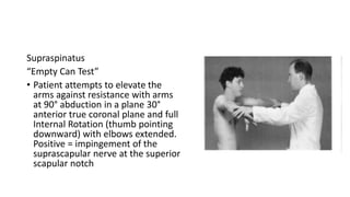
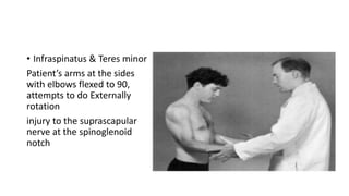
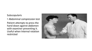
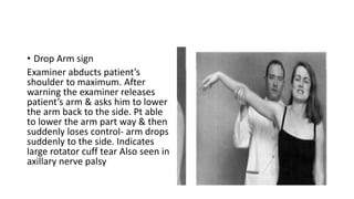
SHOULDER EXAMINATION
PRESENTER – DR.SHARAN HONGAL
EVALUATION
Inspection
Palpation
Muscle strength
Movement
Special tests
INSPECTION
• Anterior side
• Posterior side
• Lateral
• Overhead
• Axillary
ANTERIORLY
• Sternoclavicular joint
• Acromioclavicular joint
• Deltoid
Atrophy
Pain at insertion site mostly referred
from rotator cuff pathology; rarely due
to deltoid tendinitis
• Subacromial region
Swelling- bursitis
Axillary folds
Posterior side
• Scapula Position – Sprengel’s
• Spine
• Axillary folds
Borders of scapula
Lateral- prominent in LD
atrophy
Superior- prominent in
supraspinatus & trapezius
atrophy
Vertebral- prominent in
serratus anterior
weakness/winging
PALPATION
• Anteriorly
Acromioclavicular joint
Sternoclavicular joint
Clavicle
Coracoid process
Subacromial bursa
Long head Biceps tendon
• Posterior
• Medial
Acromioclavicular joint
• Examiner pushes
upward on the arm
while pushing
downward the
clavicle with the
other hand
• Sternoclavicular joint
Palpate Sternal notch
approx. 1.5 – 2.0 cm lateral
• Coracoid
2 cm inferior to junction of
lateral 1/3 and middle 1/3
• Subacromial bursa
Tenderness just anterior to
acromion subacromial bursitis
• Long head Biceps tendon
Shoulder in 10° internal
rotation
Palpate along the line running
from 1 cm to 4 cm distal to the
anterior acromion
• Laterally
Deltoid
the examiner places one hand against the
lateral aspect of the patient's distal arm at
the elbow and asks the patient to
isometrically abduct against this resistance.
The index and long fingers of the
examiner's other hand are placed with light
to moderate pressure on the patient's
deltoid approximately 1 cm lateral to the
lateral border of the acromion
• Posterior
Trapezius
Rhomboids
scapula
• Medial
Axillary sheath
MUSCLE STRENGTH
• SCAPULAR STABILIZERS
Serratus Anterior
originates from the anterior parts
of ribs and inserts on the medial
border of the scapula
Give push ups
Long thorasic nerve
Rhomboids
Spinous process of the
C7 to T5 vertebrae to
medial border of
scapula
Attention position
dorsal scapular nerve
Trapezius
Spinous process of
vertebrae c7 to t12 to
lateral 1/3 of clavicle
,acromion proess and
spine of scapula
Shrug the shoulder
Spinal accessory nerve
MOVEMENTS
FLEXION
EXTENSION
ADDUCTION ABDUCTION
NEUROMUSCULAR EXAMINATION
• Motor examination
• Sensory examination
• Deep tendon reflexes
• Axillary nerve injury
Autonomous zone-where
single nerve is supplied to non
overlapping skin
Anaesthesia in the
‘Regimental badge area’ due
to injury to axillary nerve
SPECIAL TEST
1. INSTABILITY
2. IMPINGEMENT SYNDROME
3. ROTATOR CUFF TEAR
4. BICEPS TENDON PROBLEMS
5. AC JOINT PROBLEMS
6. STIFF SHOULDER
• Appley’s scratch test
Patient attempts to touch
the opposite scapula thus
testing abduction &
External Rotation and
adduction & Internal
Rotation
Good screening test for
ROM assessment
ANTERIOR DISLOCATION
• Hamilton Ruler test – straight ruler cannot touch acromion process
and lateral epicondyle
• Duga’s test
• Callaway’s test
POSTERIOR DISLOCATION
• EXTERNAL ROTATION restricted
• Prominence in posterior deltoid
INSTABILITY
• Unidirectional- anterior, posterior, inferior
• Multidirectional (MDI) – anterior &/ or posterior with inferior
ANTERIOR
Apprehension test
• Crank test – Patient sitting; arm at
90° Abduction. With increasing
External rotation the examiner
exerts an anterior translatory force
with his thumb placed posteriorly
on the humerus & watches for
apprehension.
subluxation or dislocation
• Fulcrum test –Patient lying
supine with the scapula
supported by the edge of the
table. The arm is positioned in
90° Abduction. With increasing
External rotation the examiner
watches for apprehension.
POSTERIOR
• Jerk test- Patient in 90° forward
flexion of shoulder & elbow
flexed to 90°, examiner applies
posterior directed force by
holding the forearm.
• Jerk/Jump = dislocates out of the
glenoid posteriorly
INFERIOR
• Sulcus sign
Downward traction applied
Dimpling of the skin below the
acromion or widening of the
subacromial space on palpation
purpose of this traction is to sublux
the humeral head inferiorly in
relationship to the glenoid fossa
IMPINGEMENT
• Painful arc syndrome
In abduction arc of motion,
patient feels pain in the
range 60-120°.
• O’Brien test
The patient flexes the arm to 90°
with the elbow fully extended and
then adducts the arm 10-15°
midline. The arm is then maximally
internally rotated and the patient
resists the examiner's downward
force.
SLAP lesions
• Hawkins-Kennedy Test
patient sitting with arm at 90° forward
elevation and elbow flexed to 90°.
Examiner then quickly moves the arm
into internal rotation. +ve = Pain located
to the sub-acromial space
• Neer Impingement Sign
Examiner performs maximal passive
forward flexion with internal rotation
while stabilizing the scapula.
Pain =maneuver is thought to bring the
pathologic anterolateral acromion into
contact with the affected portion of the
rotator cuff and greater tuberosity,
thereby producing pain
ROTATOR CUFF TEAR
• supraspinatus – Empty can test
• infraspinatus - ER at arm at side with elbow flexed
• for subscapularis - Abdominal compression test
• Drop Arm sign
• External rotation lag sign
Supraspinatus
“Empty Can Test”
• Patient attempts to elevate the
arms against resistance with arms
at 90° abduction in a plane 30°
anterior true coronal plane and full
Internal Rotation (thumb pointing
downward) with elbows extended.
Positive = impingement of the
suprascapular nerve at the superior
scapular notch
• Infraspinatus & Teres minor
Patient’s arms at the sides
with elbows flexed to 90,
attempts to do Externally
rotation
injury to the suprascapular
nerve at the spinoglenoid
notch
Subscapularis
• Abdominal compression test
Patient attempts to press the
hand down against abdomen
with examiner preventing it.
Useful when Internal rotation
restricted
• Drop Arm sign
Examiner abducts patient’s
shoulder to maximum. After
warning the examiner releases
patient’s arm & asks him to lower
the arm back to the side. Pt able
to lower the arm part way & then
suddenly loses control- arm drops
suddenly to the side. Indicates
large rotator cuff tear Also seen in
axillary nerve palsy
• External rotation lag sign
Patient’s arm is externally rotated
maximally and released- arm rotates
internally spontaneously . Seen
when subscapularis is intact but
infraspinatus & teres minor is torn.
BICEPS TENDINITIS
Yergasson’s test
• The patient's elbow is flexed
and forearm pronated.
The examiner holds at the
wrist. Patient actively
supinates against resistance.
Pain located to bicipital groove
reflect biceps tendinitis or
instability
AC JOINT PROBLEMS
• Cross chest adduction
test
Patient elevates the
affected arm to 90°, then
actively adducts it.
STIFF SHOULDER
• Restriction of all range of motion,
MAINLY Abduction & EXTERNAL
ROTATION
External Rotation restriction occurs
in 2 conditions only
1. Stiff shoulder
2. Posterior dislocation
THANK YOU

Shoulder examination

  • 1.
  • 2.
  • 3.
    INSPECTION • Anterior side •Posterior side • Lateral • Overhead • Axillary
  • 4.
    ANTERIORLY • Sternoclavicular joint •Acromioclavicular joint • Deltoid Atrophy Pain at insertion site mostly referred from rotator cuff pathology; rarely due to deltoid tendinitis • Subacromial region Swelling- bursitis Axillary folds
  • 5.
    Posterior side • ScapulaPosition – Sprengel’s • Spine • Axillary folds
  • 6.
    Borders of scapula Lateral-prominent in LD atrophy Superior- prominent in supraspinatus & trapezius atrophy Vertebral- prominent in serratus anterior weakness/winging
  • 7.
    PALPATION • Anteriorly Acromioclavicular joint Sternoclavicularjoint Clavicle Coracoid process Subacromial bursa Long head Biceps tendon • Posterior • Medial
  • 8.
    Acromioclavicular joint • Examinerpushes upward on the arm while pushing downward the clavicle with the other hand
  • 9.
    • Sternoclavicular joint PalpateSternal notch approx. 1.5 – 2.0 cm lateral • Coracoid 2 cm inferior to junction of lateral 1/3 and middle 1/3
  • 10.
    • Subacromial bursa Tendernessjust anterior to acromion subacromial bursitis • Long head Biceps tendon Shoulder in 10° internal rotation Palpate along the line running from 1 cm to 4 cm distal to the anterior acromion
  • 11.
    • Laterally Deltoid the examinerplaces one hand against the lateral aspect of the patient's distal arm at the elbow and asks the patient to isometrically abduct against this resistance. The index and long fingers of the examiner's other hand are placed with light to moderate pressure on the patient's deltoid approximately 1 cm lateral to the lateral border of the acromion
  • 12.
  • 13.
    MUSCLE STRENGTH • SCAPULARSTABILIZERS Serratus Anterior originates from the anterior parts of ribs and inserts on the medial border of the scapula Give push ups Long thorasic nerve
  • 14.
    Rhomboids Spinous process ofthe C7 to T5 vertebrae to medial border of scapula Attention position dorsal scapular nerve
  • 15.
    Trapezius Spinous process of vertebraec7 to t12 to lateral 1/3 of clavicle ,acromion proess and spine of scapula Shrug the shoulder Spinal accessory nerve
  • 16.
  • 17.
    NEUROMUSCULAR EXAMINATION • Motorexamination • Sensory examination • Deep tendon reflexes
  • 18.
    • Axillary nerveinjury Autonomous zone-where single nerve is supplied to non overlapping skin Anaesthesia in the ‘Regimental badge area’ due to injury to axillary nerve
  • 19.
    SPECIAL TEST 1. INSTABILITY 2.IMPINGEMENT SYNDROME 3. ROTATOR CUFF TEAR 4. BICEPS TENDON PROBLEMS 5. AC JOINT PROBLEMS 6. STIFF SHOULDER
  • 20.
    • Appley’s scratchtest Patient attempts to touch the opposite scapula thus testing abduction & External Rotation and adduction & Internal Rotation Good screening test for ROM assessment
  • 21.
    ANTERIOR DISLOCATION • HamiltonRuler test – straight ruler cannot touch acromion process and lateral epicondyle • Duga’s test • Callaway’s test
  • 22.
    POSTERIOR DISLOCATION • EXTERNALROTATION restricted • Prominence in posterior deltoid
  • 23.
    INSTABILITY • Unidirectional- anterior,posterior, inferior • Multidirectional (MDI) – anterior &/ or posterior with inferior
  • 24.
    ANTERIOR Apprehension test • Cranktest – Patient sitting; arm at 90° Abduction. With increasing External rotation the examiner exerts an anterior translatory force with his thumb placed posteriorly on the humerus & watches for apprehension. subluxation or dislocation
  • 25.
    • Fulcrum test–Patient lying supine with the scapula supported by the edge of the table. The arm is positioned in 90° Abduction. With increasing External rotation the examiner watches for apprehension.
  • 26.
    POSTERIOR • Jerk test-Patient in 90° forward flexion of shoulder & elbow flexed to 90°, examiner applies posterior directed force by holding the forearm. • Jerk/Jump = dislocates out of the glenoid posteriorly
  • 27.
    INFERIOR • Sulcus sign Downwardtraction applied Dimpling of the skin below the acromion or widening of the subacromial space on palpation purpose of this traction is to sublux the humeral head inferiorly in relationship to the glenoid fossa
  • 28.
    IMPINGEMENT • Painful arcsyndrome In abduction arc of motion, patient feels pain in the range 60-120°.
  • 29.
    • O’Brien test Thepatient flexes the arm to 90° with the elbow fully extended and then adducts the arm 10-15° midline. The arm is then maximally internally rotated and the patient resists the examiner's downward force. SLAP lesions
  • 30.
    • Hawkins-Kennedy Test patientsitting with arm at 90° forward elevation and elbow flexed to 90°. Examiner then quickly moves the arm into internal rotation. +ve = Pain located to the sub-acromial space
  • 31.
    • Neer ImpingementSign Examiner performs maximal passive forward flexion with internal rotation while stabilizing the scapula. Pain =maneuver is thought to bring the pathologic anterolateral acromion into contact with the affected portion of the rotator cuff and greater tuberosity, thereby producing pain
  • 32.
    ROTATOR CUFF TEAR •supraspinatus – Empty can test • infraspinatus - ER at arm at side with elbow flexed • for subscapularis - Abdominal compression test • Drop Arm sign • External rotation lag sign
  • 33.
    Supraspinatus “Empty Can Test” •Patient attempts to elevate the arms against resistance with arms at 90° abduction in a plane 30° anterior true coronal plane and full Internal Rotation (thumb pointing downward) with elbows extended. Positive = impingement of the suprascapular nerve at the superior scapular notch
  • 34.
    • Infraspinatus &Teres minor Patient’s arms at the sides with elbows flexed to 90, attempts to do Externally rotation injury to the suprascapular nerve at the spinoglenoid notch
  • 35.
    Subscapularis • Abdominal compressiontest Patient attempts to press the hand down against abdomen with examiner preventing it. Useful when Internal rotation restricted
  • 36.
    • Drop Armsign Examiner abducts patient’s shoulder to maximum. After warning the examiner releases patient’s arm & asks him to lower the arm back to the side. Pt able to lower the arm part way & then suddenly loses control- arm drops suddenly to the side. Indicates large rotator cuff tear Also seen in axillary nerve palsy
  • 37.
    • External rotationlag sign Patient’s arm is externally rotated maximally and released- arm rotates internally spontaneously . Seen when subscapularis is intact but infraspinatus & teres minor is torn.
  • 38.
    BICEPS TENDINITIS Yergasson’s test •The patient's elbow is flexed and forearm pronated. The examiner holds at the wrist. Patient actively supinates against resistance. Pain located to bicipital groove reflect biceps tendinitis or instability
  • 39.
    AC JOINT PROBLEMS •Cross chest adduction test Patient elevates the affected arm to 90°, then actively adducts it.
  • 40.
    STIFF SHOULDER • Restrictionof all range of motion, MAINLY Abduction & EXTERNAL ROTATION External Rotation restriction occurs in 2 conditions only 1. Stiff shoulder 2. Posterior dislocation
  • 41.