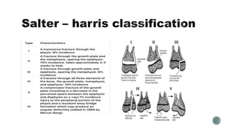
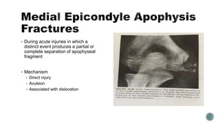
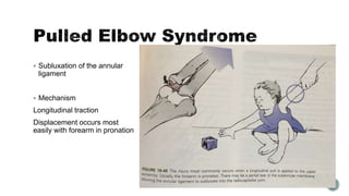
Presenter - Dr sharan hongal
 Medial Epicondyle Apophysis Fractures
 Pulled Elbow Syndrome
 Lateral Condylar Fractures
 Capitellar Fractures
 Medial Condylar Fractures
 Lateral Epicondyle Apophysis Fractures
 Trochlea Fractures
 During acute injuries in which a
distinct event produces a partial or
complete separation of apophyseal
fragment
 Mechanism
 Direct injury
 Avulsion
 Associated with dislocation
 Subluxation of the annular
ligament
 Mechanism
Longitudinal traction
Displacement occurs most
easily with forearm in pronation
 Fractures involving the lateral condyle in the immature skeleton can either cross the
physis or follow short distance into troclear cartilage
 Rarely associated with other injuries , but uncommonly injuries associated are
 Dislocation of elbow
 Radial head fracture
 Olecranon fracture
 Mechanism of injury
 Pull off theory
This injury was produced by adducting the forearm with elbow fully extended and forearm
supinated
Avulsion Injury to common extensor injury owing to varus stress exerted on the extended
elbow
 Push off theory
This injury was produced by direct blow to palm with elbow flexed causing radial head to push
lateral condyle
Axial load transmitted to forearm leading to impingement of radial head to lateral condyle
 Classification
Anatomical
Milch
 Type 1 – fracture line courses lateral to the trochlea and into capitellar-troclear groove.
It represent a salter –harris type 4 , elbow is stable because trochlea is intact
 Type 2 – fracture line extends into the apex of trochlea
It represents a salter- harris type 2 , elbow is unstable because trochlea is disrupted
Stages of displacement
 Jakob et al.
First stage – fracture is displaced and the articular surface is intact
 Second stage
Fracture extends completely through the articular surface leading to proximal fragment
to become more displaced and can allow lateral displacement of the olecranon
 Third stage
The condylar fragment is rotated and totally displaced laterally and proximally , allowing
olecranon and radial head translocation
 Weiss modiefied
 Type 1 – displaced less than
2mm
 Type 2 – displaced more than
2mm but have intact cartilaginous
hinge
 Type 3 – displace more than
2mm and no intact cartilaginous
hinge
 Fracture involve only the true articular surface of lateral condyle
 Rarely seen in children
 Classification
 Hahn – steinthal type – contains a rather large portion of cancellous bone of the lateral
condyle, lateral crista of trochlea is included
 Kocher – Lorenz is more of pure articular fracture with little if any subchondral bone
attached
 Fracture involving medial condyle has 2 components
 The intra – articular component involves troclear articular surface
 The extra – articular involves medial epiphysis and medial condyle
These are called trochlea fractures because the fracture line extend into the articular
surface of trochlea
Classification
 Milch
 Type 1 – fracture line traverses the apex of trochlea
 Type 2 – fracture line traverses through the capitulotrochlear groove
 Kilfoyle
 Type 1 – incomplete fracture that does not violate joint but may hinge open
 Type 2 – fracture line enters the joint but displacement is less than 2mm
 Type 3 – fracture line enters the joint and results in malalignment , malrotation and
displacement
 Least common fractures
 Mechanism
 Direct blow to lateral condyle of elbow
 Sometimes in children avulsion forces from the extensor muscles may be responsible for
these injuries
 If the proximal part of fracture line lies between common extensors and extensor carpi
radialis longus there eill b elittle displacement
 If fracture line enters the origin of the extensor carpi radialis longus then considerable
displacement occcurs
Fractures around elbow joint in children

Fractures around elbow joint in children

  • 1.
    Presenter - Drsharan hongal
  • 5.
     Medial EpicondyleApophysis Fractures  Pulled Elbow Syndrome  Lateral Condylar Fractures  Capitellar Fractures  Medial Condylar Fractures  Lateral Epicondyle Apophysis Fractures  Trochlea Fractures
  • 6.
     During acuteinjuries in which a distinct event produces a partial or complete separation of apophyseal fragment  Mechanism  Direct injury  Avulsion  Associated with dislocation
  • 8.
     Subluxation ofthe annular ligament  Mechanism Longitudinal traction Displacement occurs most easily with forearm in pronation
  • 9.
     Fractures involvingthe lateral condyle in the immature skeleton can either cross the physis or follow short distance into troclear cartilage  Rarely associated with other injuries , but uncommonly injuries associated are  Dislocation of elbow  Radial head fracture  Olecranon fracture
  • 12.
     Mechanism ofinjury  Pull off theory This injury was produced by adducting the forearm with elbow fully extended and forearm supinated Avulsion Injury to common extensor injury owing to varus stress exerted on the extended elbow  Push off theory This injury was produced by direct blow to palm with elbow flexed causing radial head to push lateral condyle Axial load transmitted to forearm leading to impingement of radial head to lateral condyle
  • 13.
     Classification Anatomical Milch  Type1 – fracture line courses lateral to the trochlea and into capitellar-troclear groove. It represent a salter –harris type 4 , elbow is stable because trochlea is intact  Type 2 – fracture line extends into the apex of trochlea It represents a salter- harris type 2 , elbow is unstable because trochlea is disrupted
  • 15.
    Stages of displacement Jakob et al. First stage – fracture is displaced and the articular surface is intact
  • 16.
     Second stage Fractureextends completely through the articular surface leading to proximal fragment to become more displaced and can allow lateral displacement of the olecranon
  • 17.
     Third stage Thecondylar fragment is rotated and totally displaced laterally and proximally , allowing olecranon and radial head translocation
  • 18.
     Weiss modiefied Type 1 – displaced less than 2mm  Type 2 – displaced more than 2mm but have intact cartilaginous hinge  Type 3 – displace more than 2mm and no intact cartilaginous hinge
  • 19.
     Fracture involveonly the true articular surface of lateral condyle  Rarely seen in children  Classification  Hahn – steinthal type – contains a rather large portion of cancellous bone of the lateral condyle, lateral crista of trochlea is included  Kocher – Lorenz is more of pure articular fracture with little if any subchondral bone attached
  • 21.
     Fracture involvingmedial condyle has 2 components  The intra – articular component involves troclear articular surface  The extra – articular involves medial epiphysis and medial condyle These are called trochlea fractures because the fracture line extend into the articular surface of trochlea
  • 25.
    Classification  Milch  Type1 – fracture line traverses the apex of trochlea  Type 2 – fracture line traverses through the capitulotrochlear groove  Kilfoyle  Type 1 – incomplete fracture that does not violate joint but may hinge open  Type 2 – fracture line enters the joint but displacement is less than 2mm  Type 3 – fracture line enters the joint and results in malalignment , malrotation and displacement
  • 27.
     Least commonfractures  Mechanism  Direct blow to lateral condyle of elbow  Sometimes in children avulsion forces from the extensor muscles may be responsible for these injuries  If the proximal part of fracture line lies between common extensors and extensor carpi radialis longus there eill b elittle displacement  If fracture line enters the origin of the extensor carpi radialis longus then considerable displacement occcurs