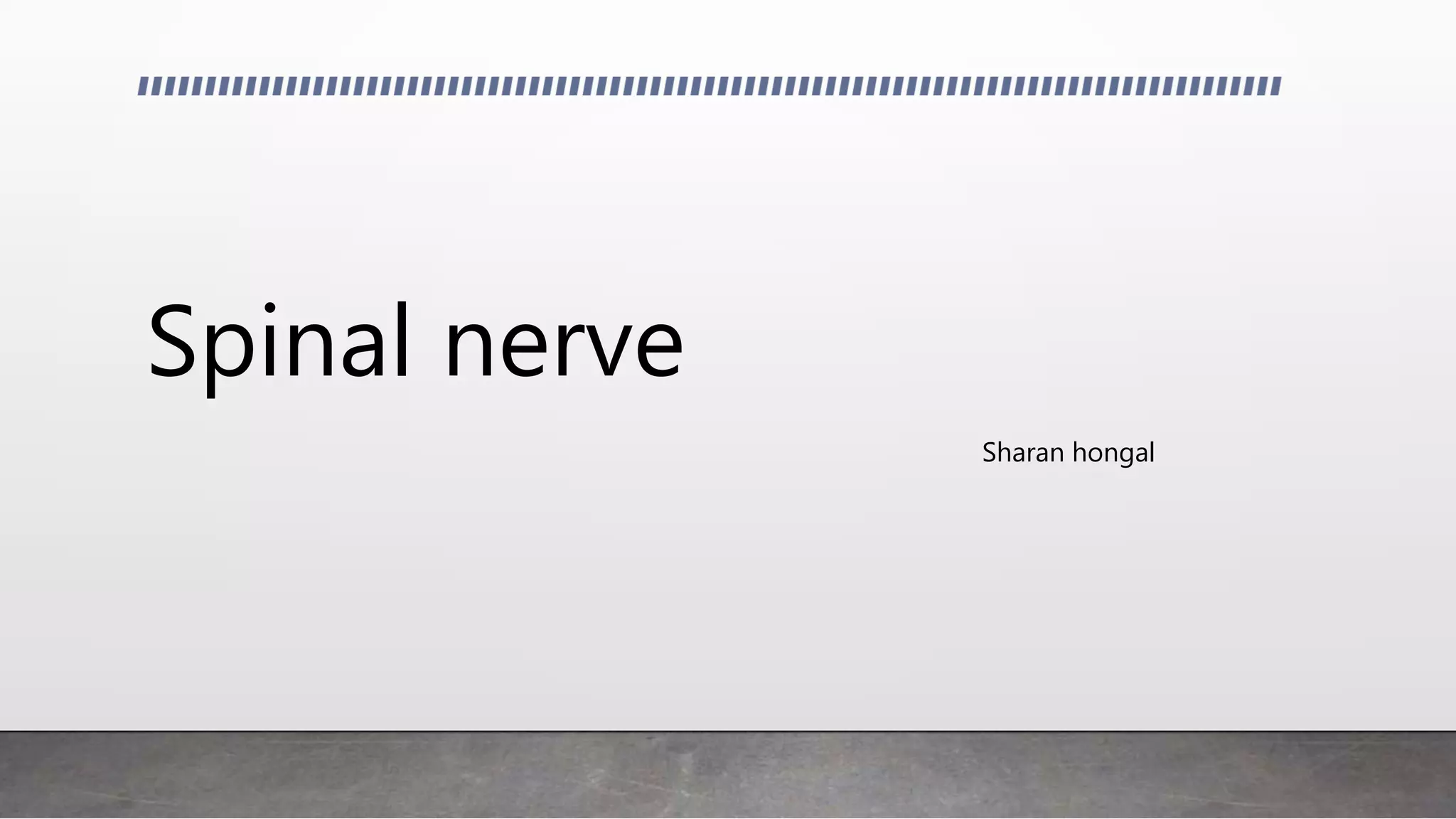
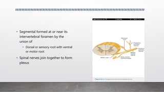
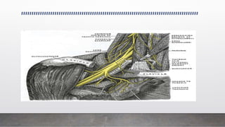
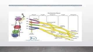
Spinal nerve
Sharan hongal
• Segmental formed at or near its
intervertebral foramen by the
union of
• Dorsal or sensory root with ventral
or motor root
• Spinal nerves join together to form
plexus
• 31 spinal nerves
• 8 cervical
• 12 thoracic
• 5 lumbar
• 5 sacral
• 1 coccygeal
• Components of spinal nerve
• Motor
• Sensory
• Sympathetic
• Nerve fiber or axon is a
direct extension of
dorsal root ganglion
cell , an anterior horn or
a post ganglionic
sympathetic nerve cell
• Either myelinated or
unmyelinated
• The segment of myelinated nerve fiber
enclosed by a single schwann cell –
internode , varies in length from .1 – 1.8
mm
• The point at which one schwann cell
ends and next begins – nodal gap or
node of ranvier
• Neuron detached from its nucleus
degenerates and is destroyed by
phagocytes this process degeneration
distal to a point of injury is called
secondary or wallerian degeneration
• The reaction proximal to the point of
detachment is called primary or traumatic
or retrograde degeneration
Seddon
• Neurapraxia
• Axonotmesis
• Neurotmesis
Sunderland
• Myelin
• Axon
• Endoneural tube
• Perineurium
• Epineurium
BRACHIAL PLEXUS
Introduction
ANATOMY
ETIOLOGY
MECHANISM OF INJURY
CLASSIFICATION
CLINICAL FEATURES
INVESTIGATIONS
MANAGEMENT
Anatomy
• Brachial plexus is a somatic nerve plexus formed by the union of anterior rami of
C5,C6,C7,C8 and T1.
• The formation of brachial plexus begins just distal to the scalenus muscles.
• Function: The brachial plexus is responsible for cutaneous and muscular
innervation of the entire upper limb, with two exceptions: the trapezius muscle
innervated by the spinal accessory nerve (CN XI) and an area of skin near the
axilla innervated by the intercostobrachial nerve.
The plexus consists of
• Roots
• Trunks
• Divisions
• cords
• and branches.
• Roots : Lower 4 cervical (C5-8) and the 1st thoracic.
• Situated between the scalenus anterior and medius muscle deep to
sternocleidomastoid muscle.
• The origin of the plexus may shift one segment either upward or downward resulting
in a PRE FIXED PLEXUS or POST FIXED PLEXUS respectively.
• In a prefixed plexus, the contribution by C4 is large and in that from T2 is often absent.
In a post fixed plexus, the contribution by T1 is large, T2 is always present, C4 is
absent, and C5 is reduced in size
• Trunks
• Derived from roots
• Located in the antero-inferior portion of post triangle of neck
• C5-6 ant primary rami unite - upper trunk.
• C8-T1 ant primary rami unite - lower trunk.
• C7 ant primary rami continues as - middle trunk.
• Each trunk ends by splitting into
• Anterior
• Posterior divisions
• CORDS: it forms 3 cords
• The Posterior Cord is formed from the three posterior divisions of the trunks
(C5-C8,T1)
• The Lateral Cord is the anterior divisions from the upper and middle trunks (C5-
C7)
• The Medial Cord is simply a continuation of the anterior division of the lower
trunk (C8,T1)
BRANCHES:
Supraclavicular branches
• Supraclavicular branches arise from roots or from trunks as follows:
From roots
1. Dorsal scapular nerve C5
2. Long thoracic nerve C5, 6 (7)
From trunks
1. Nerve to subclavius C5, 6
2. Suprascapular nerve C5, 6
Infraclavicular branches
branches come from the cords,
Lateral cord
• Lateral pectoral C5, 6, 7
• Musculocutaneous C5, 6 7
• Lateral root of median C(5), 6, 7
Medial cord
• Medial pectoral C8, T1
• Medial cutaneous of forearm C8, T1
• Medial cutaneous of arm C8, T1
• Ulnar C(7), 8, T1
• medial root of median nerve
Posterior cord
• Upper subscapular C5, 6
• Thoracodorsal C6, 7,8
• Lower subscapular C5, 6
• Axillary C5, 6
• Radial C5, 6, 7, 8, (T1)
Etiology
• penetrating wounds
• Missiles
• Stab wounds
• Injuries related to birth
• Traction applied to the plexus during falls
• Road traffic accidents
• Sports activities
COMMON ASSOCIATED INJURIES
• Fractures of the proximal humerus
• Scapula fractures
• Rib fractures
• Clavicle fractures
• Fracture of the transverse process of cervical vertebrae
• Dislocations of the shoulder, acromioclavicular and sternoclavicular joints
Erbs palsy
Site of injury: The region of the upper trunk of the brachial plexus is called Erb's
point.
Injury to the upper trunk causes Erb's Paralysis.
• Causes of injury: Undue separation of the head from the shoulder, which is commonly
encountered in
1)birth injury
2) fall on shoulder
3)during anaesthesia
• Nerve roots involved: Mainly C5 and partly C6.
• Muscles paralysed: Mainly biceps, deltoid, brachilais and brachioradialis.Partly
supraspinatus, infraspinatus and supinator
Deformity
• Arm: Hangs by the side, it is adducted and medially rotated
• Forearm: Extended and pronated
• Abduction impossible because of paralysis of deltoid & supraspinatus m/s. • ER
impossible because of paralysis of infraspinatus & teres minor m/s.
• Active flexion impossible because of paralysis biceps, brachialis & brachioradialis.
• Paralysis of supinator m/s causes pronation deformity of forearm.
• The deformity is known as "Policeman's tip hand" or "Porter's tip hand".
Klumpke palsy
Site of injury: Lower trunk of the brachial plexus.
• Cause of injury: Undue abduction of the arm, as in clutching something with the
hand after a fall from a height, or sometimes in birth injury.
•Nerve roots involved: Mainly T1 and partly C8.
• Muscles paralysed:
• Intrinsic muscles of the hand (T1)
• Ulnar flexors of the wrist and fingers (C8).
Deformity: claw hand due to the
unopposed action of the long flexors and
extensors of the fingers.
in a claw hand there is hyperextension at
the metacarpophalangeal joints and
flexion at the interphalangeal joints.
Disability:
• Claw hand
Cutaneous anaesthesia and analgesia in a narrow zone along the ulnar border of
the forearm and hand.
• Horner's syndrome: ptosis, miosis, anhydrosis, enophthalmos and loss of
ciliospinal reflex- may be associated. This is because of injury to sympathetic fibres
to the head and neck that leave the spinal cord through nerve T1.
• Vasomotor changes: The skin areas with sensory loss is warmer due to arteriolar
dilation. skin is dry due to the absence of sweating as there is loss of sympathetic
activity.
• Tropic changes: Long standing case of paralysis leads to dry and scaly skin. The
nails crack easily with atrophy of the pulp of fingers.
Clinical features
• The pain from brachial plexus injuries results from injury to the spinal cord where
the nerve rootlets are avulsed from the cord. This pain is neuropathic in nature.
The pain can last for a very long time.
• Brachial plexus injuries that occur at the level of the spinal cord often produce
greater pain than injuries more distant from the spinal cord.
• In addition, injuries nearer the spinal cord may cause a burning numbness, which
is called paresthesias or dysesthesias.
• Examination of all nerve groups controlled by the brachial plexus to identify the specific location
of the nerve injury and its severity. In addition, some patients display specific signs that help
determine the location of the nerve injury
• Narrowing of the eye pupils, drooping of the eyelid, and lack of ability for the face to sweat
(Horner's syndrome) is a sign that the injury is close to the spinal cord.
• A shooting nerve-like pain on taping along the affected nerves (Tinel sign) suggests an injury
farther from the spinal cord. Over time, if the location of the Tinel sign moves down the arm
toward the hand, it is a sign that the injury is repairing itself. During the physical examination,
assess the arm and shoulder for stability and range of motion
Imaging Studies
• Electromyogram
• Nerve Conduction Velocity
• Intraoperative Nerve Action Potential
• Myelography
• CT scan for any tumours
• MRI
Management
• In the case of closed BPI wounds and when there are no other emergent injuries,
surgical exploration and recovery may not take place immediately.
• Recommendations include
• managing pain
• starting rehabilitation.
Closed
• Barnes divided Upper & Lower Plexuses injuries caused by traction into four
groups
1. Injuries at C5 & C6
2. Injuries at C5,C6 & C7
3. Degenerative lesions of entire plexus
4. Injuries at C7,C8 & T1 (rare)
• Barnes reported that spontaneous recovery in group 1 & 2 cases
• But in case of Degenerative plexus injuries there is partial recovery.
• EMG should be done at 3 to 4 wks.
At 6 to 8wks additional studies like myelography & axon reflex evaluation can be
done if return of functions not seen.
• exploration is justified at 3 to 6 months after injury if function has not returned.
Open
• Open wounds in BPI are uncommon and vary from small penetrating injuries to
high energy injuries.
• INDICATIONS OF SURGERY:
• Injuries caused by sharp objects or missiles.
• When patient seen soon after injury & pt's general condition permits exploration &
primary repair can be done
• When patient not seen soon after injury but only after initial management, It is
best to wait for wound healing & stabilization of any other injuries.
• During this period locate neurological deficit for level of injury.
• EMG performed 3 to 4 wks after injury.
• Exploration of plexus & neurorrhaphy, autogenous interfascicular nerve grafting
or neurolysis is indicated 3 to 6wks after injury.
• Motor function recovered to a grade of 3 or better in half of pts.
• Best results obtained in upper trunk & lateral cord & posterior cord injuries.
• Poor prognosis can be expected in lower trunk injuries.
• AIMS:
• to maintain the range of motion of the extremity
• to strengthen the remaining functional muscles
• to protect the denervated dermatomes, and
• to manage pain.
Goals of surgery
• Restoration of elbow flexion
• Restoration of shoulder abduction
• Restoration of sensation of medial border of forearm & hand.
• Depending on extent of injury various surgical techniques may be required:
• Primary neurorrhaphy
• Neurolysis
• Nerve grafting
• Neurotization
Thank you

Spinal nerve

  • 1.
  • 2.
    • Segmental formedat or near its intervertebral foramen by the union of • Dorsal or sensory root with ventral or motor root • Spinal nerves join together to form plexus
  • 3.
    • 31 spinalnerves • 8 cervical • 12 thoracic • 5 lumbar • 5 sacral • 1 coccygeal • Components of spinal nerve • Motor • Sensory • Sympathetic
  • 4.
    • Nerve fiberor axon is a direct extension of dorsal root ganglion cell , an anterior horn or a post ganglionic sympathetic nerve cell • Either myelinated or unmyelinated
  • 5.
    • The segmentof myelinated nerve fiber enclosed by a single schwann cell – internode , varies in length from .1 – 1.8 mm • The point at which one schwann cell ends and next begins – nodal gap or node of ranvier
  • 6.
    • Neuron detachedfrom its nucleus degenerates and is destroyed by phagocytes this process degeneration distal to a point of injury is called secondary or wallerian degeneration • The reaction proximal to the point of detachment is called primary or traumatic or retrograde degeneration
  • 7.
  • 8.
    Sunderland • Myelin • Axon •Endoneural tube • Perineurium • Epineurium
  • 9.
  • 10.
  • 11.
    Anatomy • Brachial plexusis a somatic nerve plexus formed by the union of anterior rami of C5,C6,C7,C8 and T1. • The formation of brachial plexus begins just distal to the scalenus muscles. • Function: The brachial plexus is responsible for cutaneous and muscular innervation of the entire upper limb, with two exceptions: the trapezius muscle innervated by the spinal accessory nerve (CN XI) and an area of skin near the axilla innervated by the intercostobrachial nerve.
  • 13.
    The plexus consistsof • Roots • Trunks • Divisions • cords • and branches.
  • 14.
    • Roots :Lower 4 cervical (C5-8) and the 1st thoracic. • Situated between the scalenus anterior and medius muscle deep to sternocleidomastoid muscle. • The origin of the plexus may shift one segment either upward or downward resulting in a PRE FIXED PLEXUS or POST FIXED PLEXUS respectively. • In a prefixed plexus, the contribution by C4 is large and in that from T2 is often absent. In a post fixed plexus, the contribution by T1 is large, T2 is always present, C4 is absent, and C5 is reduced in size
  • 15.
    • Trunks • Derivedfrom roots • Located in the antero-inferior portion of post triangle of neck • C5-6 ant primary rami unite - upper trunk. • C8-T1 ant primary rami unite - lower trunk. • C7 ant primary rami continues as - middle trunk. • Each trunk ends by splitting into • Anterior • Posterior divisions
  • 16.
    • CORDS: itforms 3 cords • The Posterior Cord is formed from the three posterior divisions of the trunks (C5-C8,T1) • The Lateral Cord is the anterior divisions from the upper and middle trunks (C5- C7) • The Medial Cord is simply a continuation of the anterior division of the lower trunk (C8,T1)
  • 18.
    BRANCHES: Supraclavicular branches • Supraclavicularbranches arise from roots or from trunks as follows: From roots 1. Dorsal scapular nerve C5 2. Long thoracic nerve C5, 6 (7) From trunks 1. Nerve to subclavius C5, 6 2. Suprascapular nerve C5, 6
  • 19.
    Infraclavicular branches branches comefrom the cords, Lateral cord • Lateral pectoral C5, 6, 7 • Musculocutaneous C5, 6 7 • Lateral root of median C(5), 6, 7
  • 20.
    Medial cord • Medialpectoral C8, T1 • Medial cutaneous of forearm C8, T1 • Medial cutaneous of arm C8, T1 • Ulnar C(7), 8, T1 • medial root of median nerve
  • 21.
    Posterior cord • Uppersubscapular C5, 6 • Thoracodorsal C6, 7,8 • Lower subscapular C5, 6 • Axillary C5, 6 • Radial C5, 6, 7, 8, (T1)
  • 22.
    Etiology • penetrating wounds •Missiles • Stab wounds • Injuries related to birth • Traction applied to the plexus during falls • Road traffic accidents • Sports activities
  • 23.
    COMMON ASSOCIATED INJURIES •Fractures of the proximal humerus • Scapula fractures • Rib fractures • Clavicle fractures • Fracture of the transverse process of cervical vertebrae • Dislocations of the shoulder, acromioclavicular and sternoclavicular joints
  • 24.
    Erbs palsy Site ofinjury: The region of the upper trunk of the brachial plexus is called Erb's point. Injury to the upper trunk causes Erb's Paralysis.
  • 25.
    • Causes ofinjury: Undue separation of the head from the shoulder, which is commonly encountered in 1)birth injury 2) fall on shoulder 3)during anaesthesia • Nerve roots involved: Mainly C5 and partly C6. • Muscles paralysed: Mainly biceps, deltoid, brachilais and brachioradialis.Partly supraspinatus, infraspinatus and supinator
  • 26.
    Deformity • Arm: Hangsby the side, it is adducted and medially rotated • Forearm: Extended and pronated • Abduction impossible because of paralysis of deltoid & supraspinatus m/s. • ER impossible because of paralysis of infraspinatus & teres minor m/s.
  • 27.
    • Active flexionimpossible because of paralysis biceps, brachialis & brachioradialis. • Paralysis of supinator m/s causes pronation deformity of forearm. • The deformity is known as "Policeman's tip hand" or "Porter's tip hand".
  • 28.
    Klumpke palsy Site ofinjury: Lower trunk of the brachial plexus. • Cause of injury: Undue abduction of the arm, as in clutching something with the hand after a fall from a height, or sometimes in birth injury. •Nerve roots involved: Mainly T1 and partly C8. • Muscles paralysed: • Intrinsic muscles of the hand (T1) • Ulnar flexors of the wrist and fingers (C8).
  • 29.
    Deformity: claw handdue to the unopposed action of the long flexors and extensors of the fingers. in a claw hand there is hyperextension at the metacarpophalangeal joints and flexion at the interphalangeal joints.
  • 30.
    Disability: • Claw hand Cutaneousanaesthesia and analgesia in a narrow zone along the ulnar border of the forearm and hand. • Horner's syndrome: ptosis, miosis, anhydrosis, enophthalmos and loss of ciliospinal reflex- may be associated. This is because of injury to sympathetic fibres to the head and neck that leave the spinal cord through nerve T1.
  • 31.
    • Vasomotor changes:The skin areas with sensory loss is warmer due to arteriolar dilation. skin is dry due to the absence of sweating as there is loss of sympathetic activity. • Tropic changes: Long standing case of paralysis leads to dry and scaly skin. The nails crack easily with atrophy of the pulp of fingers.
  • 32.
    Clinical features • Thepain from brachial plexus injuries results from injury to the spinal cord where the nerve rootlets are avulsed from the cord. This pain is neuropathic in nature. The pain can last for a very long time. • Brachial plexus injuries that occur at the level of the spinal cord often produce greater pain than injuries more distant from the spinal cord. • In addition, injuries nearer the spinal cord may cause a burning numbness, which is called paresthesias or dysesthesias.
  • 33.
    • Examination ofall nerve groups controlled by the brachial plexus to identify the specific location of the nerve injury and its severity. In addition, some patients display specific signs that help determine the location of the nerve injury • Narrowing of the eye pupils, drooping of the eyelid, and lack of ability for the face to sweat (Horner's syndrome) is a sign that the injury is close to the spinal cord. • A shooting nerve-like pain on taping along the affected nerves (Tinel sign) suggests an injury farther from the spinal cord. Over time, if the location of the Tinel sign moves down the arm toward the hand, it is a sign that the injury is repairing itself. During the physical examination, assess the arm and shoulder for stability and range of motion
  • 34.
    Imaging Studies • Electromyogram •Nerve Conduction Velocity • Intraoperative Nerve Action Potential • Myelography • CT scan for any tumours • MRI
  • 35.
    Management • In thecase of closed BPI wounds and when there are no other emergent injuries, surgical exploration and recovery may not take place immediately. • Recommendations include • managing pain • starting rehabilitation.
  • 36.
    Closed • Barnes dividedUpper & Lower Plexuses injuries caused by traction into four groups 1. Injuries at C5 & C6 2. Injuries at C5,C6 & C7 3. Degenerative lesions of entire plexus 4. Injuries at C7,C8 & T1 (rare)
  • 37.
    • Barnes reportedthat spontaneous recovery in group 1 & 2 cases • But in case of Degenerative plexus injuries there is partial recovery. • EMG should be done at 3 to 4 wks. At 6 to 8wks additional studies like myelography & axon reflex evaluation can be done if return of functions not seen. • exploration is justified at 3 to 6 months after injury if function has not returned.
  • 38.
    Open • Open woundsin BPI are uncommon and vary from small penetrating injuries to high energy injuries. • INDICATIONS OF SURGERY: • Injuries caused by sharp objects or missiles. • When patient seen soon after injury & pt's general condition permits exploration & primary repair can be done • When patient not seen soon after injury but only after initial management, It is best to wait for wound healing & stabilization of any other injuries.
  • 39.
    • During thisperiod locate neurological deficit for level of injury. • EMG performed 3 to 4 wks after injury. • Exploration of plexus & neurorrhaphy, autogenous interfascicular nerve grafting or neurolysis is indicated 3 to 6wks after injury. • Motor function recovered to a grade of 3 or better in half of pts. • Best results obtained in upper trunk & lateral cord & posterior cord injuries. • Poor prognosis can be expected in lower trunk injuries.
  • 40.
    • AIMS: • tomaintain the range of motion of the extremity • to strengthen the remaining functional muscles • to protect the denervated dermatomes, and • to manage pain.
  • 41.
    Goals of surgery •Restoration of elbow flexion • Restoration of shoulder abduction • Restoration of sensation of medial border of forearm & hand.
  • 42.
    • Depending onextent of injury various surgical techniques may be required: • Primary neurorrhaphy • Neurolysis • Nerve grafting • Neurotization
  • 43.