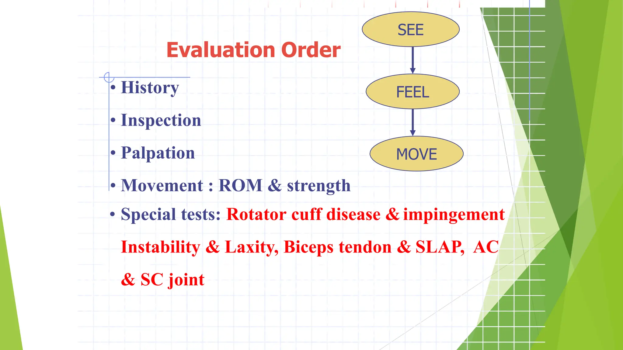
SHOULDER EXAMINATION
PAIN INSTABILITY LOSS OF MOTION
EXTRINSIC
OR
INTRINSIC
ACTIVE
OR
PASSIVE
EVALUATION PRINCIPLES
Get a History: Is this a new injury, old chronic
injury
Assessment: what is the primary problem ?
Evaluation Order
• History
• Inspection
• Palpation
• Movement : ROM & strength
• Special tests: Rotator cuff disease &impingement
Instability & Laxity, Biceps tendon & SLAP, AC
& SC joint
SEE
FEEL
MOVE
Subjective assessment
1. What is the patient’s age?
2. Does the patient support the upper limb in a protected position or hesitate
to move it?
3. If there was an injury, what exactly was the mechanism of injury?
4. Are there any movements or positions that cause the patient pain or
symptoms?
5. What is the extent and behaviour of the patients pain?
Subjective assessment
6. are there any activities that cause or increase the pain?
7. do any positions relieve the pain?
8. What is the patient unable to do functionally?
9. How long has the problem bothered the patient?
10. Is there any indication of muscle spasm, deformity, bruising, wasting,
paresthesia or numbness?
Subjective assessment
11. Does the patient complain of weakness and heaviness in the limb after
activity?
12. Is there any indication of nerve injury?
13. Which hand is dominant?
Observation
INSPECTION
Anterior side
Posterior side
Lateral
Overhead
Axillary
Sometimes too obvious
Anterior view
 Forward head posture
 Step deformity (AC joint dislocation
 Sulcus sign for shoulder instability
Wasting of deltoid muscles
Deltoid
Atrophy
Pain at insertion site-
mostly referred from
rotator cuff pathology;
rarely due to deltoid
tendinitis
Subacromial
region
Swelling- bursitis
Biceps tendon
Rupture- Popeye bulge
Posterior
side
Scapula
• Position
High – Sprengel’s Spine
• Fossae – supraspinatus &
infraspinatus atrophy
• Winging of the scapula
Sprengel’s deformity
Lennie test
Winging of the scapula
Scapular winging
Lateral: prominent in LD atrophy
Superior: prominent in
Supraspinatus & Trapezius
atrophy
Vertebral; prominent in serratus
ant. weakness/winging
Borders of scapula
PALPATION
• Tenderness
• Swelling
• Palpable gap in muscles
Acromioclavicular joint
Coracoid process
Subacromial bursa
Biceps tendon
MOVEMENTS
Active
Passive
Resistive
FLEXION- 0-
160/180°
- 0-45° 0-180°
FORWARD EXTENSION ABDUCTION-ADDUCTION
- 0-45°
CROSS
BODY
ADDUC
TION
EXTERNAL ROTATION- 0-45°
INTERNAL ROTATION- 0-55°
Shoulder impingement syndrome
Reverse scapulohumeral rhythm
Internal and external rotation
Scapulohumeral rhythm
Apley’s scratch test
Patient attempts to touch the opposite scapula thus testing
abduction & ER and adduction & IR
Good screening test for ROM assessment
Muscle strength tests
Pectoralis major
Latissimus dorsi
Deltoid
Trapezius Serratus anterior
Rhomboids
NEUROMUSCULAR EXAMINATION
 Motor examination
 Sensory examination
 Deep tendon reflexes
 Cervical spine
 Spurling test, L-Hermitte sign
Thoracic outlet syndrome
 Adson’s test, Hyperabduction test, Roos test
Brachial Plexus Injury
Brachial Neuritis
Compression Neuropathies
SPECIAL TEST
NEUROLOGICAL FUNCTION
• Upper limb neurodynamic test
SPECIAL TEST
THORACIC OUTLET SYNDROME
• Roos test (Elevated arm stress test)
• Adson maneuver
Axillary nerve injury
 Anaesthesia in the ‘Regimental badge
area’
1. INSTABILITY
2. IMPINGEMENT SYNDROME
3. ROTATOR CUFF TEAR
4. BICEPS TENDON PROBLEMS
5. AC JOINT PROBLEMS
6. STIFF SHOULDER
Chronic Instability
Instability can be-
Unidirectional- anterior, posterior, inferior
Multidirectional (MDI) – anterior &/ or
posterior + inferior
TUBS AMBRI
•Traumatic
•Unidirectional
•Bankart’s lesion
•Surgical t/t
•Atraumatic
•Multidirectional
•Bilateral
•Rehabilitation
•Inferior capsular shift
CHRONIC UNIDIRECTIONAL INSTABILITY
PROVOCATIVE TESTS
to document the presence
& direction of instability
QUANTITATIVE TESTS
To quantitate the
amount of laxity
Anterior Instability
•Crank test
•Fulcrum test
•Jobe’s relocation test
Posterior Instability
•Jerk test
•Circumduction test
•Drawer tests
•Load & shift test
for both anterior and
posterior instability
Anterior shoulder instability
 Anterior apprehension (crank) test
 Anterior drawer test
 Crank and relocation test
 Fulcrum test
 Load and shift test
Anterior apprehension crank test
Anterior drawer test
 The patient's arm is pulled anteriorly to apply a gliding force to the
glenohumeral joint.
 If an audible click is heard/ felt apprehension during the movement, the glenoid
labrum may be torn, or the joint may be sufficiently lax to allow the humeral
head to glide over the glenoid labrum rim.
Anterior drawer test
Crank and relocation test
A. Perform the crank test
B. Add fulcrum test
C. If the examiner then applies a posterior translation
stress to the head of the humerus or the arm (relocation
test), the patient commonly loses the apprehension, any
pain that is present commonly decreases, and further
lateral rotation is possible before the apprehension or
pain returns (Fowler sign or test or the Jobe relocation
test)
D. If the arm is released, pain and forward translation
indicates positive test (SLAP /Bankart lesion,
Fulcrum test
By placing a hand under the GH
joint
to act as a fulcrum, apprehension
test becomes the fulcrum test
LOAD AND SHIFT TEST
Grasp the humeral head
and stabilize the
shoulder. Seat the
humerus on the glenoid
fossa and push
anteriorly and
posteriorly to check for
instability
Load and shift test in supine lying
POSTERIOR DISLOCATION SHOULDER
 ER restricted
 Prominence in posterior deltoid LIGHT BULB
SIGN
Posterior shoulder instability
 Jerk test
 Load and shift test
 Posterior apprehension test
 Push pull test
Jerk test
 The patient sits with the arm medially rotated and forward flexed to 90°.
 The examiner grasps the patient’s elbow and axially loads the humerus in a
proximal direction.
 While maintaining the axial loading, the examiner moves the arm horizontally
(crossflexion/horizontal adduction) across the body
 A positive test for recurrent posterior instability is the production of a sudden
jerk or clunk as the humeral head slides off (subluxes) the back of the glenoid
Jerk test
Load and shift test for posterior instability
Posterior apprehension test
 The patient is in a supine lying or sitting position. The examiner elevates the
patient’s shoulder in the plane of the scapula to 90° while stabilizing the
scapula with the other hand.
 The examiner then applies a posterior force on the patient’s elbow.
 While applying the axial load, the examiner horizontally adducts and medially
rotates the arm.
 A positive result is indicated by a look of apprehension or alarm on the
patient’s face
Circumduction test
 Pt standing, examiner standing
behind & holds the arm in
extension & abduction; performs
circumduction
 Visible subluxation/ apprehension
in position of foreward flexion 160°
& adduction (position of risk)
 = instability
Push pull test
• The patient lies supine.
• The examiner holds the patient’s arm at the
wrist, abducts the arm 90°, and forward flexes
it 30°.
• The examiner places the other hand over the
humerus close to the humeral head.
• The examiner then pulls up on the arm at the
wrist while pushing down on the humerus with
the other hand
• Normally, 50% posterior translation can be
accomplished.
• If more than 50% posterior translation occurs
or if the patient becomes apprehensive or pain
results, the examiner should suspect posterior
Inferior and multidirectional shoulder
instability
 Sulcus sign
 Feagin test
Sulcus sign
• The patient stands with the arm by the side and
shoulder muscles relaxed.
• The examiner grasps the patient’s forearm
below the elbow and pulls the arm distally
• The presence of a sulcus sign indicate inferior
instability or glenohumeral laxity but should
only be considered positive for instability if the
patient is symptomatic (e.g., pain/ache on
activity)
The Feagin test
 arm abducted to 90° and the examiner pushes the humerus down
and forward
 the examiner holds the patient’s arm at the elbow (elbow straight)
abducted to 90° with one hand and arm holding the arm against the
examiner’s body.
 The other hand is placed just lateral to the acromion over the
humeral head. Ensuring the shoulder musculature is relaxed, the
examiner pushes forward and downward
 Positive signs are apprehension on the patient’s face and sulcus
Anterior impingement test
Hawkins kennedy test
Neer’s test
Yokum test
posterior impingement test
Posterior internal impingement test
Labral lesions
 Active compression test of O’brien
Hawkins-Kennedy Test
 patient sitting with arm at 90° forward
elevation and elbow flexed to 90°.
 Examiner then quickly moves the
arm into internal rotation.
 +ve = Pain located to the sub-acromial
space, Subacromial impingement, rotator
cuff tendinitis
 involves horizontally adducting the arm
across the body 10° to 20° before doing
the medial rotation (corocoid impingement
test)
Yocum test
Neer Impingement test
 Examiner performs maximal passive forward
flexion with internal rotation whilst stabilizing
the scapula.
 + = Pain located to the sub- acromial space
or anterior edge of acromion
 Subacromial impingement of supraspinatius &
anterior part of infraspinatus
Posterior internal impingement test
• To perform the test, the patient
is placed in the supine lying
position.
• The examiner passively abducts
the shoulder to 90° to 110°, with
15° to 20° extension and
maximum lateral rotation
• The test is considered positive if
it elicits localized pain in the
posterior shoulder.
LABRAL TEARS
 A Bankart lesion occurs most commonly with a traumatic anterior dislocation leading to anterior
instability
 Injury results in the labrum being detached anywhere from the 3 o’clock to the 7 o’clock position
resulting in both anterior and posterior structural injury
 the stability of the inferior glenohumeral ligament is lost.
 The SLAP lesion has the labrum detaching (pulled or peeled depending on the mechanism) from the 10
o’clock to the 2 o’clock position
 Results from a FOOSH injury, occurs during deceleration when throwing, or arises when sudden traction
is applied to the biceps
 the support of the superior glenohumeral ligament is lost.
Bankart lesions
SLAP LESIONS
Mechanism of injury for SLAP lesions
• The patient flexes the arm to 90° with the
elbow fully extended and then adducts the
arm 10-15° medial to sagittal plane.
• The arm is then maximally internally
rotated and externally rotate the patient
• resists the examiner's downward force.
• Pain on the joint line or painful clicking is
produced in the first part of the test and
decreased in the second part, the test is
positive
O’Brien test
CLUNK TEST
• The patient lies supine.
• The examiner places one hand on the posterior aspect of
the shoulder over the humeral head.
• The examiner’s other hand holds the humerus above the
elbow.
• The examiner fully abducts the arm over the patient’s
head.
• The examiner then pushes anteriorly with the hand over
the humeral head (a fist may be used to apply more
anterior pressure) while the other hand rotates the
humerus into lateral rotation
• A clunk or grinding sound indicates both a positive test
and a tear of the labrum.
• The test may also cause apprehension if anterior
• Empty can test and drop arm test - Supraspinatus
• Dropping sign, Infraspinatus test and Lateral
rotation lag sign - Infraspinatus
• Hornblower’s sign – Teres minor
• Lift off test/ abdominal compression test
/bear hug test – Subscapularis
• Speed test / Yergason’s test- Biceps
Rotator cuff tears
SUPRASPINATUS TEST
Supraspinatus “Empty Can Test”
 Pt attempts to elevate the arms against resistance with
arms at 90° abduction in a plane 30° anterior true coronal
plane and full IR (thumb pointing downward) with elbows
extended.
 Positive = supraspinatus tear
Drop Arm test
 Examiner abducts patient’s shoulder to
maximum. After warning the patient,
examiner releases pt’s arm & asks him
to lower the arm back to the side
 Pt able to lower the arm part way &
then suddenly loses control- arm drops
suddenly to the side
 Indicates large rotator cuff tear Also
seen in axillary nerve palsy
INFRASPINATUS AND TERES MINOR
TEST
Dropping sign
• The patient stands with the test arm by the side.
• The examiner stands by the test side and passively places the
patient’s elbow in 90° flexion with the arm in 45° lateral
rotation.
• The patient is then asked to isometrically laterally rotate the
arm against resistance and then relax.
• If the patient is not able to maintain the laterally rotated
position and the arm drops back to the neutral position - positive
for an infraspinatus tear.
Lateral rotation lag sign
• The patient is seated or in standing
position with the arm by the side and
the elbow flexed to 90°
• The examiner passively abducts the
arm to 90° in the scapular plane,
laterally rotates the shoulder to end
range and asks the patient to hold it
• For a positive test, the patient cannot
hold the position and the hand springs
back anteriorly toward midline,
indicating infraspinatus and teres minor
cannot hold the position due to
weakness or pain
Hornblower’s sign
 The examiner elevates the patient’s arm
to 90° in the scapular plane (scaption).
 The examiner then flexes the elbow to
90°, and the patient is asked to laterally
rotate the shoulder against resistance.
 A positive test is indicated when the
patient is unable to laterally rotate the
arm and indicates a tear of teres minor.
 The patient is standing with the arms by
the side and then is asked to bring the
hands to the mouth.
 With a massive posterior rotator cuff
tear, the patient is unable to do this
without abducting the arm first
 This abduction with hands to the mouth
is called hornblower’s sign.
Subscapularis test
 1. “Lift off test/ Gerber’s test”
 Patient standing with hand behind back with the
dorsum of the hand resting on the back. The hand is
raised off the back by maintaining or increasing
internal rotation of the humerus and extension at the
shoulder.
 Full passive internal rotation is prerequisite.
 Inability = subscapularis tear/ dysfunction
2. Abdominal compression test
• Patient attempts to press the hand
down against abdomen with
examiner preventing it.
• Useful when IR restricted.
• Inability = subscapularis tear/
dysfunction
3. Bear hug test
• The patient stands with the hand of the test
shoulder on top of the other shoulder with the
fingers extended and the elbow in front of the
body
• The examiner stands in front of the patient and
tries to lift the hand away from the shoulder
applying a perpendicular lateral rotation force
while the patient resists the movement
• The examiner’s other hand stabilizes the patient’s
elbow
• If the patient cannot hold the hand on top of the
shoulder because of weakness, it is considered a
positive test for subscapularis strain
• The patient's elbow is flexed and their
forearm pronated. The examiner holds
their arm at the wrist.
• Patient actively supinates against
resistance.
• Pain located to bicipital groove = +ve
Yergasson’s test
Speed’s test
The patient's elbow is extended, forearm
supinated and the humerus elevated to 60°
The examiner resists humeral forward flexion
Pain located to bicipital groove = +ve
AC crossover
 Pt. elevates the affected arm to
90°, then actively adducts it
 If the patient feels localized pain over the AC joint, the test
is positive
Crank test for different ligaments
• Restriction of all range of motion, esp-
Abduction & ER
• Pain on attempted movements
Adhesive capsulitis
Note –
ER restriction occurs in 2 conditions only
1. Stiff shoulder
2.Posterior dislocation Overhead athletes may
have
restriction of IR due to posterior capsular tightness
Conclusion
Clinical examination of shoulder should
be guided according to patient's age,
chief complains and professional
activities.
All tests needn’t be performed to clinch
the diagnosis.
Merely knowledge of test is not enough,
good practice is essential to perform the
tests.
Shoulder Examinations and assessment PPT

Shoulder Examinations and assessment PPT

  • 1.
  • 2.
    PAIN INSTABILITY LOSSOF MOTION EXTRINSIC OR INTRINSIC ACTIVE OR PASSIVE EVALUATION PRINCIPLES Get a History: Is this a new injury, old chronic injury Assessment: what is the primary problem ?
  • 3.
    Evaluation Order • History •Inspection • Palpation • Movement : ROM & strength • Special tests: Rotator cuff disease &impingement Instability & Laxity, Biceps tendon & SLAP, AC & SC joint SEE FEEL MOVE
  • 5.
    Subjective assessment 1. Whatis the patient’s age? 2. Does the patient support the upper limb in a protected position or hesitate to move it? 3. If there was an injury, what exactly was the mechanism of injury? 4. Are there any movements or positions that cause the patient pain or symptoms? 5. What is the extent and behaviour of the patients pain?
  • 6.
    Subjective assessment 6. arethere any activities that cause or increase the pain? 7. do any positions relieve the pain? 8. What is the patient unable to do functionally? 9. How long has the problem bothered the patient? 10. Is there any indication of muscle spasm, deformity, bruising, wasting, paresthesia or numbness?
  • 7.
    Subjective assessment 11. Doesthe patient complain of weakness and heaviness in the limb after activity? 12. Is there any indication of nerve injury? 13. Which hand is dominant?
  • 8.
  • 9.
  • 10.
    Anterior view  Forwardhead posture  Step deformity (AC joint dislocation  Sulcus sign for shoulder instability
  • 12.
  • 14.
    Deltoid Atrophy Pain at insertionsite- mostly referred from rotator cuff pathology; rarely due to deltoid tendinitis
  • 15.
  • 16.
    Posterior side Scapula • Position High –Sprengel’s Spine • Fossae – supraspinatus & infraspinatus atrophy • Winging of the scapula
  • 17.
  • 18.
  • 19.
  • 20.
  • 21.
    Lateral: prominent inLD atrophy Superior: prominent in Supraspinatus & Trapezius atrophy Vertebral; prominent in serratus ant. weakness/winging Borders of scapula
  • 22.
    PALPATION • Tenderness • Swelling •Palpable gap in muscles Acromioclavicular joint Coracoid process Subacromial bursa Biceps tendon
  • 23.
  • 26.
    FLEXION- 0- 160/180° - 0-45°0-180° FORWARD EXTENSION ABDUCTION-ADDUCTION - 0-45° CROSS BODY ADDUC TION
  • 28.
  • 29.
  • 31.
  • 33.
  • 35.
  • 36.
  • 40.
    Apley’s scratch test Patientattempts to touch the opposite scapula thus testing abduction & ER and adduction & IR Good screening test for ROM assessment
  • 41.
    Muscle strength tests Pectoralismajor Latissimus dorsi Deltoid
  • 42.
  • 44.
    NEUROMUSCULAR EXAMINATION  Motorexamination  Sensory examination  Deep tendon reflexes  Cervical spine  Spurling test, L-Hermitte sign Thoracic outlet syndrome  Adson’s test, Hyperabduction test, Roos test Brachial Plexus Injury Brachial Neuritis Compression Neuropathies
  • 45.
    SPECIAL TEST NEUROLOGICAL FUNCTION •Upper limb neurodynamic test
  • 46.
    SPECIAL TEST THORACIC OUTLETSYNDROME • Roos test (Elevated arm stress test) • Adson maneuver
  • 47.
    Axillary nerve injury Anaesthesia in the ‘Regimental badge area’
  • 49.
    1. INSTABILITY 2. IMPINGEMENTSYNDROME 3. ROTATOR CUFF TEAR 4. BICEPS TENDON PROBLEMS 5. AC JOINT PROBLEMS 6. STIFF SHOULDER
  • 51.
    Chronic Instability Instability canbe- Unidirectional- anterior, posterior, inferior Multidirectional (MDI) – anterior &/ or posterior + inferior TUBS AMBRI •Traumatic •Unidirectional •Bankart’s lesion •Surgical t/t •Atraumatic •Multidirectional •Bilateral •Rehabilitation •Inferior capsular shift
  • 52.
    CHRONIC UNIDIRECTIONAL INSTABILITY PROVOCATIVETESTS to document the presence & direction of instability QUANTITATIVE TESTS To quantitate the amount of laxity Anterior Instability •Crank test •Fulcrum test •Jobe’s relocation test Posterior Instability •Jerk test •Circumduction test •Drawer tests •Load & shift test for both anterior and posterior instability
  • 53.
    Anterior shoulder instability Anterior apprehension (crank) test  Anterior drawer test  Crank and relocation test  Fulcrum test  Load and shift test
  • 54.
  • 55.
    Anterior drawer test The patient's arm is pulled anteriorly to apply a gliding force to the glenohumeral joint.  If an audible click is heard/ felt apprehension during the movement, the glenoid labrum may be torn, or the joint may be sufficiently lax to allow the humeral head to glide over the glenoid labrum rim.
  • 56.
  • 57.
    Crank and relocationtest A. Perform the crank test B. Add fulcrum test C. If the examiner then applies a posterior translation stress to the head of the humerus or the arm (relocation test), the patient commonly loses the apprehension, any pain that is present commonly decreases, and further lateral rotation is possible before the apprehension or pain returns (Fowler sign or test or the Jobe relocation test) D. If the arm is released, pain and forward translation indicates positive test (SLAP /Bankart lesion,
  • 58.
    Fulcrum test By placinga hand under the GH joint to act as a fulcrum, apprehension test becomes the fulcrum test
  • 59.
    LOAD AND SHIFTTEST Grasp the humeral head and stabilize the shoulder. Seat the humerus on the glenoid fossa and push anteriorly and posteriorly to check for instability
  • 60.
    Load and shifttest in supine lying
  • 61.
    POSTERIOR DISLOCATION SHOULDER ER restricted  Prominence in posterior deltoid LIGHT BULB SIGN
  • 62.
    Posterior shoulder instability Jerk test  Load and shift test  Posterior apprehension test  Push pull test
  • 63.
    Jerk test  Thepatient sits with the arm medially rotated and forward flexed to 90°.  The examiner grasps the patient’s elbow and axially loads the humerus in a proximal direction.  While maintaining the axial loading, the examiner moves the arm horizontally (crossflexion/horizontal adduction) across the body  A positive test for recurrent posterior instability is the production of a sudden jerk or clunk as the humeral head slides off (subluxes) the back of the glenoid
  • 64.
  • 65.
    Load and shifttest for posterior instability
  • 66.
    Posterior apprehension test The patient is in a supine lying or sitting position. The examiner elevates the patient’s shoulder in the plane of the scapula to 90° while stabilizing the scapula with the other hand.  The examiner then applies a posterior force on the patient’s elbow.  While applying the axial load, the examiner horizontally adducts and medially rotates the arm.  A positive result is indicated by a look of apprehension or alarm on the patient’s face
  • 67.
    Circumduction test  Ptstanding, examiner standing behind & holds the arm in extension & abduction; performs circumduction  Visible subluxation/ apprehension in position of foreward flexion 160° & adduction (position of risk)  = instability
  • 68.
    Push pull test •The patient lies supine. • The examiner holds the patient’s arm at the wrist, abducts the arm 90°, and forward flexes it 30°. • The examiner places the other hand over the humerus close to the humeral head. • The examiner then pulls up on the arm at the wrist while pushing down on the humerus with the other hand • Normally, 50% posterior translation can be accomplished. • If more than 50% posterior translation occurs or if the patient becomes apprehensive or pain results, the examiner should suspect posterior
  • 69.
    Inferior and multidirectionalshoulder instability  Sulcus sign  Feagin test
  • 70.
    Sulcus sign • Thepatient stands with the arm by the side and shoulder muscles relaxed. • The examiner grasps the patient’s forearm below the elbow and pulls the arm distally • The presence of a sulcus sign indicate inferior instability or glenohumeral laxity but should only be considered positive for instability if the patient is symptomatic (e.g., pain/ache on activity)
  • 71.
    The Feagin test arm abducted to 90° and the examiner pushes the humerus down and forward  the examiner holds the patient’s arm at the elbow (elbow straight) abducted to 90° with one hand and arm holding the arm against the examiner’s body.  The other hand is placed just lateral to the acromion over the humeral head. Ensuring the shoulder musculature is relaxed, the examiner pushes forward and downward  Positive signs are apprehension on the patient’s face and sulcus
  • 73.
    Anterior impingement test Hawkinskennedy test Neer’s test Yokum test
  • 74.
    posterior impingement test Posteriorinternal impingement test
  • 75.
    Labral lesions  Activecompression test of O’brien
  • 76.
    Hawkins-Kennedy Test  patientsitting with arm at 90° forward elevation and elbow flexed to 90°.  Examiner then quickly moves the arm into internal rotation.  +ve = Pain located to the sub-acromial space, Subacromial impingement, rotator cuff tendinitis  involves horizontally adducting the arm across the body 10° to 20° before doing the medial rotation (corocoid impingement test)
  • 77.
  • 78.
    Neer Impingement test Examiner performs maximal passive forward flexion with internal rotation whilst stabilizing the scapula.  + = Pain located to the sub- acromial space or anterior edge of acromion  Subacromial impingement of supraspinatius & anterior part of infraspinatus
  • 79.
    Posterior internal impingementtest • To perform the test, the patient is placed in the supine lying position. • The examiner passively abducts the shoulder to 90° to 110°, with 15° to 20° extension and maximum lateral rotation • The test is considered positive if it elicits localized pain in the posterior shoulder.
  • 80.
    LABRAL TEARS  ABankart lesion occurs most commonly with a traumatic anterior dislocation leading to anterior instability  Injury results in the labrum being detached anywhere from the 3 o’clock to the 7 o’clock position resulting in both anterior and posterior structural injury  the stability of the inferior glenohumeral ligament is lost.  The SLAP lesion has the labrum detaching (pulled or peeled depending on the mechanism) from the 10 o’clock to the 2 o’clock position  Results from a FOOSH injury, occurs during deceleration when throwing, or arises when sudden traction is applied to the biceps  the support of the superior glenohumeral ligament is lost.
  • 81.
  • 82.
  • 83.
    Mechanism of injuryfor SLAP lesions
  • 84.
    • The patientflexes the arm to 90° with the elbow fully extended and then adducts the arm 10-15° medial to sagittal plane. • The arm is then maximally internally rotated and externally rotate the patient • resists the examiner's downward force. • Pain on the joint line or painful clicking is produced in the first part of the test and decreased in the second part, the test is positive O’Brien test
  • 85.
    CLUNK TEST • Thepatient lies supine. • The examiner places one hand on the posterior aspect of the shoulder over the humeral head. • The examiner’s other hand holds the humerus above the elbow. • The examiner fully abducts the arm over the patient’s head. • The examiner then pushes anteriorly with the hand over the humeral head (a fist may be used to apply more anterior pressure) while the other hand rotates the humerus into lateral rotation • A clunk or grinding sound indicates both a positive test and a tear of the labrum. • The test may also cause apprehension if anterior
  • 87.
    • Empty cantest and drop arm test - Supraspinatus • Dropping sign, Infraspinatus test and Lateral rotation lag sign - Infraspinatus • Hornblower’s sign – Teres minor • Lift off test/ abdominal compression test /bear hug test – Subscapularis • Speed test / Yergason’s test- Biceps Rotator cuff tears
  • 88.
  • 89.
    Supraspinatus “Empty CanTest”  Pt attempts to elevate the arms against resistance with arms at 90° abduction in a plane 30° anterior true coronal plane and full IR (thumb pointing downward) with elbows extended.  Positive = supraspinatus tear
  • 90.
    Drop Arm test Examiner abducts patient’s shoulder to maximum. After warning the patient, examiner releases pt’s arm & asks him to lower the arm back to the side  Pt able to lower the arm part way & then suddenly loses control- arm drops suddenly to the side  Indicates large rotator cuff tear Also seen in axillary nerve palsy
  • 91.
  • 92.
    Dropping sign • Thepatient stands with the test arm by the side. • The examiner stands by the test side and passively places the patient’s elbow in 90° flexion with the arm in 45° lateral rotation. • The patient is then asked to isometrically laterally rotate the arm against resistance and then relax. • If the patient is not able to maintain the laterally rotated position and the arm drops back to the neutral position - positive for an infraspinatus tear.
  • 93.
    Lateral rotation lagsign • The patient is seated or in standing position with the arm by the side and the elbow flexed to 90° • The examiner passively abducts the arm to 90° in the scapular plane, laterally rotates the shoulder to end range and asks the patient to hold it • For a positive test, the patient cannot hold the position and the hand springs back anteriorly toward midline, indicating infraspinatus and teres minor cannot hold the position due to weakness or pain
  • 94.
    Hornblower’s sign  Theexaminer elevates the patient’s arm to 90° in the scapular plane (scaption).  The examiner then flexes the elbow to 90°, and the patient is asked to laterally rotate the shoulder against resistance.  A positive test is indicated when the patient is unable to laterally rotate the arm and indicates a tear of teres minor.  The patient is standing with the arms by the side and then is asked to bring the hands to the mouth.  With a massive posterior rotator cuff tear, the patient is unable to do this without abducting the arm first  This abduction with hands to the mouth is called hornblower’s sign.
  • 95.
  • 96.
     1. “Liftoff test/ Gerber’s test”  Patient standing with hand behind back with the dorsum of the hand resting on the back. The hand is raised off the back by maintaining or increasing internal rotation of the humerus and extension at the shoulder.  Full passive internal rotation is prerequisite.  Inability = subscapularis tear/ dysfunction
  • 97.
    2. Abdominal compressiontest • Patient attempts to press the hand down against abdomen with examiner preventing it. • Useful when IR restricted. • Inability = subscapularis tear/ dysfunction
  • 98.
    3. Bear hugtest • The patient stands with the hand of the test shoulder on top of the other shoulder with the fingers extended and the elbow in front of the body • The examiner stands in front of the patient and tries to lift the hand away from the shoulder applying a perpendicular lateral rotation force while the patient resists the movement • The examiner’s other hand stabilizes the patient’s elbow • If the patient cannot hold the hand on top of the shoulder because of weakness, it is considered a positive test for subscapularis strain
  • 100.
    • The patient'selbow is flexed and their forearm pronated. The examiner holds their arm at the wrist. • Patient actively supinates against resistance. • Pain located to bicipital groove = +ve Yergasson’s test
  • 101.
    Speed’s test The patient'selbow is extended, forearm supinated and the humerus elevated to 60° The examiner resists humeral forward flexion Pain located to bicipital groove = +ve
  • 105.
    AC crossover  Pt.elevates the affected arm to 90°, then actively adducts it  If the patient feels localized pain over the AC joint, the test is positive
  • 107.
    Crank test fordifferent ligaments
  • 108.
    • Restriction ofall range of motion, esp- Abduction & ER • Pain on attempted movements Adhesive capsulitis
  • 109.
    Note – ER restrictionoccurs in 2 conditions only 1. Stiff shoulder 2.Posterior dislocation Overhead athletes may have restriction of IR due to posterior capsular tightness
  • 112.
    Conclusion Clinical examination ofshoulder should be guided according to patient's age, chief complains and professional activities. All tests needn’t be performed to clinch the diagnosis. Merely knowledge of test is not enough, good practice is essential to perform the tests.