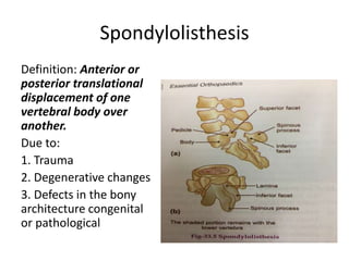
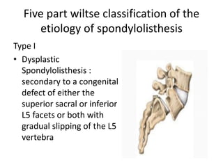
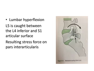
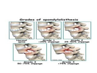
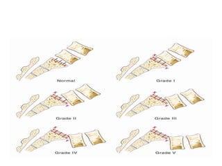
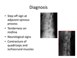
Spondylolisthesis
Dr sharan
Spondylolisthesis
Definition: Anterior or
posterior translational
displacement of one
vertebral body over
another.
Due to:
1. Trauma
2. Degenerative changes
3. Defects in the bony
architecture congenital
or pathological
Five part wiltse classification of the
etiology of spondylolisthesis
Type I
• Dysplastic
Spondylolisthesis :
secondary to a congenital
defect of either the
superior sacral or inferior
L5 facets or both with
gradual slipping of the L5
vertebra
Type II
• Isthmic or spondylolytic: the lesion is in the
isthmus or pars interarticularis,
spondylolysis :If a defect in the pars
interarticularis & no slipping.
Spondylolisthesis :If one vertebra has slipped
forward.
• Type II A: Lytic or stress
spondylolisthesis and is
most likely caused by
recurrent micro-
fractures caused by
hyperextension. It is
also called a "stress
fracture" of the pars
interarticularis and is
much more common in
males
• Type II B probably also
occurs from micro-
fractures in the pars.
However, in contrast to
Type II A, the pars
interarticularii remain
intact but stretched
out as the fracture fill
in with new bone
• Type II C is very rare
in occurrence and is
caused by an acute
fracture of the pars.
Nuclear imaging
may be needed to
establish diagnosis
Type III
• Degenerative spondylolisthesis,
and occurs as a result of the
degeneration of the lumbar facet
joints. The alteration in these
joints can allow forward or
backward vertebral displacement.
• Most often seen in older patients.
• There is no pars defect and the
vertebral slippage is never greater
than 30%
Type IV
• traumatic
spondylolisthesis, is
associated with acute
fracture of a posterior
element (pedicle,
lamina or facets)
other than the pars
interarticularis
Type V, pathologic
spondylolisthesis, occurs
because of a structural
weakness of the bone
secondary to a disease
process such as a tumor
or other bone diseases
Isthmic lysis
• The isthmic defect happens as a result of
compression caused by the lower articular
process of the superior vertebra on the
isthmic region in hyperlordosis
• The isthmus of L5 is more horizontal than
upper levels
• Lumbar hyperflexion
L5 is caught between
the L4 inferior and S1
articular surface
Resulting stress force on
pars interarticularis
• Isthemic lysis is due to
anterioposterior postural
imbalance and
secondary
spondylolisthesis is
readjacent mechanism
to adapt to postural
imbalance
• Dysplastic forms
Slippage occurs at an age when growth is not
complete
Affecting growth of involved vertebrae
Vertebral elements exposed to excessive forces
growing less than non stressed vertebrae
Traumatic Listhesis
• Traumatic listhesis is rare condition.
• Results from Acute fracture of the posterior
element other then pars interarticularis
• It is fracture dislocation of the spine involving all
three columns
• It is the shear forces which cause break in the
posterior stabilizers and the force is transmitted
at the level of Intervertebral disc resulting in
anterior or posterior displacement of the
vertebral body.
Degenrative
• Pathogenic
Degenerative destruction of posterior joints
Degenerative disc can nno longer maintain
vertebral relationship
Displacement occurs
Inferior joints of cephalad vertebrae contract and
expand due to instability of superior articular
process of inferior vertebrae
Compensatory hypertrophy
Diagnosis
• Step off sign at
adjacent spinous
process
• Tenderness on
midline
• Neurological signs
• Contracture of
quadriceps and
ischiocrural muscles
Treatment
Conservative management
• Immobilization with brace for 6 weeks
• Pain killers, anti inflammatory and injection
may limit pain but does not heal fracture
• Rehabilitation
• Sports like swimming will reduce the load on
disc
Surgical management
• Direct screw fixation
• Wire based or wire screw fixation
• Screwed hooks with compressive springe
• Hooks associated with pedicular screws
• Pedicular screws with bend rod
• Bicortical screw hooks with transverse connection
• Arthrodesis
• Isolated decompression or discectomy may
increase instability and salvage secondary
fusion
THANK YOU

spondylo listhesis

  • 1.
  • 2.
    Spondylolisthesis Definition: Anterior or posteriortranslational displacement of one vertebral body over another. Due to: 1. Trauma 2. Degenerative changes 3. Defects in the bony architecture congenital or pathological
  • 3.
    Five part wiltseclassification of the etiology of spondylolisthesis Type I • Dysplastic Spondylolisthesis : secondary to a congenital defect of either the superior sacral or inferior L5 facets or both with gradual slipping of the L5 vertebra
  • 4.
    Type II • Isthmicor spondylolytic: the lesion is in the isthmus or pars interarticularis, spondylolysis :If a defect in the pars interarticularis & no slipping. Spondylolisthesis :If one vertebra has slipped forward.
  • 5.
    • Type IIA: Lytic or stress spondylolisthesis and is most likely caused by recurrent micro- fractures caused by hyperextension. It is also called a "stress fracture" of the pars interarticularis and is much more common in males
  • 6.
    • Type IIB probably also occurs from micro- fractures in the pars. However, in contrast to Type II A, the pars interarticularii remain intact but stretched out as the fracture fill in with new bone
  • 7.
    • Type IIC is very rare in occurrence and is caused by an acute fracture of the pars. Nuclear imaging may be needed to establish diagnosis
  • 8.
    Type III • Degenerativespondylolisthesis, and occurs as a result of the degeneration of the lumbar facet joints. The alteration in these joints can allow forward or backward vertebral displacement. • Most often seen in older patients. • There is no pars defect and the vertebral slippage is never greater than 30%
  • 9.
    Type IV • traumatic spondylolisthesis,is associated with acute fracture of a posterior element (pedicle, lamina or facets) other than the pars interarticularis
  • 10.
    Type V, pathologic spondylolisthesis,occurs because of a structural weakness of the bone secondary to a disease process such as a tumor or other bone diseases
  • 11.
    Isthmic lysis • Theisthmic defect happens as a result of compression caused by the lower articular process of the superior vertebra on the isthmic region in hyperlordosis • The isthmus of L5 is more horizontal than upper levels
  • 12.
    • Lumbar hyperflexion L5is caught between the L4 inferior and S1 articular surface Resulting stress force on pars interarticularis
  • 13.
    • Isthemic lysisis due to anterioposterior postural imbalance and secondary spondylolisthesis is readjacent mechanism to adapt to postural imbalance
  • 15.
    • Dysplastic forms Slippageoccurs at an age when growth is not complete Affecting growth of involved vertebrae Vertebral elements exposed to excessive forces growing less than non stressed vertebrae
  • 17.
    Traumatic Listhesis • Traumaticlisthesis is rare condition. • Results from Acute fracture of the posterior element other then pars interarticularis • It is fracture dislocation of the spine involving all three columns • It is the shear forces which cause break in the posterior stabilizers and the force is transmitted at the level of Intervertebral disc resulting in anterior or posterior displacement of the vertebral body.
  • 18.
    Degenrative • Pathogenic Degenerative destructionof posterior joints Degenerative disc can nno longer maintain vertebral relationship Displacement occurs Inferior joints of cephalad vertebrae contract and expand due to instability of superior articular process of inferior vertebrae Compensatory hypertrophy
  • 21.
    Diagnosis • Step offsign at adjacent spinous process • Tenderness on midline • Neurological signs • Contracture of quadriceps and ischiocrural muscles
  • 22.
    Treatment Conservative management • Immobilizationwith brace for 6 weeks • Pain killers, anti inflammatory and injection may limit pain but does not heal fracture • Rehabilitation • Sports like swimming will reduce the load on disc
  • 23.
    Surgical management • Directscrew fixation • Wire based or wire screw fixation • Screwed hooks with compressive springe • Hooks associated with pedicular screws • Pedicular screws with bend rod • Bicortical screw hooks with transverse connection • Arthrodesis
  • 24.
    • Isolated decompressionor discectomy may increase instability and salvage secondary fusion
  • 25.