College Level Design:Tim Cannon
http://academic.scranton.edu/department/psych/sheep/newsheep/ welcome2.html
Edited by Dr. S.C. Wache
2.
Goal:
We will comparesheep brain structures with human brain structures.
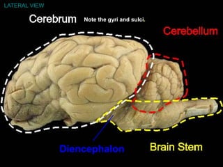
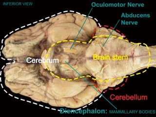
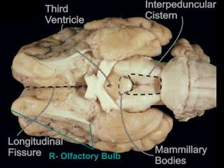
Compared to human cerebrum a human’s olfactory bulb is much shorter than
sheep olfactory bulbs compared to the sheep cerebrum. Human cerebellum is
bilobed, sheep cerebellum is not.
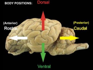
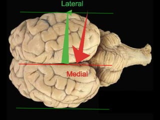
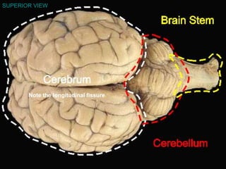
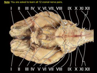
First, we will learn body positions applied to the whole sheep brain.
We will do two sections: one group will do a mid-sagittal cut, the other a
coronal cut.
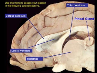
Use the mid-sagittal section to find your location through each consecutive
coronal section.
3.
Methods and Materials:
Wewill be dissecting a preserved adult sheep brain.
Our dissection kit contains a scalpel, a fine tipped pair of scissors, a blunt
metal probe, a fine tipped forceps, a blunt-tipped forceps.
It is vital to wear vinyl gloves. It is important to be careful when working with
preservatives. If necessary protect your eyes. Follow OSHA (office of safety
and Health administration) regulations.
Dispose of the dissected specimen as indicated on the MSDA (material safety
and data administration) sheets.
These are some additional web sites where you can obtain more information:
http://labs.ansci.uiuc.edu/rwjohnson/class/braintext.html
University of Scranton, Dissection of the Sheep Brain
University of Scranton, The Sheep Brain Dissection Guide
Michigan State University, Atlas of the Sheep Brain
Interactive Atlases, Digital Anatomist Project
University of Wisconsin, Global Anatomy
University of Utah, Anatomy-Histology Tutorials
Gray Cancer Institute On-line Medical Dictionary
4.
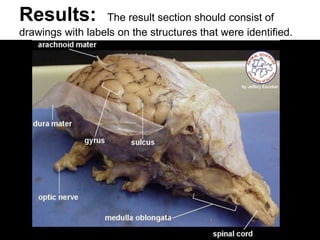
Results: The resultsection should consist of
drawings with labels on the structures that were identified.
5.
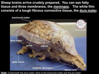
Sheep brains arrivecrudely prepared. You can see fatty
tissue and three membranes, the meninges. The white film
consists of a tough fibrous connective tissue, the dura mater.
Note: Underneath the dura mater, there is the arachnoid mater and the pia mater.
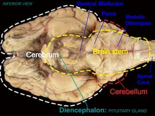
spinal cord
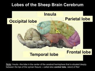
Temporal lobe
Insula
Note: Insula- the lobe in the center of the cerebral hemisphere that is situated deeply
between the lips of the sylvian fissure -- called also central lobe, island of Reil
Frontal lobe
Parietal lobe
Lobes of the Sheep Brain Cerebrum
Occipital lobe
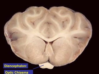
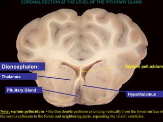
Pituitary Gland
Thalamus
Hypothalamus
CORONAL SECTIONAT THE LEVEL OF THE PITUITARY GLAND
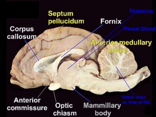
Note: septum pellucidum - the thin double partition extending vertically from the lower surface of
the corpus callosum to the fornix and neighboring parts, separating the lateral ventricles.
Septum pellucidum
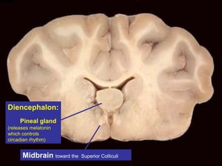
Diencephalon:
23.
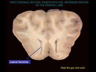
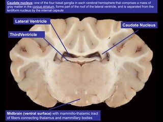
Lateral Ventricle
ThirdVentricle
Midbrain (ventralsurface) with mammillo-thalamic tract
of fibers connecting thalamus and mammillary bodies
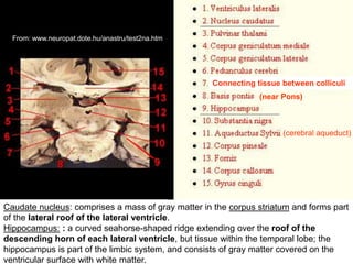
Caudate Nucleus
Caudate nucleus: one of the four basal ganglia in each cerebral hemisphere that comprises a mass of
gray matter in the corpus striatum, forms part of the roof of the lateral ventricle, and is separated from the
lentiform nucleus by the internal capsule
Caudate nucleus: comprisesa mass of gray matter in the corpus striatum and forms part
of the lateral roof of the lateral ventricle.
Hippocampus: : a curved seahorse-shaped ridge extending over the roof of the
descending horn of each lateral ventricle, but tissue within the temporal lobe; the
hippocampus is part of the limbic system, and consists of gray matter covered on the
ventricular surface with white matter.
From: www.neuropat.dote.hu/anastru/test2na.htm
(near Pons)
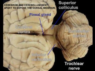
Connecting tissue between colliculi
(cerebral aqueduct)
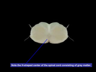
Note the H-shapedcenter of the spinal cord consisting of gray matter.
28.
Summary:
We have studiedthe structures on a preserved sheep brain specimen. We
have learned the functions of such structures using the lecture handout and
comparing to what is known about the human brain function.
We learned body positions applied to the whole sheep brain.
The mid-sagittal cut was helpful in identifying the extension of the lateral
ventricle to the third ventricle. The coronal cuts made it possible to find the
cerebral aqueduct (aqueductus sylvii) that joins the third and fourth ventricles
which house the CSF.
The coronal cut helped in the location of the hippocampus along the roof of the
descending lateral ventricle.