Downloaded 1,202 times


![ Table -1 showing the normal width of attached gingiva
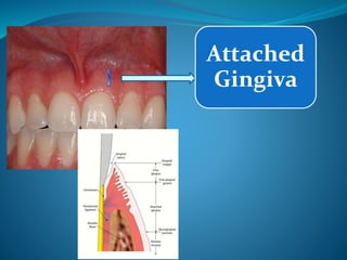
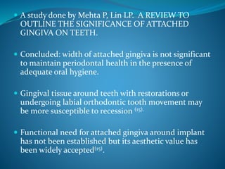
Some Studies have also shown that the width of
attached gingiva is not significant to maintain
periodontal health in the presence of adequate oral
hygiene. [8]
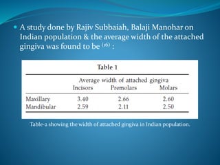
Anterior Premolars
Maxillary 3.5-4.5mm 1.9mm
Mandibular 3.3-3.9mm 1.8mm](https://image.slidesharecdn.com/seminar1-140611235032-phpapp02/85/Gingiva-Macroscopic-Features-28-320.jpg)

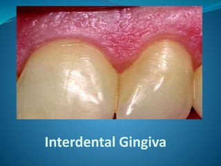
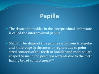


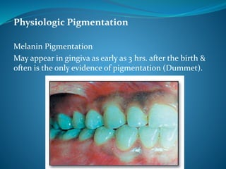


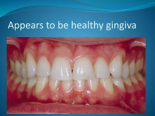


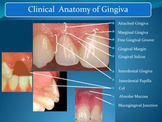
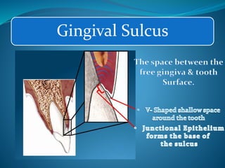
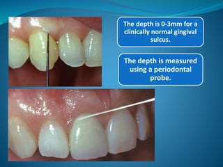
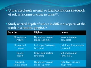
This document summarizes the macroscopic structures of healthy gingiva. It describes the key parts of gingiva including the marginal gingiva, gingival sulcus, attached gingiva, interdental gingiva, and mucogingival junction. It discusses the anatomy, functions, measurement, and clinical features of these structures. Important findings are that the width of attached gingiva varies but is typically 2mm or more to maintain periodontal health, and that the color, shape, size, contours, consistency and texture of healthy gingiva are described.


