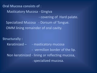
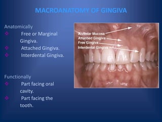
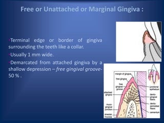
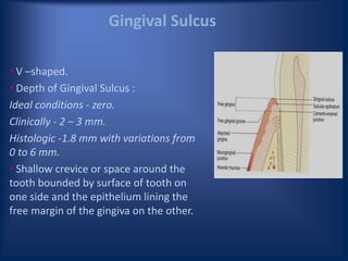
This document discusses the anatomy of the gingiva. It describes the macroanatomy, including the marginal, attached, and interdental gingiva. It then covers the microanatomy, focusing on the epithelial layers like the sulcular and junctional epithelium and the connective tissue below. Throughout, it correlates the microscopic features to normal clinical presentations of things like color, shape, and consistency.


![PERI-PROSTHETIC FRACTURE NAIL-PLATE CONSTRUCT [NPC].pptx](https://cdn.slidesharecdn.com/ss_thumbnails/drarunkumardrmohamedashrafperiprostheticfrasturenail-plateconstructnpc-260209164459-7e9d15a1-thumbnail.jpg?width=640&height=640&fit=bounds)


![ONFH[AVN HIP] -TRIPLE REGIME -A NOVAL SURGICAL CONCEPT .pptx](https://cdn.slidesharecdn.com/ss_thumbnails/onfhavnhip2026koaconcalicutdrgokuldevdrmashraf-260210064517-213ec005-thumbnail.jpg?width=640&height=640&fit=bounds)


