Night 7k to 12k Navi Mumbai Call Girl Photo 👉 BOOK NOW 9833363713 👈 ♀️ night ...
Structural anatomy and development of periodontium
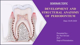
1. Presented by;-
Dr. Peri Srivani
PGT
Dept of Periodontics
DEVELOPMENT AND
STRUCTURAL ANATOMY
OF PERIODONTIUM
SEMINAR TOPIC
Date-18.09.2020
2. CONTENTS
• Introduction & Definition
• Development of Face and Oral cavity
• Development of Periodontium
• Gingiva
• Periodontal Ligament
• Cementum
• Alveolar bone
• Conclusion
• References
3. The widespread occurrence of periodontal diseases & the
realization that periodontal tissues lost to the disease can be
repaired has resulted in considerable effort to understand the
factors and cells regulating the formation, maintenance &
regeneration of the periodontium.
-Ten Cate et al Periodontology 2000 Vol 13
4. INTRODUCTION
• Greek word “peri” means “around” & “odontos” means “tooth”
• Dynamic structure composed of tissues supporting & investing the teeth.
• Consists of 4 components broadly classified into two categories.
PERIODONTIUM
Supporting
tissue
Investing tissue
Alveolar bone
Cementum
Periodontal
ligament
Gingiva
HARD TISSUE
COMPONENTS
SOFT TISSUE
COMPONENTS
PERIODONTIUM
Supporting
tissue
Investing tissue
Alveolar bone
Cementum
Periodontal
ligament
Gingiva
9. PARTS OF THE TOOTH GERM
• Enamel Organ --- Ectodermal component
• Dental Papilla Ectomesenchymal component
• Dental Sac / Follicle ( derived from Neural Crest cells)
Enamel organ Dental Papilla Dental Follicle
Enamel Dentin
Pulp
Periodontal ligament
Cementum
Alveolar bone
10. PERIODONTIUM- A Developmental and Functional unit
Periodontium is a total collective term coined to designate the totality of
tissues which anchor the teeth to the bone of the jaws, provide interdental
linkage to a row of teeth and seal the oral mucosal openings created by the
erupting teeth.
Hence root cementum, alveolar bone, periodontal ligament and gingiva
constitute and behave as a developmental, biological and functional unit.
-SCHROEDER
11. WESPI (1921/22) claimed that the Periodontium was an organic and physiological
unit because of the following reasons:-
1. After the teeth are lost, the alveolar bone is resorbed partially of completely.
2. During periodontitis, destruction is limited to tissues especially the bone which
is next to the roots of the teeth.
3. The PDL and alveolar process arise from dental follicle.
4. These tissues including the epithelial rests of Malassez follow any physiological,
pre & post eruptive movement of the tooth.
12. Experiments relevant to the existence of a periodontal unit
Landsberger 1921/23/25
Dental follicle important
for alveolar process
development
Tomes
1904,Mummery1924,
Orban1927,1928
,Scott1948, Tonge1963
Dental follicle attaches the
tooth germ to oral mucosa
Ten Cate 1969
Investing layer & Peri-
follicular mesenchyme of
dental follicle.
Ten Cate et al 1971,
Freeman1975
Root cementum & PDL are
formed by cells derived from
the dental follicle proper
Lefkowitz & Mardfin 1954
Structural separation of the
dental follicle from the peri-
follicular mesenchyme&
future connective tissue
Hoffman 1967
PDL like tissue forming
around the tooth germ
isografts very much
similar in width &
structure to PDL
developing in-situ
15. GINGIVA
• Term “gingiva” originated from
the Latin word “ gigno” meaning
“to give birth”.
• “gingevere” meaning “gums”
• Masticatory mucosa.
• Covers the alveolar processes of
the jaws and surround the neck
of the teeth.
16. DEFINITIONS
The part of the oral mucosa that covers the alveolar process of jaws and surrounds the
neck of teeth.
-CARRAANZA
Part of masticatory mucosa covering the alveolar processes of the cervical portions of
teeth.
-LINDHE
The fibrous investing layer covered by keratinised epithelium that immediately
surrounds a tooth and is contiguous with its periodontal ligament and the mucosal
tissues of the mouth.
-AAP-Glossary of Periodontics
It is a combination of epithelial and connective tissue & is defined as that portion of oral
mucous membrane which in complete post eruptive dentition of a healthy young
individual surrounds and is attached to the teeth and alveolar process.
-SCHROEDER
18. HYPOTHESIS TO EXPLAIN MODE OF EPITHELIAL
ATTACHMENT TO TOOTH SURFACE
GOTTLIEB
• Gingiva is
organically
united to tooth
surface by
epithelial
attachment.
WARHAUG
1952
• Concept of
epithelial cuff,
gingival tissues
are closely
adapted but not
organically
united.
STERN 1962
• Supported by
SCHROEDER &
LISGARTEN,
hemidesmosom
es.
19. MARGINAL
GINGIVA
• Collar like fashion
• 1mm wide
• Free Gingival groove-
30 to 40% ; 1.5 to
2mm coronal to CEJ
• Gingival Zenith
ATTACHED
GINGIVA
• Firm , resilient , tightly
bound to periosteum
• Stippling, Keratinised
• Between Marginal
gingiva and
Alveolar/Palatal
mucosa
INTERDENTAL
PAPILLA
• Occupies gingival
embrasures
• Laterally and tip-
marginal gingiva
• Centrally-Attached
gingiva
• Shape of Interdental
papilla
ANATOMICAL PARTS OF GINGIVA
20.
21. WIDTH OF ATTACHED GINGIVA
Labial region Maximum width Minimum width
Maxillary region Incisor region
3.5 to 4.5 mm
Premolar region
1.9 mm
Mandibular region Incisor region
3.3 to 3.9 mm
Premolar region
1.8 mm
23. TYPES OF GINGIVAL EPITHELIUM
Outer or Oral
Epithelium
Sulcular
Epithelium
Junctional
Epithelium
24. ORAL EPITHELIUM
• Covers the crest and outer
surface of the marginal
gingiva and the surface of
the attached gingiva.
• Orthokeratinised or
Parakeratinised or
combination.
• 4 layers
• Average- 0.2 to 0.3 mm in
thickness
25. SULCULAR EPITHELIUM
• Thin, non keratinised stratified
squamous epithelium lining the
gingival sulcus.
• Coronal limit of JE to crest of
gingival margin.
• Has the potential to keratinise
when exposed to oral cavity.
• Semi-permeable membrane.
26. JUNCTIONAL EPITHELIUM
• Collar-like band of stratified
squamous non keratinised epithelium.
• Early life 3 to 4 layers
• Increase in age 10 to 20 layers.
• Tapers from coronal end to apical
termination, located at CEJ in healthy
tissue.
• 0.25 to 1.35 mm
• 3 zones Apical-Germination
Middle-Adhesion
Coronal-Permeability
27. FUNCTIONS OF JUNCTIONAL EPITHELIUM
Attachment to tooth
Epithelial barrier
against plaque
bacteria
Rapid turnover of
cells
Repair
Endocytic capacity
IL-1, 6,8 ,TNF
Anti-microbial
Substances
Movement of GCF
31. • In health- Regular, repetitive, and layered pattern.
• In inflammation- Irregular vascular plexus, looped, dilated, convoluted.
• Branches of Anterior, Middle and Posterior Superior alveolar arteries, Nasopalatine
artery & Greater Palatine artery supply the facial gingiva and palatal mucosa of the
maxillary arch.
• Branches of Inferior Alveolar artery, Lingual Artery supply the facial and lingual
gingiva of the mandibular arch respectively.
• Venous supply accompany the arterial supply.
32. LYMPHATIC DRAINAGE OF GINGIVA
Lymphatic drainage from
connective tissue papilla
Collecting network outside
the periosteum
Regional lymph nodes
especially Submaxillary
lymph nodes
Lymphatics beneath the
junctional epithelium
Periodontal ligament
Accompany blood
vessels
REGION DRAINING INTO
Maxillary Anterior & Posterior Buccal gingiva Submandibular lymph nodes
Mandibular Posterior buccal and lingual gingiva Submandibular lymph nodes
Mandibular Anterior gingiva Submental lymph nodes
Third molar region Jugulodigastric lymph nodes
33. NERVE SUPPLY OF GINGIVA
MAXILLARY REGION
Trigeminal nerve
Maxillary Nerve.
Facial and Buccal gingiva --
branches of Anterior, Middle
& Posterior Superior Alveolar
Nerve.
Palatally -- branches of
Nasopalatine nerve (anterior
to canine) & Greater Palatine
Nerve (posterior to Canines).
34. MANDIBULAR
REGION
Trigeminal Nerve
Mandibular Nerve.
Facial gingiva till second
premolars --branches of
Inferior Alveolar Nerve.
Buccal gingiva of molar
region-- branches of Buccal
Nerve.
Lingual gingiva --
branches of Lingual Nerve.
36. PERIODONTAL LIGAMENT
Composed of a complex vascular and highly
cellular connective tissue that surround the tooth
root & connects it to the inner wall of the alveolar
bone.
OTHER NAMES:-
• Desmodont
• Gomphosis
• Pericementum
• Dental periosteum
• Alveodental ligament
• Periodontal Membrane
37. Soft richly vascular and cellular connective tissue which surrounds the roots of
the teeth and joins the root cementum with the socket wall.
- LINDHE
The periodontal ligament occupies the space which is located between the
cementum and the periodontal surface of the alveolar bone and extends
coronally to the most apical part of the lamina propria of the gingiva.
-ORBAN
It is the soft specialised connective tissue situated between the cementum
covering the root of the tooth and bone forming the socket wall.
-TEN CATE A.R 1971
DEFINITIONS
38. • Hour-glass shaped.
• Width of PDL – 0.15 to 0.38 mm.
Age (years) Width of PDL
(mm)
11-16 0.21
32-52 0.18
51-67 0.15
Functional status
of teeth
Width of PDL
(mm)
At time of eruption 0.1-0.5
At function 0.2-0.35
Hypofunction 0.1-0.15
ACC TO TEN CATE
THICKNESS OF PDL
39. DEVELOPMENT OF PERIODONTAL LIGAMENT
HERS cells initiating root development
Dental follicle cells
Perifollicular mesenchyme
Increase in cell activity
Type-1 collagen; Stem cells giving rise to other
cells
40. Starts prior to root
development.
Dentogingival
fibres formed first
Alveolar crest
fibres
Oblique fibres
Horizontal fibres
Apical fibres
Final PDL
architecture
DEVELOPMENT OF PRINCIPAL FIBRES OF PDL
41. TYPES OF PRINCIPAL PDL FIBERS
Holmstrup
1996
Alveolar
crestal
group
Horizontal
group
Oblique
group
Apical
group
Transseptal
group
Inter-
radicular group
42.
43. FIBER GROUP FUNCTION
Alveolar crest Resist lateral movements
Resist tilting, Intrusion, Extrusion
Horizontal Resist horizontal and tipping movts
Oblique Resist vertical movts & intrusion
Apical Resist luxation , tipping
Transseptal Interconnection between adjacent tooth and gingiva
Inter-radicular Resist tooth tipping, torqueing, luxation
44. PERIODONTAL LIGAMENT SPACE
• Volume- 30 to 100 mm3 in single-rooted teeth
60 to 150 mm3 in multi-rooted teeth
• 1 to 2.5% blood vessels
4 to 6.5% interstitial tissue
• 1 mm3 of cervical root surface area= 28000
principal collagen fibres.
• Sharpey’s fibres,
• Collagen.
45. • Density
&Diameter
variation
• Width of
PDL in
relation to
age
&function
• Changes
in PDL
with age
• Normal
chewing
thrust
0.412secs
& effect on
PDL Picton
(1964)
Gotze
(1965)
Aiyoshi
& Inoue
(1963)
Klein 1928
Kornfield
1931
Coolidge
1937
STUDIES ON PERIODONTAL LIGAMENT
47. Elastin Tissue stretching & compression
Fibronectin Cellular Adhesion
Laminin Attachment to Type IV collagen
Osteocalcin/Bone Gla protein Inhibits H/A precipitation
Bone Sialoprotein (BSP) 2 Attachment factor for bone cells
BSP-1 / Osteopontin Facilitates mineralisation
Tenascin Epi-Mesenchymal interaction
Osteonectin / SPARC Mineralization, Anti-adhesive
PROTEIN FUNCTION
NON-COLLAGENOUS PROTEINS OF PDL
48. FIBERS OF PDL
• Collagen Type I,III,V,XII predominant.
• All principal fibers.
COLLAGEN
• Microfibrils in elastin.
ELAUNIN
• Type III
• Basement membrane of blood vessels and epithelial cells lying on
PDL.
RETICULAR
• SHACKLE FORD 1971
• SLOAN- optical effect of arrangement of collagen
INDIFFEREN
T FIBER
PLEXUS
• Microfibrils connecting cementum with peripheral blood vessels.
• 3% of all fibers , acid-resistant
OXYTALAN
49. PERIODONTAL LIGAMENT HOMEOSTASIS
• Secretion of certain molecules & factors.
• Regulation of mineralisation.
• Width of PDL can increase upto 50% during function.
FACTORS FUNCTION
MSX-2 Prevents osteogenic differentiation
Bone sialoprotein & Osteopontin Mineralisation Balance
Matrix ‘Gla’ protein Inhibit mineralisation
RGD-cementum attachment protein PDL width maintained
TGF-beta isoforms Inhibit Osteoblasts
Prostaglandins Prevent mineralisation
51. • Crevicular capillary loops – 6 to 30 µm
• Basket-like network , more towards alveolar bone.
• Superior and Inferior Alveolar Arteries
• Molars > Incisors & Maxilla> Mandible
• Single rooted teeth- gingival >middle>apical.
• Multi rooted teeth- Apical third=middle third.
• Mesial and Distal surfaces > Facial and Lingual surfaces.
• 0.5 µm labio-palatal pulsation of teeth with each heart beat. FROHLICH (1964)
VENOUS DRAINAGE
Shunts called glomeruli , 28µm average diameter.
52. Lymphatic channel
network of PDL
Intraosseous pathway
through socket or
cribriform plate
Submaxillary &
Submandibular
Lymph nodes
LYMPHATIC DRAINAGE OF PDL
53. NERVE SUPPLY OF PERIODONTAL LIGAMENT
• Maxilla --- Anterior , Middle & Posterior Superior Alveolar Nerve
• Mandible – Inferior Alveolar Nerve
• Functionally 2 types of nerve fibres Sensory and Autonomic fibres.
Sensory
fibres
Nociception, Mechanoception,
Touch , Pressure, Pain,
Proprioception
Autonomic
fibres
Associated with PDL vessels
54. TENCATE–
LIGAMENT
INNERVATION
General Anatomic Configuration
Regional variation in termination of
neural elements
Types of Neural termination
BYERS
1985
Free nerve endings –Pain
sensation
Ruffini-like-mechanoceptors
in apical area
Coiled Meissner corpuscles-
Tactile- Midroot area
Krause end type bulbs-
Temperature, Pressure
&vibration
57. CEMENTUM
HISTORY AND LITERATURE:-
• Latin word- CEMENT means CAEMENTUM or Quarry Stone.
• Mineralised component of tooth as well as periodontium covering the entire surface of the
anatomic roots of the tooth.
• First examined by – Frankel (1835), Raschkow (1835), Students of Jan Evangelista
Purkinje and Anders Adolf Retzius (1837).
• Comparative anatomy studies by Richard Owen and others over the latter half of 19th
century.
• Anatomical studies of the periodontium performed by G.V.Black and others in the late
19th and early 20th centuries.
58. DEFINITIONS
Cementum is the calcified, avascular mesenchymal tissue that
forms the outer covering of the anatomic root.
-CARRANZA
Cementum is a mineralised connective tissue in part not unlike
bone, that covers the entire surface of anatomic roots of tooth.
-SCHROEDER
Cementum is a hard, avascular connective tissue that covers the
roots of the teeth.
-TEN CATE
59. PHYSICAL CHARACTERISTICS
• Colour - light yellow
• Distinguished from enamel by lack of lustre and darker hue, but lighter than dentin.
• Clinically not possible to distinguish cementum from dentin based on hue.
• Permeable to various molecules such as dyes.
• Permeability increases with age.
• Canaliculi of cellular cementum contiguous with dentinal tubules in some areas.
60. THICKNESS OF CEMENTUM
• Cemental deposition is directly related to ageing of teeth rather than the result of
masticatory function. –KELLNER(1931) AND KRONFIELD(1927)
• Thickness depends on tooth shape, eruptional, functional history and age.-
SCHROEDER
• Rapid deposition in apical area to compensate for occlusal attrition.
• Distal surfaces > mesial surfaces.
• Midroot level > Apical area > Cervical area.
• The thickness increases 3 times (200-215µm) between 11-70 years
• Intermediate layer of cellular cementum contributes to most of cemental thickness
and is mostly unrelated to actual Sharpey’s fibres attachment.
61. LOCATION THICKNESS
Coronal half of root surface 16-60 µm
Apical third & furcation area 150-200 µm
TOOTH SITE SPECIFIC
MAXIMUM
Maxillary incisors and canines Labially Apically
Palatally Cervically
Mandibular incisors and canine Labially Cervically
Lingually Apically
• Concave root aspects facing the furcations of maxillary and
mandibular 1st molars show thicker cementum than adjacent root
aspects. – BOWER (1979)
62. CEMENTOGENESIS
Pre-functional
developmental stage-
Primary cementum
Formed during the root development,
before eruption of tooth into the oral
cavity
Primary distribution of cementum
varieties is determined for each root.
•3.5 to 7.5 years
Functional development
stage- Secondary cementum
Starts when the tooth is about to
reach the occlusal level & associated
with the attachment of the root to the
surrounding bone and continues
throughout life
Alterations in the distribution and
appearance of cementum varieties
on the root surface with time due to
biologic responsiveness of
cementum
63. Outer and inner
enamel epithelium
join to form the
HERS
Secretes enamel
proteins or certain
epithelial products
Induces the
ectomesenchymal
pulp cells to
differentiate into
ODONTOBLASTS
Secretes a layer of
PREDENTIN
Ectomesenchymal cells
from the inner portion of
dental follicle come in
contact with predentin
Reciprocal inductive
signal from predentin
or surrounding
HERS
Differentiated into
CEMENTOBLASTS
and CEMENTUM is
secreted
Some cells from the
fragmented root
sheath form
Epithelial Cell Rests
of Malassez
Some cells remain
attached to the
forming root surface
resulting in Enamel
Pearl.
64. CLASSIFICATION OF CEMENTUM
Location on Teeth
Cellularity
Presence/Absence of collagen fibers
Location, Structure ,Function, Rate of formation ,
Function, Biochemical composition & degree of
mineralization
66. TYPE LOCATION THICKNESS FUNCTION
Acellular Afibrillar
cementum
Spurs and patches
over enamel and
dentin
1-15 µm No known
function
Acellular extrinsic
fiber cementum
Cervical margin to
Apical 1/3rd
30 to 230µm Anchorage
Cellular Intrinsic
fiber cementum
Middle to Apical
1/3rd
Furcation
Fills resorption
lacunae
Adaptation
Repair
Cellular Mixed
Stratified
Cementum
Apical portion
Furcation
100-1000µm Adaptation
Intermediate
Cementum
Apical half of
roots of premolar
& molar
Poorly defined
hyaline zone
Seals
root dentin
67. BIOCHEMICAL COMPOSITION
INORGANIC
45 -50%
ORGANIC
50-55%
Non-collagenous
proteins
Collagen
• Type-I = 90%
• Type-III, V,
VI, XII, XIV
• Bone sialoprotein
• Osteopontin
• Osteonectin
• Osteocalcin
• Cementum Adhesion
protein
• Fibronectin &Tenascin
• Proteoglycans
• Alkaline phosphatase
• Less
mineralized
than dentin
• Calcium
hydroxyapatite
• Trace elements
• Fluoride
• 0.5 -0.9% Mg
• 0.1-0.3% S
68. CEMENTOENAMEL JUNCTION
The relation between Cementum & Enamel at the Cervical region of teeth
Cementum
overlapping the
enamel
60-65%
Connective tissue
comes in direct
contact with enamel
epithelium
End to End
relation
30%
Sharp line
Space between
Enamel &
Cementum
5-10%
Delayed separation
of Enamel
epithelium from
dentin
69. CEMENTODENTINAL JUNCTION
• Dentin surface upon which the cementum
is deposited.
• Firm attachment.
• Wide zone of 2-3 µm
• Collagen associated with GAGs like
Chondroitin sulphate & Dermatan
sulphate resulting in increased water
content which contributes to the stiffness.
• Occlusal load distribution
71. FUNCTIONS OF CEMENTUM
ANCHORAGE
Medium for
attachment of
collagen fibers &
Sharpey’s fibers
REPAIR
Repair resorptions
& fractures.
Cellular cementum
ADAPTATION
Apical deposition
of cementum
Width of PDL
maintained
73. ALVEOLAR BONE
• Bone is a dynamic structure.
• Adapting to physiologic changes.
ALVEOLAR BONE It is defined as that part of
maxilla and mandible that forms and supports the
sockets of the teeth.
-LINDHE , ORBAN
• Radiographically, seen as radiopaque line around
root.
74. DEVELOPMENT OF ALVEOLAR BONE
Dental follicle
Groove into oral
cavity
Tooth
development
Ectomesenchymal
cells
Osteoid
deposition
Tooth crypt
Deposition &
Resorption
Incorporated into
maxilla &
mandible
Associated with
tooth
75. CLASSIFICATION OF ALVEOLAR BONE
Acc to functional adaptaion
Linlow’s classification 1970
Lekholm & Zarb classification 1985
Misch’s classification based on Bone
density
76. Based on Functional adaptation
Alveolar bone
proper
Supporting
alveolar bone
Cancellous
Bone
Cortical Plates
• Inner socket wall of
thin compact bone
• Lamina dura in
radiographs
• Cribriform plate-
openings .
• Bone between these two
cortical plates
• Spongy bone
• Heavy Trabeculae & bone
marrow space
• Type-I & Type-II
• Outer and inner compact
cortical plates
• Maxilla Thin ; Mandible
thick
• Fuse in Anterior region
77.
78. LINLOW-1970
CLASS-I CLASS-II CLASS-III
• Evenly spaced
trabeculae
• Small cancellated
spaces
• Slightly larger than
cancellated spaces
• Less uniformity of
osseous pattern
• Large marrow
filled spaces
between bone
trabeculae
79. LEKHOLM & ZARB - 1985
QUALITY-1 QUALITY-2 QUALITY-3 QUALITY-4
• Homogenous
compact bone
• Outer Thick
compact bone
• Core of dense
trabecular
bone
• Outer Thin
cortical bone
• Core of dense
trabecular
bone (
favourable
strength )
• Outer Thin
cortical bone
• Core of low
density
trabecular
bone
80. MISCH BONE DENSITY
D1 D2 D3 D4
Dense cortical bone Thick dense to
porous cortical
bone surrounding
coarse trabecular
bone
Thin porous cortical
bone surrounding
fine trabecular bone
Fine trabecular
bone
Anterior mandible
Posterior Mandible
Ant mandible
Posterior mandible
Anterior maxilla
Anterior maxilla
Posterior maxilla
Posterior maxilla
81. GROSS MORPHOLOGY OF ALVEOLAR BONE
Maxilla or
Mandible
Alveolar
process
Basal bone
Interradicular
septa
Interdental
septa
Trabecular
bone
Outer & Inner
cortical plates
Alveolar
bone proper
Lamellar
bone
Bundle bone
83. COMPOSITION OF BONE
Cellular Extracellular
Non-collagenous
proteins
Collagen
Osteoclastic
cells
Osteogenic
cells
Inorganic Organic
• Osteoblasts
• Osteocytes
• Osteoproge
nitor cells
• Bone-
lining Cells
• Osteocalcin
• Osteopontin
• BSP
• Osteonectin
• Proteoglycans
• TRAMP
Type 1,3,5,12
Calcium
Hydroxyapatite
Mg , Na , F , K
Osteoclasts
84. BONE REMODELING
• Simultaneous process of resorption &
deposition of bone resulting in net
balance between the two processes.
( Coupling )
MEDIATORS:-
Parathyroid hormone , Calcitonin
Vit D metabolism
Cytokines , IL-1,IL-6 , TNF α & β
INF-γ
RANKL & OPG
Prostaglandins, Aspirin
Estrogen
Bisphosphonates, statins
85. VASCULAR AND NERVE SUPPLY
Maxilla Mandible
Blood supply Anterior, Middle,
Posterior Superior
Alveolar Arteries
Inferior alveolar
Artery
Nerve supply Anterior, Middle,
Posterior Superior
Alveolar Nerves
Inferior Alveolar
Nerve
86. CONCLUSION
• Both the soft tissue & hard tissue components function together harmoniously to
maintain the structural & functional integrity of the periodontium.
• Reciprocal Induction between the Oral ectoderm and mesenchymal cells derived from
the neural crest cells form the major pathway for the development of periodontium.
• Various histochemical molecules favor the differentiation of the progenitor cells in the
dental follicle resulting in the development of periodontium.
• Various studies proved that the developed periodontium has the potential to repair or
regenerate itself in the form of Epithelial cell Rests of Malassez, progenitor cells &
stem cells which can be induced to differentiate into the cells of the periodontium.
87. REFERENCES
• Newmann & Carranza’s Clinical periodontology, 13th edition.
• The Periodontium by Hubert E. Schroeder, 1st edition.
• Orban’s Oral Histology and Embryology, 14th edition.
• Ten Cate’s Oral Histology, 9th edition.
• Wheeler’s Dental Anatomy, 11th edition.
• Periodontology 2000 , Volume 13
• Color Atlas of Dental medicine Periodontology by Herbert E.Wolf ,
Edith M & Klaus H. Rateitschak , Thomas M. Hassell ; 3rd revised &
expanded edition.