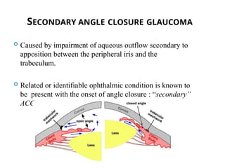
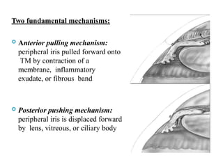
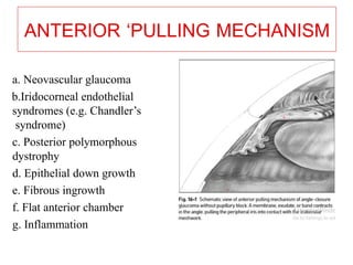
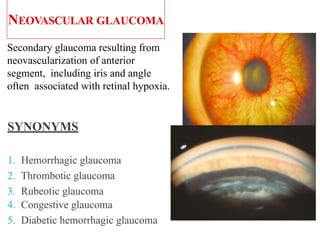
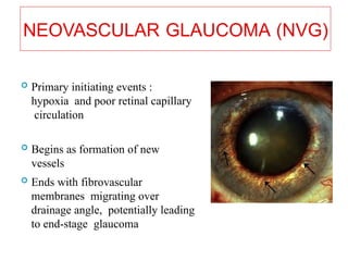
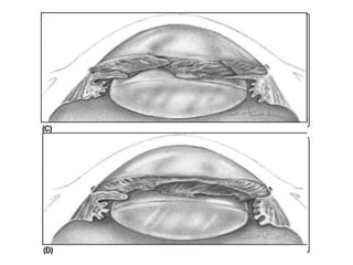
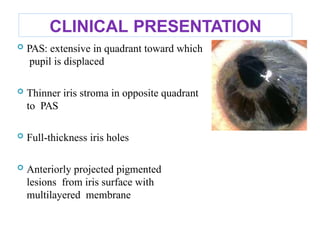
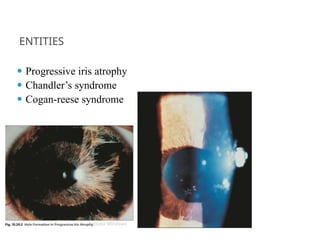
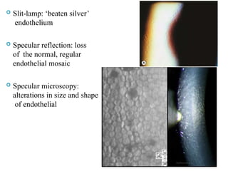
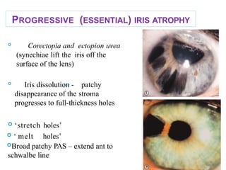
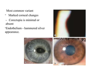
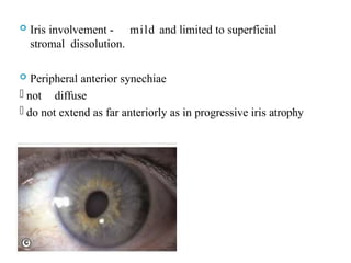
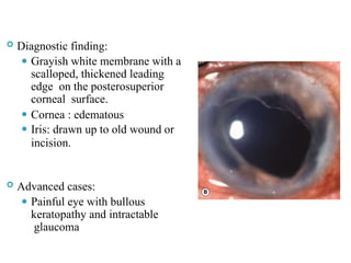
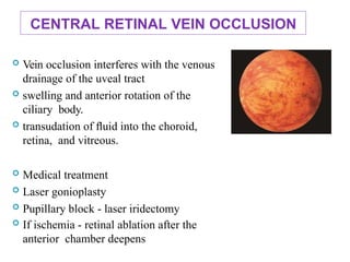
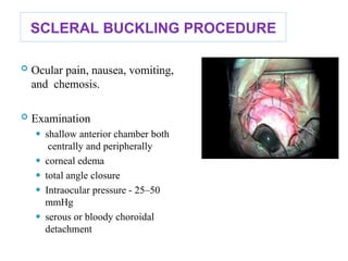
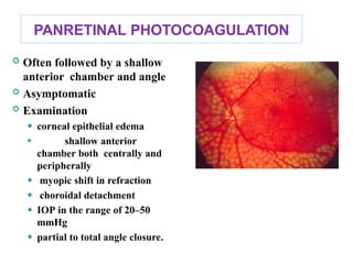
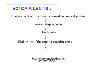
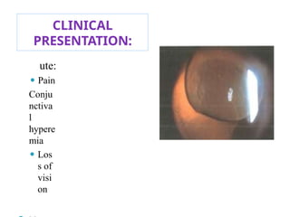
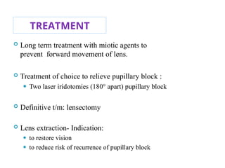
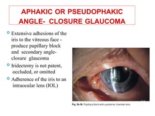
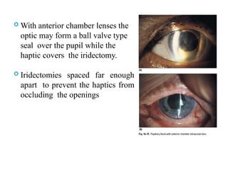
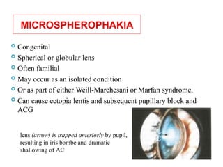
SECONDARY ANGLE CLOSURE
GLAUCOMA
PRESENTED BY,
DR. RAHUL GUPTA
1ST
YR. RESIDENT
DEPT. OF oPHTHALMOLOGY
7TH
JAN, 2024
CONTENTS
2
 Definition
 Mechanism
⚫ Anterior pulling mechanism
⚫ Posterior pulling mechanism
 Diagnosis
 Treatment
SECONDARY ANGLE CLOSURE GLAUCOMA
 Caused by impairment of aqueous outflow secondary to
apposition between the peripheral iris and the
trabeculum.
 Related or identifiable ophthalmic condition is known to
be present with the onset of angle closure : “secondary”
ACG
3
Two fundamental mechanisms:
 Anterior pulling mechanism:
peripheral iris pulled forward onto
TM by contraction of a
membrane, inflammatory
exudate, or fibrous band
 Posterior pushing mechanism:
peripheral iris is displaced forward
by lens, vitreous, or ciliary body
CLASSIFICATION
1. Anterior pulling mechanism:
Contraction of an inflammatory, hemorrhagic, or vascular
membrane, band, or exudate in the angle
PAS
Forward displacement of lens-iris interface
Often accompanied by swelling and anterior rotation of ciliary body
5
ANTERIOR ‘PULLING MECHANISM
a. Neovascular glaucoma
b.Iridocorneal endothelial
syndromes (e.g. Chandler’s
syndrome)
c. Posterior polymorphous
dystrophy
d. Epithelial down growth
e. Fibrous ingrowth
f. Flat anterior chamber
g. Inflammation
6
NEOVASCULAR GLAUCOMA
Secondary glaucoma resulting from
neovascularization of anterior
segment, including iris and angle
often associated with retinal hypoxia.
SYNONYMS
1. Hemorrhagic glaucoma
2. Thrombotic glaucoma
3. Rubeotic glaucoma
4. Congestive glaucoma
5. Diabetic hemorrhagic glaucoma
9
NEOVASCULAR GLAUCOMA (NVG)
 Primary initiating events :
hypoxia and poor retinal capillary
circulation
 Begins as formation of new
vessels
 Ends with fibrovascular
membranes migrating over
drainage angle, potentially leading
to end-stage glaucoma 10
HISTORY
1906  Coates , NVI in CRVO termed as RUBEOSIS IRIDIS
1937  Kurtz , NVA leading to PAS formation
1963  Weiss et al, coined the term NEOVASCULAR GLAUCOMA
ETIOLOGY
Diabetic retinopathy (M.C.C)
CRVO/CRAO
Ocular ischemic disease
Carotid artery occlusive disease
• 36% cases : arise from CRVO
• 32% : proliferative
diabetic retinopathy
• 13% : carotid artery
occlusive disease 13
PATHOGENESIS
 Theory of angiogenesis factor:
⚫ Michaelson 1948
Postulated existence of “X factor”, a vasoformative factor
which controlled development of new vessels
⚫ Folkman et al
Isolated a soluble substance from a solid neoplasm
capable of producing neovascularisaton and popularised
the term “tumor angiogenesis factor”
 VEGF (Vascular endothelial growth factor)
⚫ Potent angiogenic stimulator
⚫ Has role in both normal and pathologic angiogenesis
⚫ Also known as “vascular permeability factor” as induces
vascular hyperpermeability and endothelial cell
proliferation
⚫ Four isoforms:
1. VEGF121
2. VEGF165 -
3. VEGF189
4. VEGF206
most abundant form in most tissues
 Lens and vitreous acts as mechanical barriers and also
releases
vaso inhibitory factors
 So any complicated cataract surgery like PCR,
APHAKIA can lead to more predisposition
 VEGF is synthesised by all tissues in retina, mainly by
MULLERS CELL.
VEGF conc 50-100 times more in aqueous humour in
NVG
Muller cells –primary source in conditions of
retinal ischemia
Retinal capillary or venous
obstruction Hypoxia of retinal cells
Production of vasoformative factor
Diffuses anteriorly
Stimulates iris angiogenesis
 Theory of vasoinhibitory factors (VIF):
⚫ Source of VIF : Vitreous, lens and retinal pigment
epithelial cells
⚫ Risk increased by vitrectomy and lensectomy
CLINICAL
PRESENTATION
 Pain; Reduced vision
 Congested eye
 A/C reaction
 Elevated IOP < 40 mmHg
 Mid-dilated, non
reactive pupil
 Rubeosis irides (NVI)
 Neovascularization of the angle
(NVA)
 Ectropion uveae
Features
Normal vessels New vessels
Location Iris stroma Pupillary margins
Angles
Arrangemen
t
Regular Irregular
Appearance Tortuou
s
Thin
Course Radial Arbourising
Character Not fenestrated Fenestrate
d
Scleral spur Not cross Crosses
Flouroscein No leakage leakag
e
(A)Pre-glaucoma stage with new vessels appearing at pupillary margin
and in angle.
(B)Open-angle glaucoma stage with new vessels spreading
and fibrovascular tissue covering angle.
(C)Heavy neovascularization and extensive peripheral
anterior
synechiae.
25
Stages of neovascular
glaucoma.
DIAGNOSIS:
 Gonioscopy: vitally important to detect in early stage as NVA
can occur without NVI
 Strong association with CRVO
Treatment of choice
 Clear media : Panretinal photocoagulation
 Cloudy media
⚫ Prevent laser therapy
⚫ Panretinal cryotherapy - alternative to Vitrectomy to clear
the media with endophotocoagulation
⚫ Or subsequent panretinal photocoagulation
 Anti -VEGF
33
IRIDOCORNEAL ENDOTHELIAL (ICE)
SYNDROME
 Group of disorders
characterized by abnormal
corneal endothelium
 Usually includes-
⚫ iris atrophy
⚫ corneal edema
⚫ secondary angle-closure
glaucoma without pupillary
block
Cause:
Abnormal corneal endothelium forming a membrane over
anterior surface of iris and angle structures
Contraction of this membrane
Distorts iris and closes angle
In Variants:
 In early to mid adult life
 whites > blacks
 women > men
 U/L > B/L (fellow eye may have subclinical abnormalities)
PATHOGENESIS
corneal
contracts
 Defect site: corneal endothelium –
dysfunction
edema
 Corneal endothelium elaborates a membrane
forms PAS glaucoma
 Ischemia may be a secondary phenomenon
‘melt holes.’
 Few postulations:
⚫ Abnormal proliferation of neural crest cells or a fetal crest of
epithelial cells
⚫ Inflammatory endothelial proliferation
⚫ Electron micrographic, immunohistochemical, and serologic
studies - herpes simplex virus and EBV as cause 38
CLINICAL PRESENTATION
 PAS: extensive in quadrant toward which
pupil is displaced
 Thinner iris stroma in opposite quadrant
to PAS
 Full-thickness iris holes
 Anteriorly projected pigmented
lesions from iris surface with
multilayered membrane
ENTITIES
⚫ Progressive iris atrophy
⚫ Chandler’s syndrome
⚫ Cogan-reese syndrome
 Slit-lamp: ‘beaten silver’
endothelium
 Specular reflection: loss
of the normal, regular
endothelial mosaic
 Specular microscopy:
alterations in size and shape
of endothelial
42
PROGRESSIVE (ESSENTIAL) IRIS ATROPHY

Corectopia and ectopion uvea
(synechiae lift the iris off the
surface of the lens)

Iris dissolution - patchy
disappearance of the stroma
progresses to full-thickness holes
 ‘stretch holes’
 ‘ melt holes’
Broad patchy PAS – extend ant to
schwalbe line
43
Most common variant
- Marked corneal changes
- Corectopia is minimal or
absent
-Endothelium - hammered silver
appearance.
 Iris involvement - mild and limited to superficial
stromal dissolution.
 Peripheral anterior synechiae
- not diffuse
- do not extend as far anteriorly as in progressive iris atrophy
THE IRIS NAEVUS (COGAN–REESE)
SYNDROME
 diffuse naevus which covers the anterior iris or iris nodules .
 Iris atrophy - absent - 50% of case
 mild to moderate although corectopia may be severe.
TREATMENT
• Hypertonic solutions or soft contact lenses
• If corneal edema produces pain or reduced vision if
IOP
is reduced - penetrating keratoplasty

 Filtering surgery or glaucoma drainage devices used.
Goniotomy- short term
sucess
POSTERIOR POLYMORPHOUS
DYSTROPHY
 Disease of corneal endothelium sometimes
associated with glaucoma
 B/L
 Inheritence: autosomal dominant trait (AR also
reported)
 Defect on: long arm of chromosome 20, three different
forms PPCD 1-3
HISTOPATHOLOGY
 Thin descemet’s membrane covered by multiple layers of
collagen
 Cell layers resembles endothelium, epithelium
or fibroblasts
 Some cases: membrane in angle and on anterior
surface of iris
PATHOGENESIS
 Cause - It is still controversial
 Analogous to ICE syndrome
 Some postulates:
Dysplastic corneal endothelium produces BM-like
material
Onto iris Contraction of
Iris atrophy, corectopia, and iridocorneal
Extends into angle
membrane
adhesions
 developmental disorder
 Viral infection (herpes simplex)metaplasia of the
corneal endothelium
CLINICAL
PRESENTATION
 Variable
 Most typical physical finding: cluster
or linear arrangement of vesicles in
posterior cornea surrounded by a
gray haze
 Deep corneal stroma and DM :
⚫ Band-like thickenings
⚫ White patches
⚫ Peau d’orange appearance
⚫ excrescences projecting on AC
 Associated
⚫corneal edema
⚫iris atrophy
⚫mild corectopia
⚫iridocorneal adhesions
 Mostly non-progressive, asymptomatic
 Good vision throughout life
 Few cases: progressive corneal changes: corneal
edema
 Glaucoma seen in 10–15%
 Differential diagnosis:
⚫ Fuchs’ corneal dystrophy
⚫ Congenital hereditary corneal dystrophy
⚫ Axenfeld’s syndrome
⚫ Congenital glaucoma:
55
TREATMENT
 Most : need no treatment
 For edematous cornea
⚫ Hypertonic solutions
⚫ Soft contact lenses
⚫ Penetrating keratoplasty (as needed)
 glaucoma - medication
- filtering surgery
EPITHELIAL DOWNGROWTH
Pathophysiology
 Due to entry of epithelial membrane via wound
 Proliferation over corneal endothelium, TM, anterior
iris surface, and vitreous face
 Membrane in angle contracts
 PAS
 Severe angle-closure glaucoma without pupillary block
 Most common cause - Cataract surgery (ICCE)
- penetrating keratoplasty
- glaucoma surgery
- penetrating trauma,
- unsuccessful removal of epithelial
cysts of anterior segment
CLINICAL
PRESENTATION
 low-grade persistent
postoperative inflammation:
⚫ conjunctival injection
⚫ photophobia
⚫ Cells in AC
⚫ discomfort
 Evidence of current or past
wound leak
61
 Diagnostic finding:
⚫ Grayish white membrane with a
scalloped, thickened leading
edge on the posterosuperior
corneal surface.
⚫ Cornea : edematous
⚫ Iris: drawn up to old wound or
incision.
 Advanced cases:
⚫ Painful eye with bullous
keratopathy and intractable
glaucoma 62
TREATMENT
 Difficult and unrewarding
 Techniques used are to close fistula and then to excise or
destroy epithelium
 For corneal portion of membrane: destroyed with
cryotherapy
or chemical cauterization.
 Iris membrane: excised
 Cryotherapy applied to any remaining membrane on ciliary
body and retina
 En-bloc excision of all involved tissues 63
FIBROVASCULAR INGROWTH
 Occurs in an eye with open wound after
penetrating trauma or surgery
 Specially if associated with
⚫ Hemorrhage
⚫ Inflammation
⚫ Incarcerated tissue
 Membrane is seen as interlacing pattern of gray
fibers(woven cloth)
Attributing factor:
 Invading fibroblasts to subconjunctival connective tissue,
corneal stroma, limbal tissue, metaplastic endothelium
Invading tissue grows over corneal endothelium, anterior
iris surface, vitreous face, and angle
Contraction of membrane
Formation of PAS
 Other factors contributing to glaucoma:
⚫ Uveitis
⚫ Pupillary block
⚫ Underlying trauma
⚫ Less virulent in course than epithelial
ingrowth
FLAT ANTERIOR CHAMBER
After penetrating trauma or surgery
Formation of PAS
SACG without pupillary block
 Development of synechiae
⚫ Duration of flat AC
⚫ Degree of inflammation
 SACG common - AC remain flat for 5 days or more
after cataract extraction 67
IRIDOSCHISIS
 Patchy dissolution of iris in
which the ant stroma separates
from post stroma and muscle
layer
 rare condition
 elderly
 bilateral.
 Slit lamp biomicroscopy
⚫ Shallow anterior chamber
⚫ usually involves the inferior iris
⚫ severity ranges from intrastromal
atrophy to disintegrated iris fibrils
 Anterior stroma splits into
strands
 Project into AC
 Touch cornea
 Gonioscopy - narrow occludable angle – may be
associated with PAS.
 Treatment
⚫ Peripheral laser iridotomy.
⚫ Subsequent treatment is aimed at limiting
glaucomatous damage.
2. POSTERIOR ‘PUSHING MECHANISM
a. Ciliary block glaucoma (malignant glaucoma)
b. Intraocular tumors
c. Nanophthalmos
d. Suprachoroidal hemorrhage
e. Intravitreal air injection (e.g., retinal
pneumopexy)
f. Ciliochoroidal effusions (e.g., panretinal
photocoagulation)
a. Inflammation (e.g., posterior scleritis)
b. Central retinal vein occlusion
g. Scleral buckling procedure
h. Retrolental fibroplasias 72
POSTERIOR PUSHING (OR ROTATIONAL)
MECHANISM
 Peripheral iris is displaced by lens, vitreous, or ciliary body often
accompanied by swelling and anterior rotation of the ciliary
body
1. Swelling of ciliary body
Rotates forward about its attachment at scleral spur
Loosens the zonules
Diminishes diameter of ciliary ring
Displaces the root of iris
Which further acts to close angle.
73
MECHANISMS
2. swelling of anterior uveal tract (inflammation or vascular
congestion)
Narrowing of ciliary ring
Reduced tension on lens zonules
Allows lens to come forward
Displaces peripheral iris
3. Swelling of ciliary body is often
accompanied by accumulation of
suprachoroidal and supraciliary fluid
further rotates ciliary body and iris root into
angle
CILIARY BLOCK GLAUCOMA
 Syn:
⚫ Aqueous misdirection
⚫ Malignant glaucoma
⚫ Hyaloid block glaucoma
⚫ Posterior aqueous entrapment
 Described by von graefe 1869
 Definition: A shallow or flat AC with an inappropriately high
IOP despite a patent iridectomy
 Affects primarily patients who have narrow anterior chamber
angles 76
 Precipitating factors:
⚫ As a complication of a filtering procedure in eyes
with pre-existing ACG or shallow AC
⚫ Laser iridotomy
⚫ Miotic usage
⚫ Infectious endophthalmitis
77
PATHOGENESIS…
 Posterior misdirection of aqueous flow by a relatively
impermeable hyaloid membrane into or behind
vitreous
body
AC
Increase in vitreous volume Shallower
Increase in IOP
Provocating factors:
 Small, crowded anterior segment
 Angle closure
 Swelling and inflammation of ciliary processes
 Anterior rotation of ciliary body
 Movement of lens-iris diaphragm forward 79
OCULAR MANIFESTATIONS
 red, painful eye: commonly after surgery for AACG
 Timing:
⚫ Immediate- during surgery
⚫ Months to years later
⚫ Often corresponds to cessation by cycloplegic therapy or
initiation due to miotic drops
Slit-lamp examination
 Shallow or flat anterior chamber(central and
peripheral)
 Asymmetry of AC with respect to fellow eye
 No iris bombé
80
Aqueous misdirection: flat anterior chamber despite a patent iridectomy. 81
DIAGNOSIS
 Clinical suspicion after ruling out
⚫ Pupillary block
⚫ Suprachoroidal hemorrhage
⚫ Serous choroidal effusions
⚫ Or other causes of a flat anterior chamber
 High-resolution ultrasound biomicroscopy:
⚫ Anterior rotation of ciliary body against peripheral iris
⚫ Forward displacement of posterior chamber intraocular
lens
⚫ Shallow central AC 83
TREATMENT
 1st line: medical (cycloplegics and mydriatics)
 Atropine 1% and phenylephrine 2.5% QID : move lens-
iris
diaphragm back and relax ciliary muscle
 Decrease aqueous production by:
⚫ Topical β-blockers
⚫ Oral or topical CAI
⚫ α-agonists
 Shrink the vitreous volume. :
⚫ Isosorbide 1.5 mg/kg orally
⚫ Mannitol 2 g/kg intravenously over a 45-minute
85
2nd line of treatment: laser therapy:
 Neodymium:yttrium-aluminum-garnet (Nd:YAG)
⚫ To create a large PI and anterior hyaloid rupture
⚫ To release trapped aqueous from vitreous
⚫ Re-establish normal aqueous flow
 Peripheral placement :
⚫ Enable anterior migration aqueous
⚫ Maximize likelihood of resolution of malignant
glaucoma.
 Pars plana vitrectomy
⚫ Failed medical or laser therapy
⚫ Phakic eyes for which laser treatment is not a good
option:
 Narrow angle in fellow eye - laser peripheral
iridectomy performed before any other surgical
procedures.
INTRAOCULAR TUMORS
 Ocular malignant melanoma
Mechanisms:
 Direct extension of tumor into TM
 Seeding of tumor cells into outflow channels
 Obstruction of meshwork by pigment or pigment-
laden macrophages
 Neovascularization
 PAS
 Iridocyclitis
 Hyphema

Iris melanoma.
Invading angle
Melanomas of choroid
and ciliary body:
•Displace lens–iris diaphragm
•Angle-closure glaucoma without
pupillary block
Leiomyoma pushing the peripheral
iri9s1 forward and closing off chamber
angle.
Adenomas and
leiomyomas:
Pushes iris forward and
cause angle-closure
glaucoma.
 Retinoblastoma: frequently associated with glaucoma
 Mechanisms
 Neovascularization
 Angle seeding
 Iridocyclitis
 Hyphema
 By rapidly developing as posterior mass thus
displacing lens–iris diaphragm
 the mechanism in 27% of cases with elevated
IOP.
NANOPHTHALMOS
 Normal shaped but small sized eye
 sporadic or inherited in an AD/AR
pattern
 B/L
 M =F
 Prevalence: 0.06% and 0.1%
Features:
 Short AP length (20 mm)
 Small corneal diameter
 Lens: normal (even somewhat large, in size)
 Volume of lens: volume of eye =10–25% (normal=3–
4%)
 AC (central / peripheral): shallow
 Iris: anteriorly displaced
 Sclera: thick
 High hyperopia
OCULAR DIMENSIONS OF NANOPHTHALMIC
EYES
 Develop ACG :4th – 6th decades of life
 Can progress to total synechial closure
Angle closure precipitated by:
 Development of a choroidal effusion
 Rotates ciliary body anteriorly
 Displaces peripheral iris
 Loosens zonules
 Allows lens to move forward
SUPRACHOROIDAL HEMORRHAGE
 non-expulsive suprachoroidal hemorrhage
 rapidly developing posterior mass
 produce angle-closure glaucoma without pupillary
block.
 most often seen after filtering operations in aphakic
eyes
POSTERIOR SEGMENT INFLAMMATORY
DISEASE
 Posterior scleritis - increased IOP in 12–46%
 Mechanisms
⚫ increased viscosity of the aqueous humor
⚫ inflammation of the outflow channels
⚫ obstruction of the trabecular meshwork by inflammatory
cells and debris
⚫ PAS
⚫ Neovascularization
⚫ Elevated episcleral venous pressure
⚫ also be associated with choroidal effusion
⚫ secondary angle-closure glaucoma without pupillary block
⚫ swelling and anterior rotation of the ciliary body
CLINICAL FEATURES
⚫ Shallow anterior chamber both centrally and peripherally,
⚫ partial to total angle closure
⚫ sectorial or circumferential choroidal effusion
⚫ IOP - normal, high, or even low depending on the
rate of aqueous hum
Treatment
⚫ Medical management
- systemic non-steroidal anti-inflammatory agents
- topical cycloplegic agents
- topical and systemic corticosteroids
- control IOP
99
CENTRAL RETINAL VEIN OCCLUSION
 Vein occlusion interferes with the venous
drainage of the uveal tract
 swelling and anterior rotation of the
ciliary body.
 transudation of fluid into the choroid,
retina, and vitreous.
 Medical treatment
 Laser gonioplasty
 Pupillary block - laser iridectomy
 If ischemia - retinal ablation after the
anterior chamber deepens
100
SCLERAL BUCKLING PROCEDURE
 Ocular pain, nausea, vomiting,
and chemosis.
 Examination
⚫ shallow anterior chamber both
centrally and peripherally
⚫ corneal edema
⚫ total angle closure
⚫ Intraocular pressure - 25–50
mmHg
⚫ serous or bloody choroidal
detachment 101
MECHANISM
 displaces the lens, iris and ciliary body
 encircling band - temporary interference with the venous
drainage of the uveal tract
 swelling and anterior rotation of the ciliary body and
accumulation of supraciliary and suprachoroidal fluid.
 buckle may directly compress one or more vortex veins,
leading to vascular congestion and angle-closure glaucoma..
102
PANRETINAL PHOTOCOAGULATION
 Often followed by a shallow
anterior chamber and angle
 Asymptomatic
 Examination
⚫ corneal epithelial edema
⚫ shallow anterior
chamber both centrally and
peripherally
⚫ myopic shift in refraction
⚫ choroidal detachment
⚫ IOP in the range of 20–50
mmHg
⚫ partial to total angle closure.
103
 Anterior chamber- deepens spontaneously over a few days
to a few weeks
 Mechanism
⚫ interference with the venous drainage of the uveal tract
⚫ leading to choroidal detachment
⚫ swelling and anterior rotation of the ciliary body.
 Medical therapy
104
SECONDARY ANGLE
CLOSURE WITH PUPILLARY
BLOCK
Lens-induced angle closure
PHACOMORPHIC GLAUCOMA
 Abnormal lens either
compromises the lens–iris
channel (pupillary block) or
mechanically pushes the
peripheral iris forward into
the angle structures.
 intumescent cataracts - crowd
the anterior chamber
 swelling, dislocation or
subluxation –laser iridotomy
106
Intumescent and swollen lens
 Increased pupillary block - develop slowly with an age-
related cataract or rapidly with a traumatic, swollen
cataract.
 Unilateral and resembles PACG
 Definitive treatment - cataract extraction.
 Iridotomy
107
ECTOPIA LENTIS :
Displacement of lens from its normal anatomical position
Forward displacement
Iris bombe
Shallowing of the anterior chamber angle
Pupillary block
Secondary angle closure.
109
CLINICAL
PRESENTATION:
ute:
⚫ Pain
Conju
nctiva
l
hypere
mia
⚫ Los
s of
visi
on 110
TREATMENT
 Long term treatment with miotic agents to
prevent forward movement of lens.
 Treatment of choice to relieve pupillary block :
⚫ Two laser iridotomies (180° apart) pupillary block
 Definitive t/m: lensectomy
 Lens extraction- Indication:
⚫ to restore vision
⚫ to reduce risk of recurrence of pupillary block 111
APHAKIC OR PSEUDOPHAKIC
ANGLE- CLOSURE GLAUCOMA
 Extensive adhesions of the
iris to the vitreous face -
produce pupillary block
and secondary angle-
closure glaucoma
 Iridectomy is not patent,
occluded, or omitted
 Adherence of the iris to an
intraocular lens (IOL)
112
 With anterior chamber lenses the
optic may form a ball valve type
seal over the pupil while the
haptic covers the iridectomy.
 Iridectomies spaced far enough
apart to prevent the haptics from
occluding the openings
113
MICROSPHEROPHAKIA
 Congenital
 Spherical or globular lens
 Often familial
 May occur as an isolated condition
 Or as part of either Weill-Marchesani or Marfan syndrome.
 Can cause ectopia lentis and subsequent pupillary block and 114
ACG
lens (arrow) is trapped anteriorly by pupil,
resulting in iris bombe and dramatic
shallowing of AC
TREATMENT
115
 Cycloplegia
⚫ tighten zonule, flatten lens, and pull it posteriorly,
breaking pupillary block
 Miotics may worses the condition by
⚫ Rotating ciliary body forward
⚫ loosening the zonule
⚫ Allowing lens to become more globular
References
116
# Becker-Shaffer's Diagnosis and Therapy of the Glaucomas;
8th Ed.
# American Academy of Ophthalmology BCSC. Section
10; 2015-2016
# Shields Textbook of Glaucoma; 6th Ed.
# Yanoff and Duker Ophthalmology 5th
Ed
Secondary angle closure glaucoma dr. rahul.pptx

Secondary angle closure glaucoma dr. rahul.pptx

  • 1.
    SECONDARY ANGLE CLOSURE GLAUCOMA PRESENTEDBY, DR. RAHUL GUPTA 1ST YR. RESIDENT DEPT. OF oPHTHALMOLOGY 7TH JAN, 2024
  • 2.
    CONTENTS 2  Definition  Mechanism ⚫Anterior pulling mechanism ⚫ Posterior pulling mechanism  Diagnosis  Treatment
  • 3.
    SECONDARY ANGLE CLOSUREGLAUCOMA  Caused by impairment of aqueous outflow secondary to apposition between the peripheral iris and the trabeculum.  Related or identifiable ophthalmic condition is known to be present with the onset of angle closure : “secondary” ACG 3
  • 4.
    Two fundamental mechanisms: Anterior pulling mechanism: peripheral iris pulled forward onto TM by contraction of a membrane, inflammatory exudate, or fibrous band  Posterior pushing mechanism: peripheral iris is displaced forward by lens, vitreous, or ciliary body
  • 5.
    CLASSIFICATION 1. Anterior pullingmechanism: Contraction of an inflammatory, hemorrhagic, or vascular membrane, band, or exudate in the angle PAS Forward displacement of lens-iris interface Often accompanied by swelling and anterior rotation of ciliary body 5
  • 6.
    ANTERIOR ‘PULLING MECHANISM a.Neovascular glaucoma b.Iridocorneal endothelial syndromes (e.g. Chandler’s syndrome) c. Posterior polymorphous dystrophy d. Epithelial down growth e. Fibrous ingrowth f. Flat anterior chamber g. Inflammation 6
  • 7.
    NEOVASCULAR GLAUCOMA Secondary glaucomaresulting from neovascularization of anterior segment, including iris and angle often associated with retinal hypoxia. SYNONYMS 1. Hemorrhagic glaucoma 2. Thrombotic glaucoma 3. Rubeotic glaucoma 4. Congestive glaucoma 5. Diabetic hemorrhagic glaucoma 9
  • 8.
    NEOVASCULAR GLAUCOMA (NVG) Primary initiating events : hypoxia and poor retinal capillary circulation  Begins as formation of new vessels  Ends with fibrovascular membranes migrating over drainage angle, potentially leading to end-stage glaucoma 10
  • 9.
    HISTORY 1906  Coates, NVI in CRVO termed as RUBEOSIS IRIDIS 1937  Kurtz , NVA leading to PAS formation 1963  Weiss et al, coined the term NEOVASCULAR GLAUCOMA
  • 11.
    ETIOLOGY Diabetic retinopathy (M.C.C) CRVO/CRAO Ocularischemic disease Carotid artery occlusive disease • 36% cases : arise from CRVO • 32% : proliferative diabetic retinopathy • 13% : carotid artery occlusive disease 13
  • 12.
    PATHOGENESIS  Theory ofangiogenesis factor: ⚫ Michaelson 1948 Postulated existence of “X factor”, a vasoformative factor which controlled development of new vessels ⚫ Folkman et al Isolated a soluble substance from a solid neoplasm capable of producing neovascularisaton and popularised the term “tumor angiogenesis factor”
  • 13.
     VEGF (Vascularendothelial growth factor) ⚫ Potent angiogenic stimulator ⚫ Has role in both normal and pathologic angiogenesis ⚫ Also known as “vascular permeability factor” as induces vascular hyperpermeability and endothelial cell proliferation ⚫ Four isoforms: 1. VEGF121 2. VEGF165 - 3. VEGF189 4. VEGF206 most abundant form in most tissues
  • 14.
     Lens andvitreous acts as mechanical barriers and also releases vaso inhibitory factors  So any complicated cataract surgery like PCR, APHAKIA can lead to more predisposition  VEGF is synthesised by all tissues in retina, mainly by MULLERS CELL. VEGF conc 50-100 times more in aqueous humour in NVG
  • 16.
    Muller cells –primarysource in conditions of retinal ischemia Retinal capillary or venous obstruction Hypoxia of retinal cells Production of vasoformative factor Diffuses anteriorly Stimulates iris angiogenesis
  • 17.
     Theory ofvasoinhibitory factors (VIF): ⚫ Source of VIF : Vitreous, lens and retinal pigment epithelial cells ⚫ Risk increased by vitrectomy and lensectomy
  • 18.
    CLINICAL PRESENTATION  Pain; Reducedvision  Congested eye  A/C reaction  Elevated IOP < 40 mmHg  Mid-dilated, non reactive pupil  Rubeosis irides (NVI)  Neovascularization of the angle (NVA)  Ectropion uveae
  • 19.
    Features Normal vessels Newvessels Location Iris stroma Pupillary margins Angles Arrangemen t Regular Irregular Appearance Tortuou s Thin Course Radial Arbourising Character Not fenestrated Fenestrate d Scleral spur Not cross Crosses Flouroscein No leakage leakag e
  • 20.
    (A)Pre-glaucoma stage withnew vessels appearing at pupillary margin and in angle. (B)Open-angle glaucoma stage with new vessels spreading and fibrovascular tissue covering angle. (C)Heavy neovascularization and extensive peripheral anterior synechiae. 25 Stages of neovascular glaucoma.
  • 21.
    DIAGNOSIS:  Gonioscopy: vitallyimportant to detect in early stage as NVA can occur without NVI  Strong association with CRVO Treatment of choice  Clear media : Panretinal photocoagulation  Cloudy media ⚫ Prevent laser therapy ⚫ Panretinal cryotherapy - alternative to Vitrectomy to clear the media with endophotocoagulation ⚫ Or subsequent panretinal photocoagulation  Anti -VEGF 33
  • 22.
    IRIDOCORNEAL ENDOTHELIAL (ICE) SYNDROME Group of disorders characterized by abnormal corneal endothelium  Usually includes- ⚫ iris atrophy ⚫ corneal edema ⚫ secondary angle-closure glaucoma without pupillary block
  • 23.
    Cause: Abnormal corneal endotheliumforming a membrane over anterior surface of iris and angle structures Contraction of this membrane Distorts iris and closes angle
  • 24.
    In Variants:  Inearly to mid adult life  whites > blacks  women > men  U/L > B/L (fellow eye may have subclinical abnormalities)
  • 25.
    PATHOGENESIS corneal contracts  Defect site:corneal endothelium – dysfunction edema  Corneal endothelium elaborates a membrane forms PAS glaucoma  Ischemia may be a secondary phenomenon ‘melt holes.’  Few postulations: ⚫ Abnormal proliferation of neural crest cells or a fetal crest of epithelial cells ⚫ Inflammatory endothelial proliferation ⚫ Electron micrographic, immunohistochemical, and serologic studies - herpes simplex virus and EBV as cause 38
  • 27.
    CLINICAL PRESENTATION  PAS:extensive in quadrant toward which pupil is displaced  Thinner iris stroma in opposite quadrant to PAS  Full-thickness iris holes  Anteriorly projected pigmented lesions from iris surface with multilayered membrane
  • 28.
    ENTITIES ⚫ Progressive irisatrophy ⚫ Chandler’s syndrome ⚫ Cogan-reese syndrome
  • 29.
     Slit-lamp: ‘beatensilver’ endothelium  Specular reflection: loss of the normal, regular endothelial mosaic  Specular microscopy: alterations in size and shape of endothelial 42
  • 30.
    PROGRESSIVE (ESSENTIAL) IRISATROPHY  Corectopia and ectopion uvea (synechiae lift the iris off the surface of the lens)  Iris dissolution - patchy disappearance of the stroma progresses to full-thickness holes  ‘stretch holes’  ‘ melt holes’ Broad patchy PAS – extend ant to schwalbe line 43
  • 31.
    Most common variant -Marked corneal changes - Corectopia is minimal or absent -Endothelium - hammered silver appearance.
  • 32.
     Iris involvement- mild and limited to superficial stromal dissolution.  Peripheral anterior synechiae - not diffuse - do not extend as far anteriorly as in progressive iris atrophy
  • 33.
    THE IRIS NAEVUS(COGAN–REESE) SYNDROME  diffuse naevus which covers the anterior iris or iris nodules .  Iris atrophy - absent - 50% of case  mild to moderate although corectopia may be severe.
  • 34.
    TREATMENT • Hypertonic solutionsor soft contact lenses • If corneal edema produces pain or reduced vision if IOP is reduced - penetrating keratoplasty   Filtering surgery or glaucoma drainage devices used. Goniotomy- short term sucess
  • 35.
    POSTERIOR POLYMORPHOUS DYSTROPHY  Diseaseof corneal endothelium sometimes associated with glaucoma  B/L  Inheritence: autosomal dominant trait (AR also reported)  Defect on: long arm of chromosome 20, three different forms PPCD 1-3
  • 36.
    HISTOPATHOLOGY  Thin descemet’smembrane covered by multiple layers of collagen  Cell layers resembles endothelium, epithelium or fibroblasts  Some cases: membrane in angle and on anterior surface of iris
  • 37.
    PATHOGENESIS  Cause -It is still controversial  Analogous to ICE syndrome  Some postulates: Dysplastic corneal endothelium produces BM-like material Onto iris Contraction of Iris atrophy, corectopia, and iridocorneal Extends into angle membrane adhesions  developmental disorder  Viral infection (herpes simplex)metaplasia of the corneal endothelium
  • 38.
    CLINICAL PRESENTATION  Variable  Mosttypical physical finding: cluster or linear arrangement of vesicles in posterior cornea surrounded by a gray haze  Deep corneal stroma and DM : ⚫ Band-like thickenings ⚫ White patches ⚫ Peau d’orange appearance ⚫ excrescences projecting on AC
  • 39.
     Associated ⚫corneal edema ⚫irisatrophy ⚫mild corectopia ⚫iridocorneal adhesions
  • 40.
     Mostly non-progressive,asymptomatic  Good vision throughout life  Few cases: progressive corneal changes: corneal edema  Glaucoma seen in 10–15%  Differential diagnosis: ⚫ Fuchs’ corneal dystrophy ⚫ Congenital hereditary corneal dystrophy ⚫ Axenfeld’s syndrome ⚫ Congenital glaucoma: 55
  • 41.
    TREATMENT  Most :need no treatment  For edematous cornea ⚫ Hypertonic solutions ⚫ Soft contact lenses ⚫ Penetrating keratoplasty (as needed)  glaucoma - medication - filtering surgery
  • 42.
    EPITHELIAL DOWNGROWTH Pathophysiology  Dueto entry of epithelial membrane via wound  Proliferation over corneal endothelium, TM, anterior iris surface, and vitreous face  Membrane in angle contracts  PAS  Severe angle-closure glaucoma without pupillary block
  • 44.
     Most commoncause - Cataract surgery (ICCE) - penetrating keratoplasty - glaucoma surgery - penetrating trauma, - unsuccessful removal of epithelial cysts of anterior segment
  • 45.
    CLINICAL PRESENTATION  low-grade persistent postoperativeinflammation: ⚫ conjunctival injection ⚫ photophobia ⚫ Cells in AC ⚫ discomfort  Evidence of current or past wound leak 61
  • 46.
     Diagnostic finding: ⚫Grayish white membrane with a scalloped, thickened leading edge on the posterosuperior corneal surface. ⚫ Cornea : edematous ⚫ Iris: drawn up to old wound or incision.  Advanced cases: ⚫ Painful eye with bullous keratopathy and intractable glaucoma 62
  • 47.
    TREATMENT  Difficult andunrewarding  Techniques used are to close fistula and then to excise or destroy epithelium  For corneal portion of membrane: destroyed with cryotherapy or chemical cauterization.  Iris membrane: excised  Cryotherapy applied to any remaining membrane on ciliary body and retina  En-bloc excision of all involved tissues 63
  • 48.
    FIBROVASCULAR INGROWTH  Occursin an eye with open wound after penetrating trauma or surgery  Specially if associated with ⚫ Hemorrhage ⚫ Inflammation ⚫ Incarcerated tissue  Membrane is seen as interlacing pattern of gray fibers(woven cloth)
  • 49.
    Attributing factor:  Invadingfibroblasts to subconjunctival connective tissue, corneal stroma, limbal tissue, metaplastic endothelium Invading tissue grows over corneal endothelium, anterior iris surface, vitreous face, and angle Contraction of membrane Formation of PAS
  • 50.
     Other factorscontributing to glaucoma: ⚫ Uveitis ⚫ Pupillary block ⚫ Underlying trauma ⚫ Less virulent in course than epithelial ingrowth
  • 51.
    FLAT ANTERIOR CHAMBER Afterpenetrating trauma or surgery Formation of PAS SACG without pupillary block  Development of synechiae ⚫ Duration of flat AC ⚫ Degree of inflammation  SACG common - AC remain flat for 5 days or more after cataract extraction 67
  • 52.
    IRIDOSCHISIS  Patchy dissolutionof iris in which the ant stroma separates from post stroma and muscle layer  rare condition  elderly  bilateral.
  • 53.
     Slit lampbiomicroscopy ⚫ Shallow anterior chamber ⚫ usually involves the inferior iris ⚫ severity ranges from intrastromal atrophy to disintegrated iris fibrils  Anterior stroma splits into strands  Project into AC  Touch cornea
  • 54.
     Gonioscopy -narrow occludable angle – may be associated with PAS.  Treatment ⚫ Peripheral laser iridotomy. ⚫ Subsequent treatment is aimed at limiting glaucomatous damage.
  • 55.
    2. POSTERIOR ‘PUSHINGMECHANISM a. Ciliary block glaucoma (malignant glaucoma) b. Intraocular tumors c. Nanophthalmos d. Suprachoroidal hemorrhage e. Intravitreal air injection (e.g., retinal pneumopexy) f. Ciliochoroidal effusions (e.g., panretinal photocoagulation) a. Inflammation (e.g., posterior scleritis) b. Central retinal vein occlusion g. Scleral buckling procedure h. Retrolental fibroplasias 72
  • 56.
    POSTERIOR PUSHING (ORROTATIONAL) MECHANISM  Peripheral iris is displaced by lens, vitreous, or ciliary body often accompanied by swelling and anterior rotation of the ciliary body 1. Swelling of ciliary body Rotates forward about its attachment at scleral spur Loosens the zonules Diminishes diameter of ciliary ring Displaces the root of iris Which further acts to close angle. 73
  • 57.
    MECHANISMS 2. swelling ofanterior uveal tract (inflammation or vascular congestion) Narrowing of ciliary ring Reduced tension on lens zonules Allows lens to come forward Displaces peripheral iris
  • 58.
    3. Swelling ofciliary body is often accompanied by accumulation of suprachoroidal and supraciliary fluid further rotates ciliary body and iris root into angle
  • 59.
    CILIARY BLOCK GLAUCOMA Syn: ⚫ Aqueous misdirection ⚫ Malignant glaucoma ⚫ Hyaloid block glaucoma ⚫ Posterior aqueous entrapment  Described by von graefe 1869  Definition: A shallow or flat AC with an inappropriately high IOP despite a patent iridectomy  Affects primarily patients who have narrow anterior chamber angles 76
  • 60.
     Precipitating factors: ⚫As a complication of a filtering procedure in eyes with pre-existing ACG or shallow AC ⚫ Laser iridotomy ⚫ Miotic usage ⚫ Infectious endophthalmitis 77
  • 61.
    PATHOGENESIS…  Posterior misdirectionof aqueous flow by a relatively impermeable hyaloid membrane into or behind vitreous body AC Increase in vitreous volume Shallower Increase in IOP Provocating factors:  Small, crowded anterior segment  Angle closure  Swelling and inflammation of ciliary processes  Anterior rotation of ciliary body  Movement of lens-iris diaphragm forward 79
  • 62.
    OCULAR MANIFESTATIONS  red,painful eye: commonly after surgery for AACG  Timing: ⚫ Immediate- during surgery ⚫ Months to years later ⚫ Often corresponds to cessation by cycloplegic therapy or initiation due to miotic drops Slit-lamp examination  Shallow or flat anterior chamber(central and peripheral)  Asymmetry of AC with respect to fellow eye  No iris bombé 80
  • 63.
    Aqueous misdirection: flatanterior chamber despite a patent iridectomy. 81
  • 64.
    DIAGNOSIS  Clinical suspicionafter ruling out ⚫ Pupillary block ⚫ Suprachoroidal hemorrhage ⚫ Serous choroidal effusions ⚫ Or other causes of a flat anterior chamber  High-resolution ultrasound biomicroscopy: ⚫ Anterior rotation of ciliary body against peripheral iris ⚫ Forward displacement of posterior chamber intraocular lens ⚫ Shallow central AC 83
  • 65.
    TREATMENT  1st line:medical (cycloplegics and mydriatics)  Atropine 1% and phenylephrine 2.5% QID : move lens- iris diaphragm back and relax ciliary muscle  Decrease aqueous production by: ⚫ Topical β-blockers ⚫ Oral or topical CAI ⚫ α-agonists  Shrink the vitreous volume. : ⚫ Isosorbide 1.5 mg/kg orally ⚫ Mannitol 2 g/kg intravenously over a 45-minute 85
  • 66.
    2nd line oftreatment: laser therapy:  Neodymium:yttrium-aluminum-garnet (Nd:YAG) ⚫ To create a large PI and anterior hyaloid rupture ⚫ To release trapped aqueous from vitreous ⚫ Re-establish normal aqueous flow  Peripheral placement : ⚫ Enable anterior migration aqueous ⚫ Maximize likelihood of resolution of malignant glaucoma.
  • 67.
     Pars planavitrectomy ⚫ Failed medical or laser therapy ⚫ Phakic eyes for which laser treatment is not a good option:  Narrow angle in fellow eye - laser peripheral iridectomy performed before any other surgical procedures.
  • 68.
    INTRAOCULAR TUMORS  Ocularmalignant melanoma Mechanisms:  Direct extension of tumor into TM  Seeding of tumor cells into outflow channels  Obstruction of meshwork by pigment or pigment- laden macrophages  Neovascularization  PAS  Iridocyclitis  Hyphema
  • 69.
     Iris melanoma. Invading angle Melanomasof choroid and ciliary body: •Displace lens–iris diaphragm •Angle-closure glaucoma without pupillary block
  • 70.
    Leiomyoma pushing theperipheral iri9s1 forward and closing off chamber angle. Adenomas and leiomyomas: Pushes iris forward and cause angle-closure glaucoma.
  • 71.
     Retinoblastoma: frequentlyassociated with glaucoma  Mechanisms  Neovascularization  Angle seeding  Iridocyclitis  Hyphema  By rapidly developing as posterior mass thus displacing lens–iris diaphragm  the mechanism in 27% of cases with elevated IOP.
  • 72.
    NANOPHTHALMOS  Normal shapedbut small sized eye  sporadic or inherited in an AD/AR pattern  B/L  M =F  Prevalence: 0.06% and 0.1%
  • 73.
    Features:  Short APlength (20 mm)  Small corneal diameter  Lens: normal (even somewhat large, in size)  Volume of lens: volume of eye =10–25% (normal=3– 4%)  AC (central / peripheral): shallow  Iris: anteriorly displaced  Sclera: thick  High hyperopia
  • 74.
    OCULAR DIMENSIONS OFNANOPHTHALMIC EYES
  • 75.
     Develop ACG:4th – 6th decades of life  Can progress to total synechial closure Angle closure precipitated by:  Development of a choroidal effusion  Rotates ciliary body anteriorly  Displaces peripheral iris  Loosens zonules  Allows lens to move forward
  • 76.
    SUPRACHOROIDAL HEMORRHAGE  non-expulsivesuprachoroidal hemorrhage  rapidly developing posterior mass  produce angle-closure glaucoma without pupillary block.  most often seen after filtering operations in aphakic eyes
  • 77.
    POSTERIOR SEGMENT INFLAMMATORY DISEASE Posterior scleritis - increased IOP in 12–46%  Mechanisms ⚫ increased viscosity of the aqueous humor ⚫ inflammation of the outflow channels ⚫ obstruction of the trabecular meshwork by inflammatory cells and debris ⚫ PAS ⚫ Neovascularization ⚫ Elevated episcleral venous pressure ⚫ also be associated with choroidal effusion ⚫ secondary angle-closure glaucoma without pupillary block ⚫ swelling and anterior rotation of the ciliary body
  • 78.
    CLINICAL FEATURES ⚫ Shallowanterior chamber both centrally and peripherally, ⚫ partial to total angle closure ⚫ sectorial or circumferential choroidal effusion ⚫ IOP - normal, high, or even low depending on the rate of aqueous hum Treatment ⚫ Medical management - systemic non-steroidal anti-inflammatory agents - topical cycloplegic agents - topical and systemic corticosteroids - control IOP 99
  • 79.
    CENTRAL RETINAL VEINOCCLUSION  Vein occlusion interferes with the venous drainage of the uveal tract  swelling and anterior rotation of the ciliary body.  transudation of fluid into the choroid, retina, and vitreous.  Medical treatment  Laser gonioplasty  Pupillary block - laser iridectomy  If ischemia - retinal ablation after the anterior chamber deepens 100
  • 80.
    SCLERAL BUCKLING PROCEDURE Ocular pain, nausea, vomiting, and chemosis.  Examination ⚫ shallow anterior chamber both centrally and peripherally ⚫ corneal edema ⚫ total angle closure ⚫ Intraocular pressure - 25–50 mmHg ⚫ serous or bloody choroidal detachment 101
  • 81.
    MECHANISM  displaces thelens, iris and ciliary body  encircling band - temporary interference with the venous drainage of the uveal tract  swelling and anterior rotation of the ciliary body and accumulation of supraciliary and suprachoroidal fluid.  buckle may directly compress one or more vortex veins, leading to vascular congestion and angle-closure glaucoma.. 102
  • 82.
    PANRETINAL PHOTOCOAGULATION  Oftenfollowed by a shallow anterior chamber and angle  Asymptomatic  Examination ⚫ corneal epithelial edema ⚫ shallow anterior chamber both centrally and peripherally ⚫ myopic shift in refraction ⚫ choroidal detachment ⚫ IOP in the range of 20–50 mmHg ⚫ partial to total angle closure. 103
  • 83.
     Anterior chamber-deepens spontaneously over a few days to a few weeks  Mechanism ⚫ interference with the venous drainage of the uveal tract ⚫ leading to choroidal detachment ⚫ swelling and anterior rotation of the ciliary body.  Medical therapy 104
  • 84.
    SECONDARY ANGLE CLOSURE WITHPUPILLARY BLOCK Lens-induced angle closure
  • 85.
    PHACOMORPHIC GLAUCOMA  Abnormallens either compromises the lens–iris channel (pupillary block) or mechanically pushes the peripheral iris forward into the angle structures.  intumescent cataracts - crowd the anterior chamber  swelling, dislocation or subluxation –laser iridotomy 106
  • 86.
    Intumescent and swollenlens  Increased pupillary block - develop slowly with an age- related cataract or rapidly with a traumatic, swollen cataract.  Unilateral and resembles PACG  Definitive treatment - cataract extraction.  Iridotomy 107
  • 87.
    ECTOPIA LENTIS : Displacementof lens from its normal anatomical position Forward displacement Iris bombe Shallowing of the anterior chamber angle Pupillary block Secondary angle closure. 109
  • 88.
  • 89.
    TREATMENT  Long termtreatment with miotic agents to prevent forward movement of lens.  Treatment of choice to relieve pupillary block : ⚫ Two laser iridotomies (180° apart) pupillary block  Definitive t/m: lensectomy  Lens extraction- Indication: ⚫ to restore vision ⚫ to reduce risk of recurrence of pupillary block 111
  • 90.
    APHAKIC OR PSEUDOPHAKIC ANGLE-CLOSURE GLAUCOMA  Extensive adhesions of the iris to the vitreous face - produce pupillary block and secondary angle- closure glaucoma  Iridectomy is not patent, occluded, or omitted  Adherence of the iris to an intraocular lens (IOL) 112
  • 91.
     With anteriorchamber lenses the optic may form a ball valve type seal over the pupil while the haptic covers the iridectomy.  Iridectomies spaced far enough apart to prevent the haptics from occluding the openings 113
  • 92.
    MICROSPHEROPHAKIA  Congenital  Sphericalor globular lens  Often familial  May occur as an isolated condition  Or as part of either Weill-Marchesani or Marfan syndrome.  Can cause ectopia lentis and subsequent pupillary block and 114 ACG lens (arrow) is trapped anteriorly by pupil, resulting in iris bombe and dramatic shallowing of AC
  • 93.
    TREATMENT 115  Cycloplegia ⚫ tightenzonule, flatten lens, and pull it posteriorly, breaking pupillary block  Miotics may worses the condition by ⚫ Rotating ciliary body forward ⚫ loosening the zonule ⚫ Allowing lens to become more globular
  • 94.
    References 116 # Becker-Shaffer's Diagnosisand Therapy of the Glaucomas; 8th Ed. # American Academy of Ophthalmology BCSC. Section 10; 2015-2016 # Shields Textbook of Glaucoma; 6th Ed. # Yanoff and Duker Ophthalmology 5th Ed

Editor's Notes

  • #6 1. Iridocorneal Endothelial Syndrome (ICE) syndrome is a unique ophthalmic disorder that involves an irregular corneal endothelium that can lead to varying degrees of corneal edema, iris atrophy, and secondary angle-closure glaucoma. ICE syndrome is a group of disorders with three clinical variants: Chandler Syndrome Essential / Progressive Iris Atrophy Iris Nevus / Cogan-Reese Syndrome 2. PPMD is a subtype of congenital hereditary corneal dystrophies, which often manifest as bilateral, non-inflammatory corneal opacities that may result in corneal edema of the stroma and degradation directly affecting vision. Due to abnormal developmental differentiation, the endothelial layer transforms into cells similar to stratified, squamous epithelium, thus causing an abnormal basement membrane and thickening of Descemets membrane. 3. Epithelial downgrowth occurs when non-keratinized stratified squamous epithelial cells from the conjunctiva or cornea migrate through a wound and proliferate onto a variety of intraocular structures. Typically, the epithelium grows as a sheet, proliferating over the cornea, iris, trabecular meshwork, ciliary body, crystalline/artificial lens, vitreous body, and/or retina