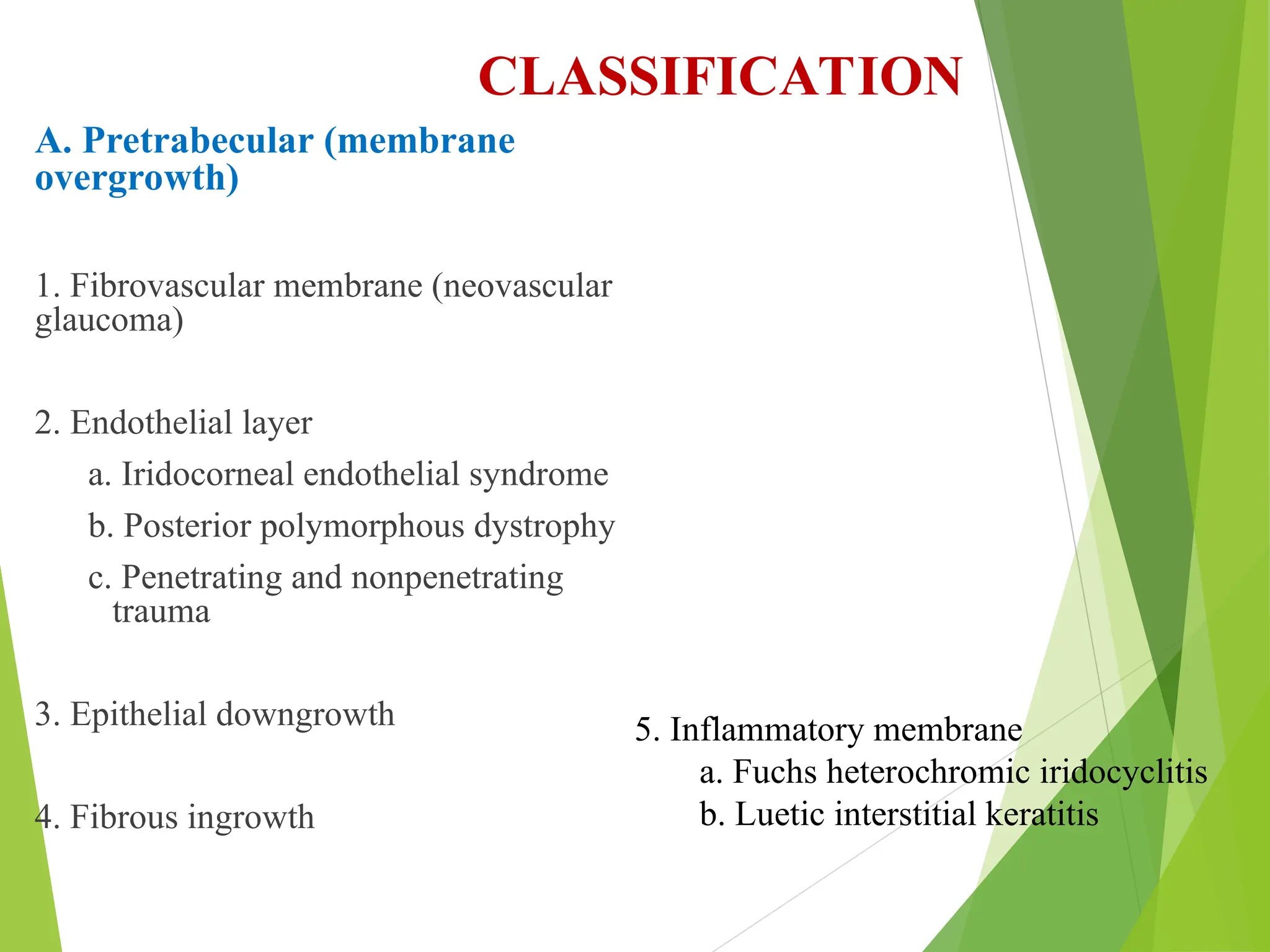
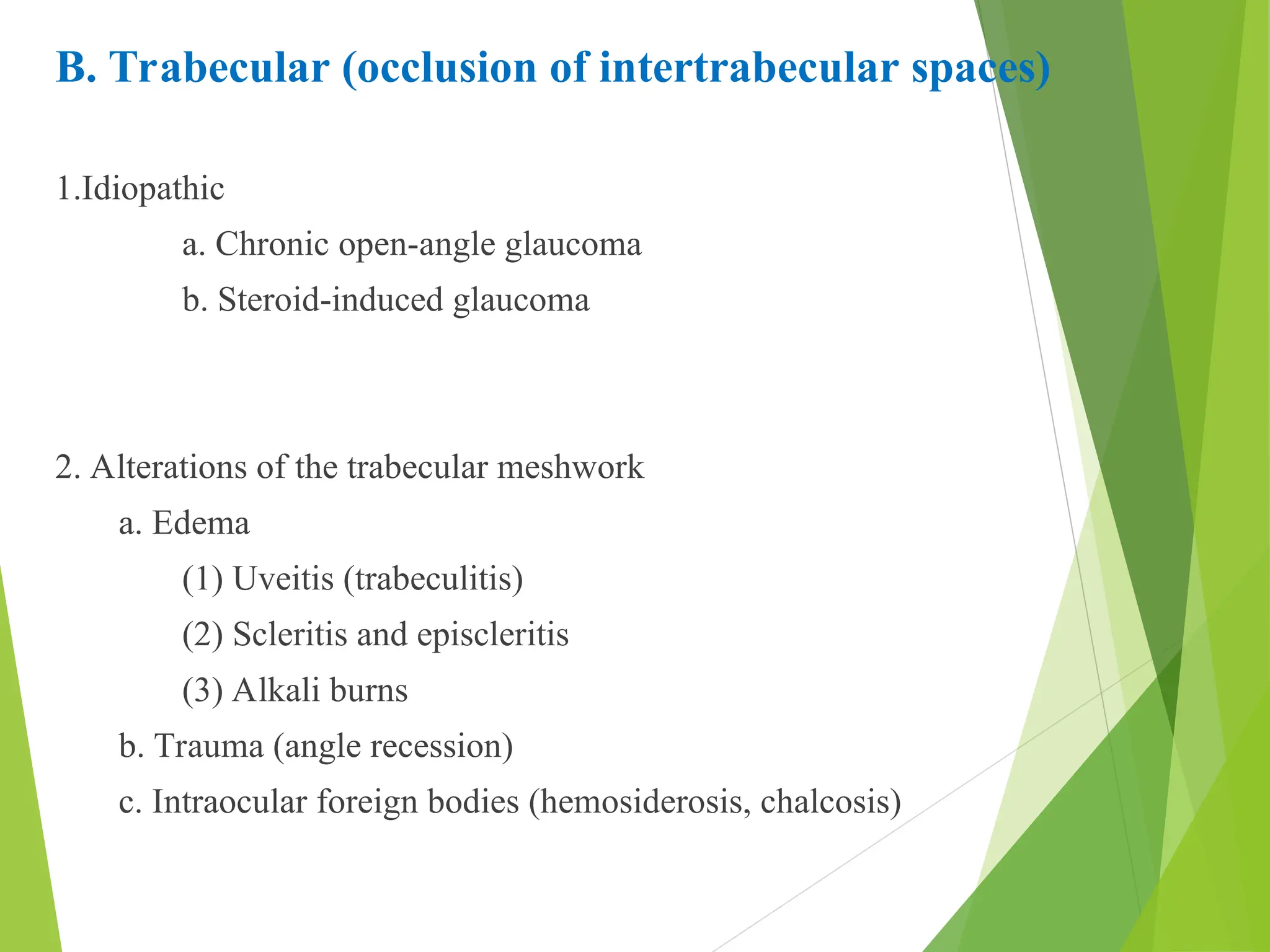
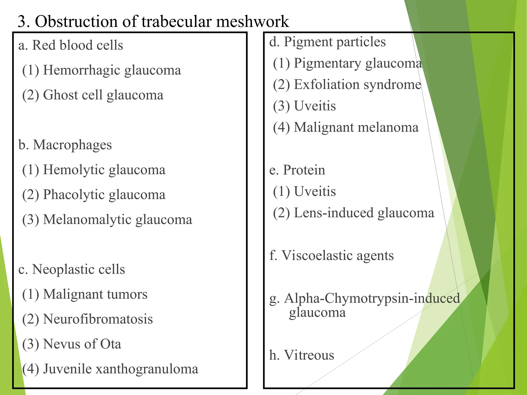
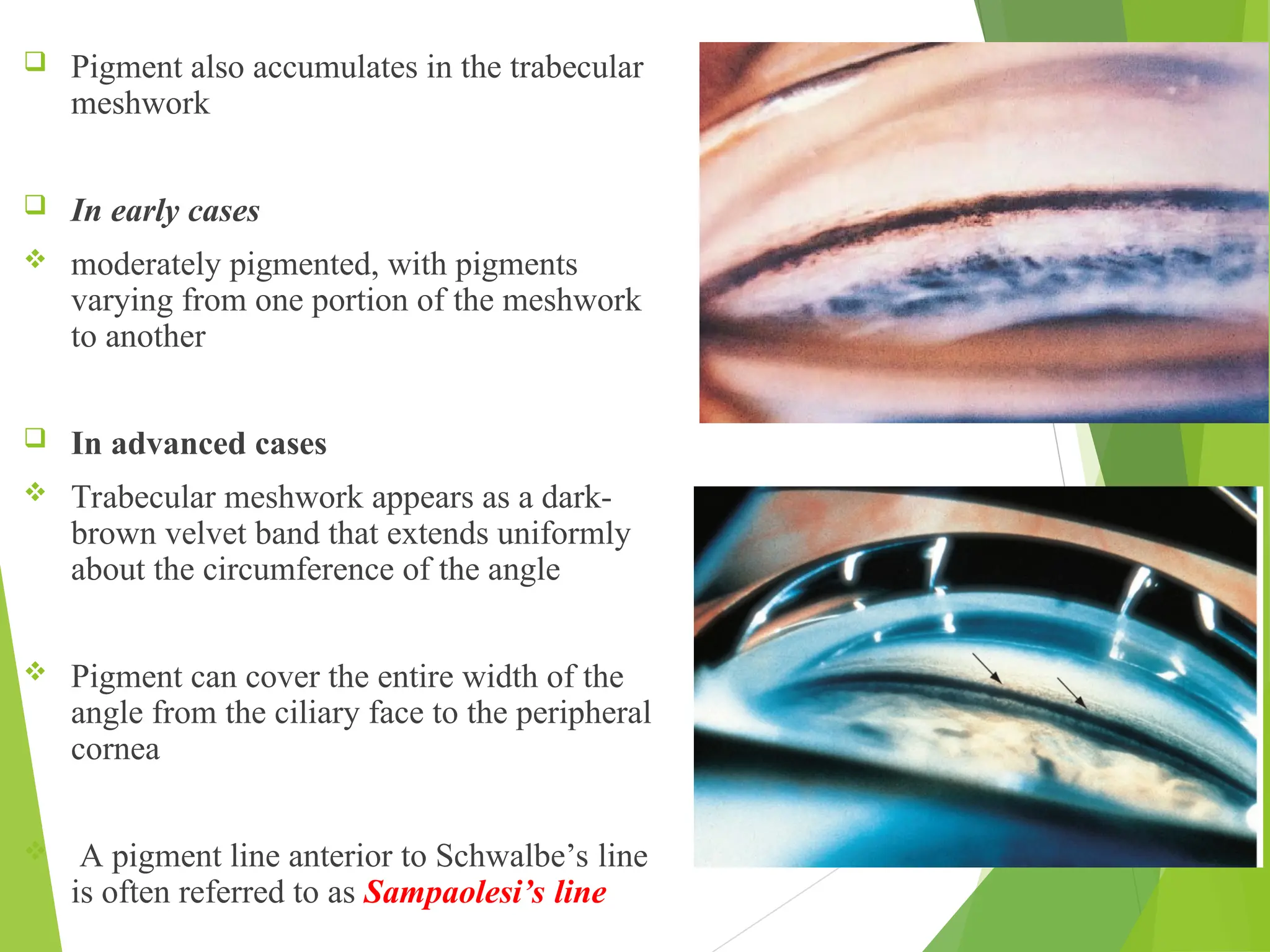
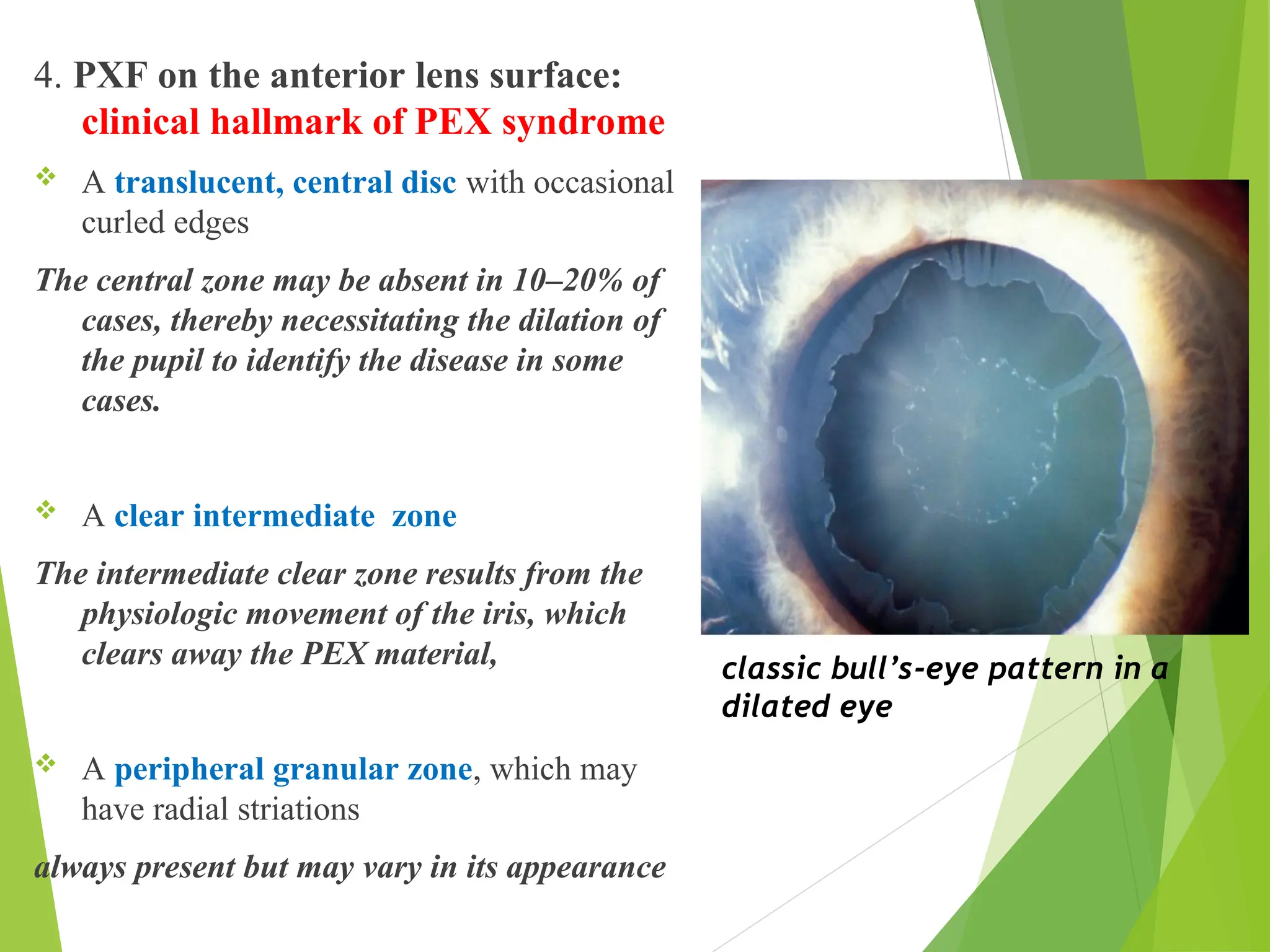
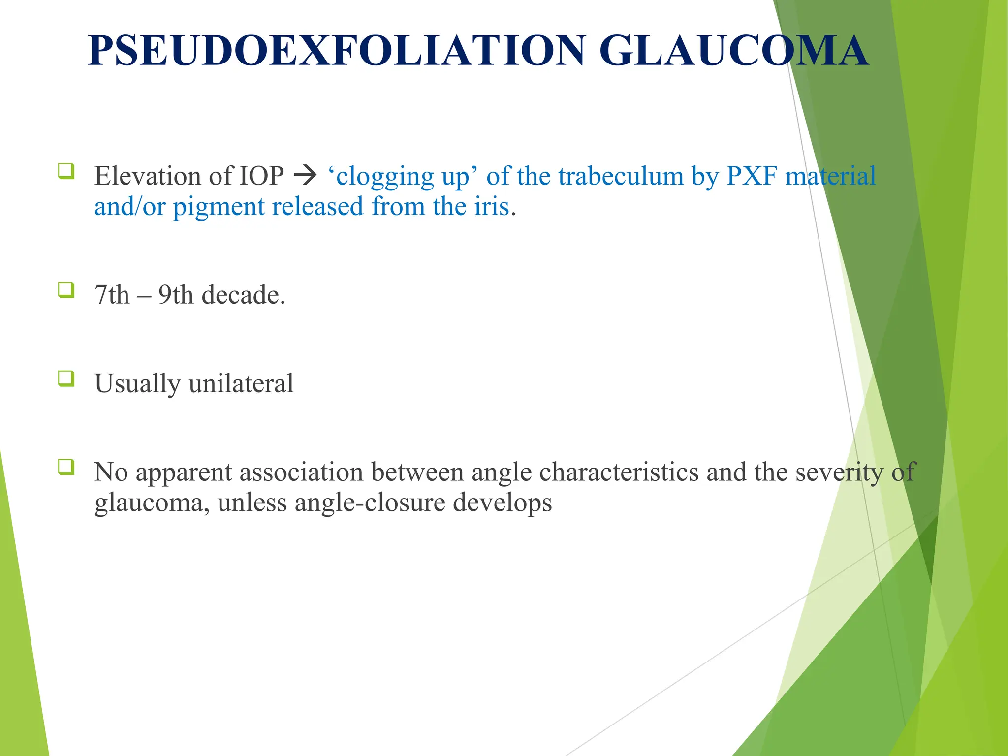
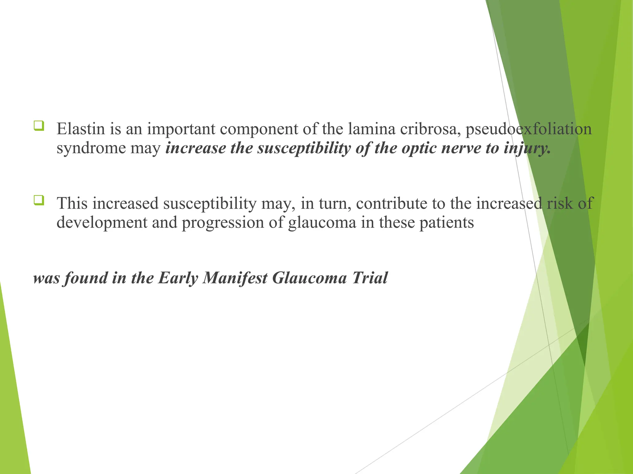
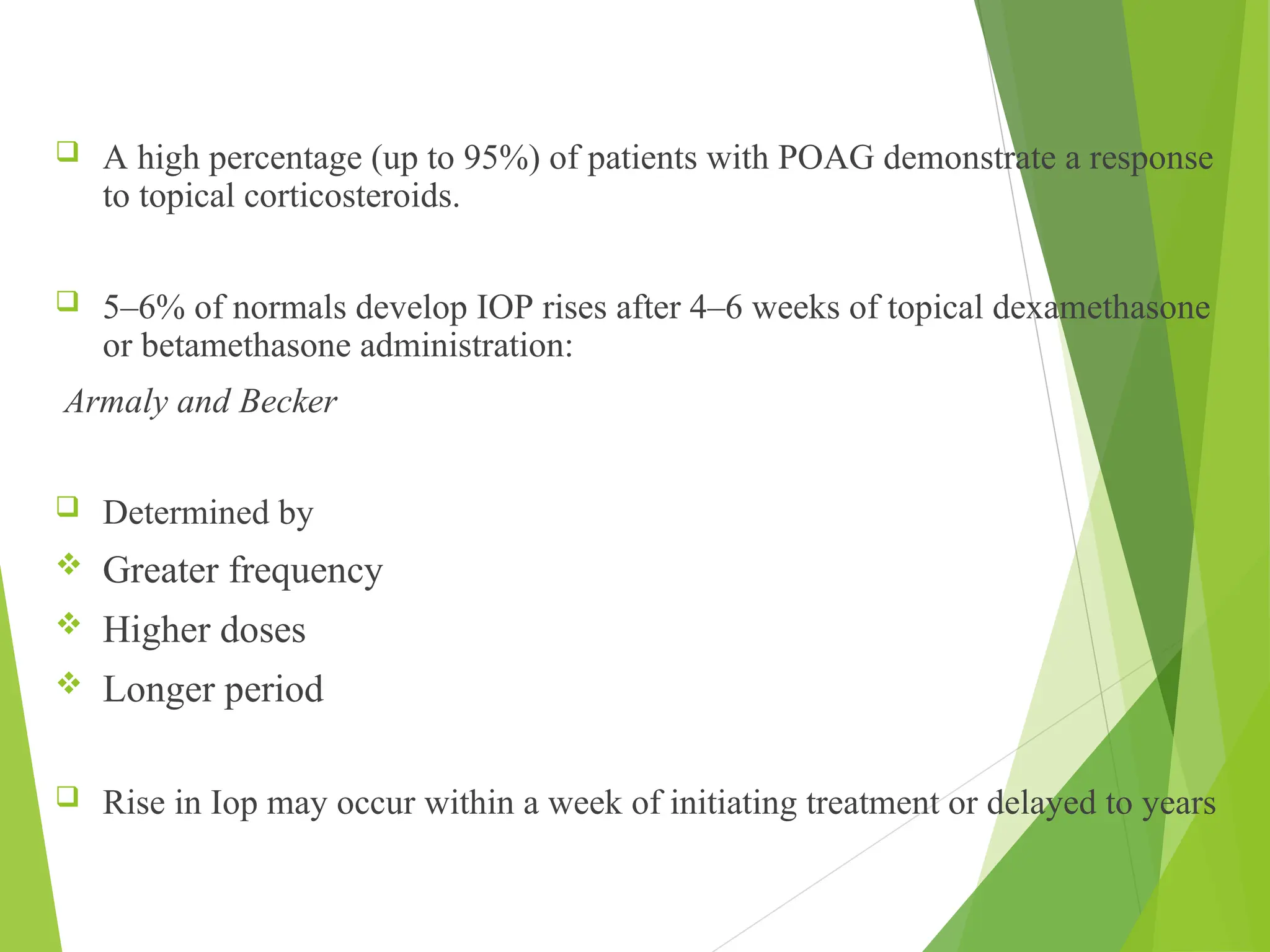
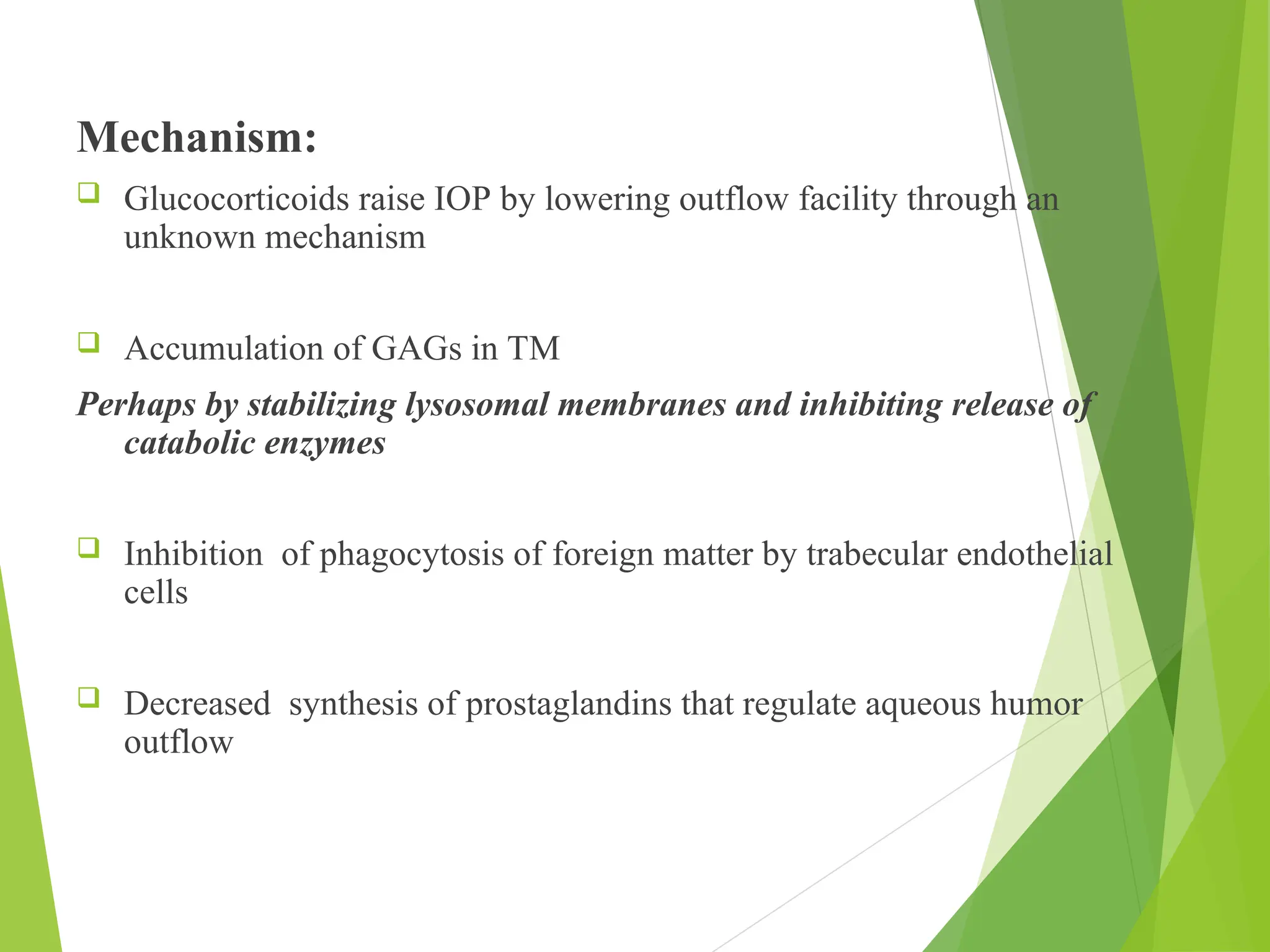
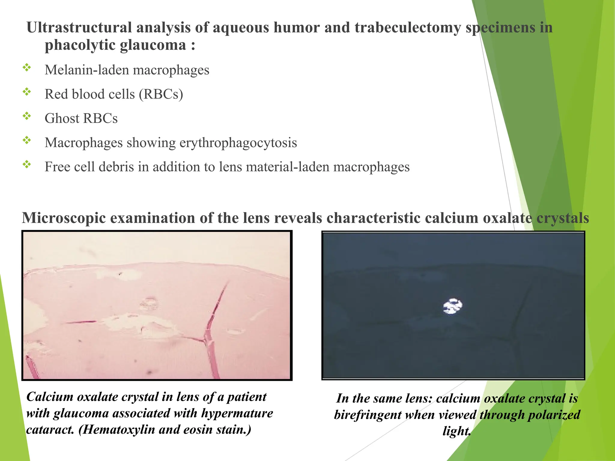
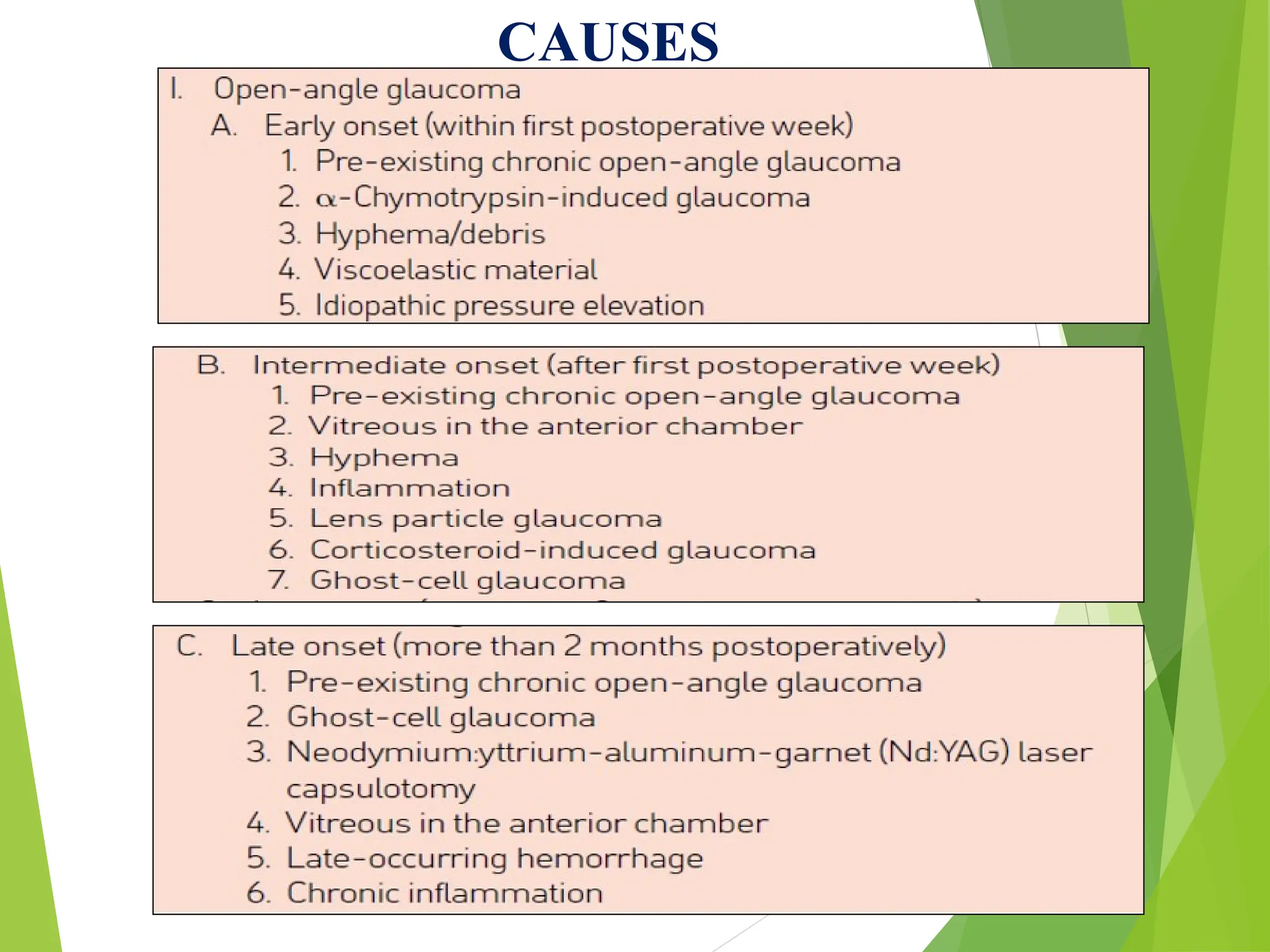
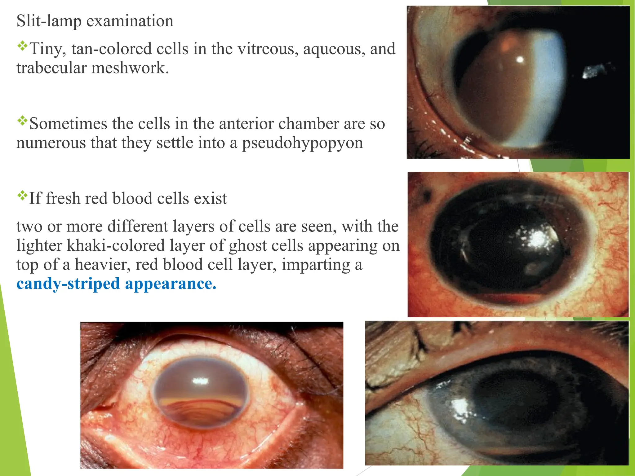
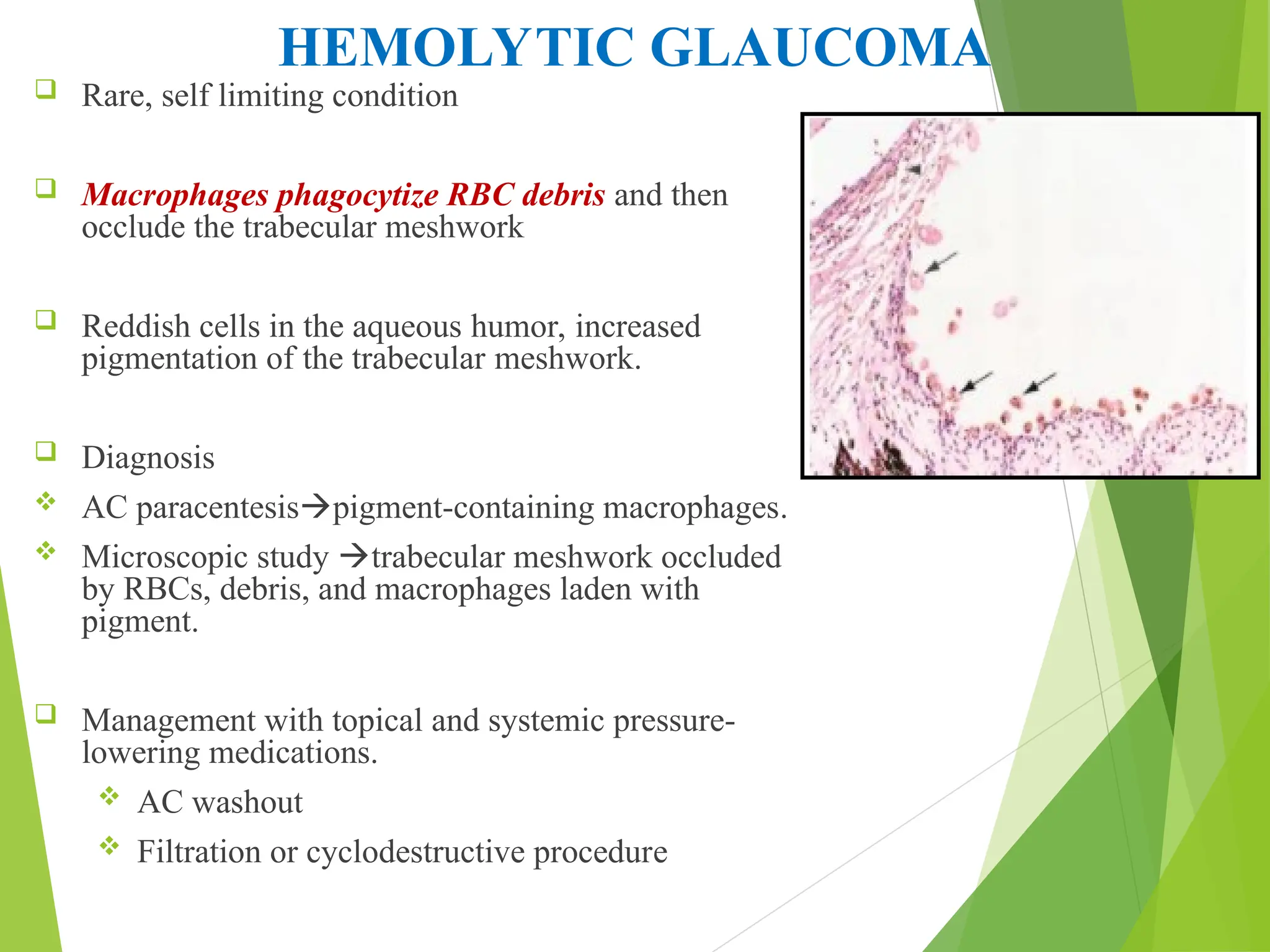
SECONDARY OPEN ANGLE
GLAUCOMA
1
Dr suchana Sharma
Department of ophthalmology
Dhulikhel hospital
11th
October, 2020
Secondary glaucoma per se is not a disease entity, but a group of
disorders in which rise of intraocular pressure is associated with
some primary ocular or systemic disease.
INTRODUCTION
CLASSIFICATION
A. Pretrabecular (membrane
overgrowth)
1. Fibrovascular membrane (neovascular
glaucoma)
2. Endothelial layer
a. Iridocorneal endothelial syndrome
b. Posterior polymorphous dystrophy
c. Penetrating and nonpenetrating
trauma
3. Epithelial downgrowth
4. Fibrous ingrowth
5. Inflammatory membrane
a. Fuchs heterochromic iridocyclitis
b. Luetic interstitial keratitis
B. Trabecular (occlusion of intertrabecular spaces)
1.Idiopathic
a. Chronic open-angle glaucoma
b. Steroid-induced glaucoma
2. Alterations of the trabecular meshwork
a. Edema
(1) Uveitis (trabeculitis)
(2) Scleritis and episcleritis
(3) Alkali burns
b. Trauma (angle recession)
c. Intraocular foreign bodies (hemosiderosis, chalcosis)
a. Red blood cells
(1) Hemorrhagic glaucoma
(2) Ghost cell glaucoma
b. Macrophages
(1) Hemolytic glaucoma
(2) Phacolytic glaucoma
(3) Melanomalytic glaucoma
c. Neoplastic cells
(1) Malignant tumors
(2) Neurofibromatosis
(3) Nevus of Ota
(4) Juvenile xanthogranuloma
d. Pigment particles
(1) Pigmentary glaucoma
(2) Exfoliation syndrome
(3) Uveitis
(4) Malignant melanoma
e. Protein
(1) Uveitis
(2) Lens-induced glaucoma
f. Viscoelastic agents
g. Alpha-Chymotrypsin-induced
glaucoma
h. Vitreous
3. Obstruction of trabecular meshwork
C. Posttrabecular
1. Obstruction of Schlemm canal
a. Collapse of canal
b. Obstruction of Schlemm canal
(e.g., sickled red blood cells)
2. Elevated episcleral venous pressure
a. Carotid-cavernous fistula
b. Cavernous sinus thrombosis
c. Retrobulbar tumors
d. Thyrotropic exophthalmos
e. Superior vena cava obstruction
f. Mediastinal tumors
g. Sturge-Weber syndrome
h. Idiopathic
PIGMENTARY DISPERSION SYNDROME
AND PIGMENTARY GLAUCOMA
 As a normal feature of maturation and aging
 A variable amount of uveal pigment is chronically released and dispersed into
the anterior ocular segment.
 This is best appreciated by observing the trabecular meshwork,
Which is nonpigmented in the infant eye but becomes progressively pigmented to
various degrees with the passage of years because of the accumulation of the
dispersed pigment in the aqueous outflow system.
 Several ocular conditions are associated with an unusually heavy
dispersion of pigment, which may be significantly involved in the
increased resistance to aqueous outflow
which differed from other forms of pigment dispersion by typical clinical
and histopathologic features
 They referred to the condition as PIGMENTARY GLAUCOMA
 When the typical findings are encountered without associated glaucoma,
the term PIGMENT DISPERSION SYNDROME (PDS)
 Epidemiology:
 Accounts for 1.0–2.5% of glaucomas (Western countries)
 usually bilateral
 Age: young adults (generally)
With increasing age, the signs of pigment dispersion may decrease result of
normal growth of the lens
 inducing a physiologic pupillary block
 anterior movement of the iris.
 Loss of accommodation may also occur.
As pigment dispersion is reduced, the deposited pigment may fade from the
corneal endothelium, trabecular meshwork, or anterior surface of the iris
 Sex : M>F (female( mean age: 46yrs): affected, usually are a decade
older than male(35 yrs))
The reason for the male predilection appears to be the sex difference in
anterior chamber depth, which one study showed to be 3.22 ± 0.42
mm in men and 2.88 ± 0.38 mm in women
 Race: white>black and Asians
Heavy pigmentation and compactness of iris stroma in black individual
prevents posterior sagging of mid- peripheral iris
 Refractive condition: strong positive association with myopia
 Most are sporadic, rarely familial
PATHOGENESIS
MECHANISM OF PIGMENT DISPERSION
An inherent weakness or degeneration in the iris pigment epithelium was first
proposed as a cause of PDS by Scheie and Fleischauer in 1958
Histopathologic observations in the iris pigment epithelium
Focal atrophy and hypopigmentation
An apparent delay in melanogenesis
Hyperplasia of the dilator muscle
The additional observation of retinal pigment epithelial dystrophy in two brothers
with pigmentary glaucoma
raises the possibility of an inherited defect of pigment epithelium in the anterior
and posterior ocular segments
 Campbell proposed an alternative mechanical theory
 The peripheral radial defects of the iris corresponded in location and number to
anterior packets of lens zonules
 Suggested that a background bowing of the peripheral iris led to the
mechanical rubbing of the lens zonules against the iris pigment epithelium
with the subsequent dispersion of pigment
 Supported by biometric and photogrammetric studies of anterior chamber
dimensions,
 Deeper anterior chambers and flatter lenses in the involved eyes of unilateral
cases
 A deeper-than-normal midperipheral chamber depth with corresponding
concavity of the iris in eyes with the PDS
 Ultrasonographic biomicroscopy studies in PDS shown that the radial width of the iris
compared with the size of the anterior segment is larger than normal
Larger size results in a floppier iris, which may predispose to iridozonular contact when
combined with the posterior iris insertion.
Mechanism by which the peripheral iris is bowed backward??
Laser iridotomy relieves the posterior bowing, which led to the concept of REVERSE
PUPILLARY BLOCK
 Aqueous is moved into the anterior chamber against the normal pressure gradient, possibly by the
movement of the peripheral iris in response to movement of the eye (e.g., blinking) or accommodation.
 Once in the anterior chamber, the aqueous is prevented from returning to the posterior chamber by a one
way valve effect between the iris and lens
 Resulting in a relatively greater pressure in the anterior chamber and subsequent backward bowing of
the peripheral iris.
Elongated anterior zonules that may be pigmented encroaching in the
central visual axis
Pigment release from the pigmented epithelium located at the pupillary ruff
and the central iris, which are close to the elongated zonules
Although subtle and easily missed on biomicroscopy, long anterior zonules
may be more common than suspected but not previously recognized as a distinct
entity associated with pigment dispersion
Transmission electron microscopy shows central anterior lens capsule covered by an irregular zonule
lamella with pigment granules and degenerative lens epithelium in pigmented long anterior zonules
MECHANISM OF INTRAOCULAR PRESSURE
ELEVATION
Trabecular cells engulf melanin, which eventually leads to cell injury and death
from phagocytic overload.
Because melanoprotein is only partially digested, it is retained in intracellular
storage vacuoles, where it generates deleterious oxygen free radicals.
The trabecular cell loss leaves the collagen beams denuded and vulnerable to
fusion, with obliteration of the aqueous channels.
CLINICAL FEATURES
 Loss of pigment from the iris is
detected as a series of radial, spoke
like, midperipheral transillumination
defects
Result of contact between zonular fibers
and posterior iris pigment epithelium
 Number: 1-2 to 65-70
 Corneal endothelium pigment
depositions: vertically oriented spindle
called krukenberg’s spindle
Caused by aqueous convection current
and phagocytosis of pigment by
corneal endothelium
 Anterior chambers: very deep
 Pigment also accumulates in the trabecular
meshwork
 In early cases
 moderately pigmented, with pigments
varying from one portion of the meshwork
to another
 In advanced cases
 Trabecular meshwork appears as a dark-
brown velvet band that extends uniformly
about the circumference of the angle
 Pigment can cover the entire width of the
angle from the ciliary face to the peripheral
cornea
 A pigment line anterior to Schwalbe’s line
is often referred to as Sampaolesi’s line
 Pigment is also deposited on the zonular
fibers , post hyaloid face and equatorial
region of lens (Zentmayer’s ring or
Scheie’s line)
 Iris pigments
dull or even a heterochromic appearance if
the pigment dispersion is asymmetric in
the two eyes
 Peripheral iris: concave
 Approximately 15% of individuals with pigment dispersion syndrome
progress to glaucoma or elevated IOP requiring treatment.
 Pigmentary glaucoma is 3 times more common in men than in women
 Other features are same as POAG,except
 Large diurnal IOP fluctuations(can exceed up to 50 mm of Hg): sufficient
to cause corneal edema, blurring, and halo vision
 sudden release of pigment with severe IOP elevations after pupillary
dilation or exercise
blocked by topical pilocarpine therapy
 Initial treatment is typically medical therapy
 Laser trabeculoplasty is effective in pigmentary glaucoma, especially in the
early stages of the disease.
 With selective laser trabeculoplasty, however, patients with heavily pigmented
trabecular meshwork may have significant post-laser IOP elevations
 Because the heavy trabecular meshwork pigmentation allows increased
absorption of laser energy
 Lower-energy settings are recommended during laser trabeculoplasty to
avoid an acute rise in IOP after treatment
DIFFERENTIAL DIAGNOSIS
Any condition producing
pigmentation of TM :
 Normal eyes with aging
 POAG
 Uveitis
 Cysts of the iris and ciliary body
 Pigmented intraocular tumors
 Previous surgery (including laser
surgery)
 Trauma
 Angle-closure glaucoma,
 Amyloidosis
 Diabetes mellitus
 Herpes zoster
 Megalocornea
 Radiation
 Siderosis
 Hemosiderosis
 Mostly confused with exfoliation
syndrome
 Iris atrophy : central and
geographic
 Pigment accumulation in
TM consists of larger
particles
EXFOLIATION SYNDROME
(PSEUDOEXFOLIATION SYNDROME)
 Is an age-related generalized disorder of the extracellular matrix
associated with excessive synthesis and progressive deposition of a
fibrillar material in the ocular tissues as well as in the skin and connective
tissue of various visceral organs
 Histologically, this material has been found in and on the lens epithelium and
capsule, pupillary margin, ciliary epithelium, iris pigment epithelium, iris
stroma, iris blood vessels, and subconjunctival tissue
 Occurs when several ocular tissues synthesize an abnormal protein. This
protein may obstruct the trabecular meshwork and cause glaucoma
 When an eye with PXF develops secondary open-angle glaucoma:
pseudoexfoliation glaucoma (PXG)
 Prevalence is closely linked to age, reaching a maximum in the seventh to ninth
decades of life
 PXF :F>M but the combination of exfoliation syndrome and glaucoma occurs
equally in both sexes.
 unilateral or bilateral, and over half of unilateral cases become bilateral over a 20-
year period
 Risk of glaucoma in eyes with PXF is 5% at 5 years and 15% at 10 years
 Particularly common in Scandinavia
 Most cases appear to be sporadic, X-linked inheritance pattern
 Mutations in LOXL1 gene at locus 15q22, coding for elastic fiber components of
extracellular matrix
PATHOGENESIS
Reduced /abnormal synthesis of
elastic fibers
PATHOGENESIS
 A grey-white fibrillary extracellular material
 Composed of a protein core surrounded by GAGs
 Produced by abnormal BM of ageing epithelial cells in:
 Trabeculum
 Equatorial lens capsule
 Iris
 Ciliary body
 Deposited on anterior lens capsule, zonules, ciliary body, iris, trabeculum,
anterior vitreous face and conjunctiva
 Exfoliative fibrillopathy: skin and visceral organs
 PXFS is associated with an increasing number of vascular disorders, hearing loss
and alzheimer disease
CLINICAL FEATURES
1. Cornea : PXF on endothelium
 Flakes of exfoliative material and pigment
accumulation: usually diffuse/ in the form of
a vertical spindle Krukenberg spindle.
 Specular microscopy significantly lower-
than-normal cell density in eyes with the
exfoliation syndrome and changes in cell
size and shape
2.Anterior chamber: Mild aqueous flare
(breakdown of the iris blood-aqueous
barrier)
3. Iris : PXF on pupillary margin
 white flecks on the pupillary margin of
the iris, with loss of pigment at the
pupillary ruff
 Sphincter atrophy : ‘moth-eaten’
transillumination defects at the
pupillary margin
4. PXF on the anterior lens surface:
clinical hallmark of PEX syndrome
 A translucent, central disc with occasional
curled edges
The central zone may be absent in 10–20% of
cases, thereby necessitating the dilation of
the pupil to identify the disease in some
cases.
 A clear intermediate zone
The intermediate clear zone results from the
physiologic movement of the iris, which
clears away the PEX material,
 A peripheral granular zone, which may
have radial striations
always present but may vary in its appearance
classic bull’s-eye pattern in a
dilated eye
Precapsular film has been noted on the anterior lens capsule of many older
individuals: precursor
 Pupil often dilates poorly, likely because of infiltration of fibrillar material into
the iris stroma.
 Phacodonesis and iridodonesis can be seen and are due to the weak zonular
fibers.
 frank subluxation or dislocation of the natural or implant lens
 Great care must be taken during cataract surgery to reduce the risk of zonular
dehiscence, vitreous loss, lens dislocation, and other complications intraoperatively
and postoperatively
Gonioscopy
 Trabecular hyperpigmentation: usually most marked inferiorly/ uneven
distribution (as compared to PDS )
 Sampaolesi line: scalloped band of pigment running on to or anterior to Schwalbe
line
 PXF deposits in the trabeculum :‘ dandruff-like’ appearance.
PSEUDOEXFOLIATION GLAUCOMA
 Elevation of IOP  ‘clogging up’ of the trabeculum by PXF material
and/or pigment released from the iris.
 7th – 9th decade.
 Usually unilateral
 No apparent association between angle characteristics and the severity of
glaucoma, unless angle-closure develops
 Elastin is an important component of the lamina cribrosa, pseudoexfoliation
syndrome may increase the susceptibility of the optic nerve to injury.
 This increased susceptibility may, in turn, contribute to the increased risk of
development and progression of glaucoma in these patients
was found in the Early Manifest Glaucoma Trial
Exfoliation glaucoma differs from POAG
Often: unilateral
Greater pigmentation of TM
lOP: higher with greater diurnal fluctuations
50–60 mmHg or higher on presentation, yet most patients have no pain,
indicating the chronicity of the IOP elevation
Overall prognosis: worse.
Laser trabeculoplasty: very effective
Lens extraction does not alleviate condition
DIFFERENTIAL DIAGNOSIS
Pigment dispersion syndrome
Bilateral, more common in young myopic males than females, and displays characteristic TIDs
and angle pigmentation findings
Capsular delamination (true exfoliation)
Is a splitting of the anterior lens capsule without deposition of PEX material.
 It can occur secondary to heat, trauma, irradiation, or inflammation and is rare.
Glassblower disese
Primary amyloidosis
Evaluation of this material has shown it to be distinctly different from PEX material.
CORTICOSTEROID GLAUCOMA
 Patients who experience a transient or sustained pressure rise after
corticosteroid instillation : STEROID RESPONDER
 If glaucomatous damage ensues (optic nerve or on visual field testing):
STEROID GLAUCOMA
 Approximately one-third of the population without glaucoma demonstrates an
IOP elevation of between 6 and 15 mm Hg in response to corticosteroids
 Small percentage (4%–6%) has a signficant IOP elevation of more than 15 mm
Hg.
Normal population divide into 3 groups based on their IOP responses to topical
betamethasone in 4- 6 weeks.
High responders- >30 mmHg
Moderate responders- 22 – 30 mmHg
Non-responders- no change in IOP.
 A high percentage (up to 95%) of patients with POAG demonstrate a response
to topical corticosteroids.
 5–6% of normals develop IOP rises after 4–6 weeks of topical dexamethasone
or betamethasone administration:
Armaly and Becker
 Determined by
 Greater frequency
 Higher doses
 Longer period
 Rise in Iop may occur within a week of initiating treatment or delayed to years
Mechanism:
 Glucocorticoids raise IOP by lowering outflow facility through an
unknown mechanism
 Accumulation of GAGs in TM
Perhaps by stabilizing lysosomal membranes and inhibiting release of
catabolic enzymes
 Inhibition of phagocytosis of foreign matter by trabecular endothelial
cells
 Decreased synthesis of prostaglandins that regulate aqueous humor
outflow
Risk groups:
First-degree relative with COAG
High myopia
Previous steroid response
Type 1 diabetes mellitus
Connective tissue disease (e.g., rheumatoid arthritis)
Penetrating keratoplasty, particularly in eyes with Fuchs endothelial dystrophy or
keratoconus
Prolonged use of topical, periocular(depot/repository preparations), intravitreal,
inhaled, or systemic corticosteroids.
Excessive levels of endogenous corticosteroids (eg, Cushing syndrome)
Following LASIK use of steroidincrease IOP  vision threatening corneal
opacity
MANAGEMENT
 The cause of the elevation in lOP is not always corticosteroid
underlying ocular disease such as anterior uveitis.
 Stop corticosteroid lOP usually decreases with a time
If Steroids imp for patient’s life then weakest possible drug at lowest
dose
 Anti glaucoma medications
 Laser trabeculoplasty
 Filtering surgery
 However, unmasked POAG or secondary open-angle inflammatory
glaucoma may remain.
 Excision of corticosteroid-producing tumor or hyperplastic tissue.
 Cycloplegic drugs can increase lOP in individuals with open
angles.
 Routine dilation for ophthalmoscopy may increase IOP, those at
greater risk include
 patients with POAG
 exfoliation syndrome
 pigment dispersion syndrome
 those on miotic therapy.
LENS-INDUCED GLAUCOMA
OPEN ANGLE ANGLE CLOSURE
Phacolytic Glaucoma
lens protein leaks from an intact cataract
and obstructs the trabecular meshwork
Phacomorphic:
a swollen lens causes increased pupillary
block and secondary angle closure.
Lens particle:
lens material liberated by trauma or
surgery obstructs the outflow channel
Ectopia lentis:
a dislocated lens causes increased
pupillary block and secondary angle
closure.
Phacoantigenic/
phacoanaphylactic glaucoma:
sensitization to lens protein produces
granulomatous inflammation and
occasionally secondary glaucoma
A. PHACOLYTIC GLAUCOMA
Mechanism:
Lens protein: normally sequestered within lens capsule.
 Aging = development of cataract = altered protein composition of lens
(heavier molecular weight soluble proteins )
 Leakage of these soluble molecules through intact capsule = obstruct
TM
HMW-rare in childhood lenses, which may explain why phacolytic
glaucoma rarely occurs in children
MACROPHAGE THEORY-
Lens protein also stimulates inflammation and a
macrophage response
Macrophages engulf lens protein
macrophages, laden with phagocytosed lens
material, block the trabecular meshwork to produce
the acute glaucoma
Supported by-
Demonstration of macrophages in the aqueous
and trabecular meshwork
Electron microscopic study- macrophages found
to have phagocytized, degenerated lens material
Against the macrophage theory
observation that lens-laden macrophages in the
anterior chamber do not invariably lead to elevated
IOP
CLINICAL FEATURES
 Age of presentation: older patients with history
of poor vision for month to years
 Typically present with acute onset of
Monocular pain and Redness
 Further decrease in vision
Examination
 Ciliary injection
 Corneal edema
 Heavy cell and flare
 The cells appear larger than white blood cells
and somewhat iridescent.
 The cells may precipitate on the corneal
endothelium, but no true keratic precipitates or
hypopyon is seen
hypermature cataract, lens protein-
containing macrophages floating in the
aqueous, and a pseudohypopyon;
dense milky aqueous with
pseudohypopyon
 Lens:
 mature/hypermature/morgagnian cataract
 Rarely immature with a zone of liquefied cortex
 Severe IOP elevation
 Open angles
Conjunctival hyperemia
Microcystic corneal edema
Mature cataract
Prominent AC reaction
Note lens protein deposits on endothelium and
layering in the angle, creating a pseudohypopyon
 hypermature cataract with
wrinkling of the anterior lens
capsule, which results from loss of
cortical volume.
 Extensive posterior synechiae are
present, which suggests previous
inflammation.
Ultrastructural analysis of aqueous humor and trabeculectomy specimens in
phacolytic glaucoma :
 Melanin-laden macrophages
 Red blood cells (RBCs)
 Ghost RBCs
 Macrophages showing erythrophagocytosis
 Free cell debris in addition to lens material-laden macrophages
Microscopic examination of the lens reveals characteristic calcium oxalate crystals
Calcium oxalate crystal in lens of a patient
with glaucoma associated with hypermature
cataract. (Hematoxylin and eosin stain.)
In the same lens: calcium oxalate crystal is
birefringent when viewed through polarized
light.
 Rarely phacolytic glaucoma has a subacute course
 With intermittent leakage of protein producing recurrent episodes of glaucoma,
hyperemia, and inflammation.
 This appearance is more likely if the cataract has been dislocated into the
vitreous.
 The diagnosis of phacolytic glaucoma is usually made on clinical grounds.
 If the diagnosis is in doubt, an anterior chamber paracentesis should be
performed
Detect macrophages engorged with lens material.
Treatment :
 Definitive treatment : cataract extraction
 Medical Rx for IOP and inflammation before surgery
 Phacolytic glaucoma due to dislocated lens: the lens removed by vitrectomy
instruments
 If dislocated lens floating in AC:
 Stream of irrigation fluid
 Removed through a limbal incision
B.LENS PARTICLE GLAUCOMA
Mechanism:
 Disruption of lens capsule by penetrating trauma or surgery--Liberates
lens material– directly Obstruct TM
 Contribution from cellular reaction to the lens particles
Glaucoma depends on
 Amount of lens material liberated
 Inflammatory response
 Ability of TM to clear foreign matter
 Presents soon after precipitating event
 Rarely long after surgery or trauma
CLINICAL FINDINGS
PRESENTS WITH
Significant pain, Redness, Decreased vision
EXAMINATION
Corneal edema
Elevated IOP
Heavy cell and flare
Chunky white particles in aqueous humor
A hypopyon may be present, as may fluffy cortical material
Condition lasting for long: PAS and posterior synechiae
Gonioscopy may reveal open angle with obvious lens particles overlying
the trabecular meshwork.
 Diagnosis of lens-particle glaucoma is suggested by the sequence of events.
 It is more difficult to diagnose delayed cases or cases with spontaneous
rupture of the lens capsule
which may be confused with phacolytic glaucoma, phacoanaphylactic gl­
aucoma
Management:
 Medical therapy: to control IOP until residual lens material resorbs
 Medications to decrease aqueous formation
 Mydriatics to inhibit posterior synechia formation
 Topical corticosteroids to reduce inflammation
 If IOP cannot be controlled, surgical removal of Lens material is necessary
Sometimes the entire lens mass can be teased from the eye after
intracameral infusion of alpha-chymotrypsin.
P
PHACOANTIGENIC GLAUCOMA/
HACOANTIGENIC GLAUCOMA/
PHACOANAPHYLAXIS
PHACOANAPHYLAXIS
 Rare condition
 Patients become sensitized to their own lens protein
 Typically develops after penetrating trauma or extracapsular cataract
extraction
 autoimmune granulomatous inflammation
 The mechanism causing the reaction seems to be an Type III
hypersensitivity reaction mediated by IgG and the complement system
Histopathology:
 Granulomatous inflammation of the lens with
 Polymorphonuclear leukocytes
 Lymphocyte
 Epithelioid cells
 Giant cells
 Occasionally the inflammation involves the trabecular meshwork and
leads to a rise in IOP
CLINICAL FEATURES
 History of- disruption of the lens capsule by cataract surgery or
penetrating injury
 Latent period of 1 to 14 days(distinguishing feature )
 Marked congestion
 Moderate anterior chamber reaction with KP on both the corneal
endothelium and the anterior lens surface.
 Residual lens material in the anterior chamber
 Aqueous cell and flare and sterile hypopyon
 Synechial formation
 Low –grade vitritis,
 Glaucomatous optic neuropathy rare
Management
 Reduce inflammation
 Control IOP
 If unsuccessful
 residual lens material should be removed
GLAUCOMA AFTER CATARACT SURGERY
 Transient rise in IOP = 33- 100%
Depending on the method of extraction and the surgeon involved
 This pressure rise may be undetected because it occurs several hours after
surgery
 The pressure may return to near-normal levels by the next morning or
whenever the patient is seen for the first postoperative visit
 Elevated IOP abates spontaneously over 2-4 days
60
CAUSES
MECHANISM
1. Inflammation release of active substances, including Prostaglandins
secondary aqueous humor formation
2. A watertight wound closure with multiple fine sutures limiting the ‘safety valve’
leak of aqueous humor.
3. Deformation of the limbal area, reducing trabecular outflow.
 On gonioscopy, Kirsch and co-workers noted a white ridge internal to limbal
cataract wounds.
 This ridge, attributed to tight sutures and to operative edema and swelling, is
associated with reduced trabecular function
4. Obstruction of the trabecular meshwork by pigment, blood, lens particles,
inflammatory cells, and viscoelastic substances.
ALPHA-CHYMOTRYPSIN GLAUCOMA
 Alpha Chymotrypsin fragments zonules and was used widely to
facilitate intracapsular cataract extraction
 Elevated iop 1–5 days
 Self limited lasting for 2–4 days
 Zonular fragments obstruct the outflow channels + inflammation +
direct toxic effect on the meshwork
 Quite rarely today because intracapsular cataract extraction with alpha
chymotrypsin is rarely used .
 During the period of elevated IOP, patients are treated with topical and
systemic glaucoma medications as needed
 Prophylactic timolol and acetazolamide useful
 Incidence and severity of the pressure rise can be reduced by using a
 lesser concentration of the drug (1:10 000 instead of 1:5000)
 in a lower volume (0.25–0.5 ml instead of 2 ml).
 The anterior chamber should be irrigated before lens extraction to remove
zonular fragments.
GLAUCOMA FROM VISCOELASTIC SUBSTANCES
 Viscoelastic substances: sodium hyaluronate used in cataract surgery
 Protect corneal endothelium
 Facilitate IOL insertion
 Frequently causes: marked postoperative IOP elevations
 Mechanism :Obstruct the trabecular meshwork.
 Low-viscosity sodium hyaluronate produces a greater rise in IOP than high-
viscosity sodium hyaluronate.
 Chondroitin sulfate and methylcellulose less likely to elevate IOP
 Cellular and particulate matter in the aqueous humor appear suspended and
almost immobile if large amounts of viscoelastic is present
 But IOP elevations may occur even in the absence of clinically detectable
viscoelastic.
 It is sometimes possible to see tiny ruby-like globs of hemorrhage on the iris
surface or suspended in the anterior chamber.
These isolated blood droplets indicate the presence of retained viscoelastic
 IOP maximum in 12–16 hours and then abates spontaneously - next 72
hours.
 Corneal edema, deep anterior chambers
 Treated with topical and systemic glaucoma medications and hyperosmotic
agents
 The postoperative IOP rise can apparently be limited by removing as much
of the sodium hyaluronate as possible at the end of the surgery
GLAUCOMA WITH PIGMENT DISPERSION
FROM INTRAOCULAR LENSES
 Pigment dispersion and elevated IOP have been described with posterior
chamber IOLs and, in a few cases, with iris fixation lenses
 In most instances lenes are
 Decentered
 Tilted
 excessively mobile
 Too small
 Reversed in position
MECHANISM
All leading to excess friction between optic or haptic and iris pigment
epithelium.
Pigments accumulate trabecular meshwork obstruct aqueous
humor outflow.
Pigment epithelial loss of iris is geographic atrophic appearance.
Iris: grey atrophic appearance
 IOP: rises days to months after cataract surgery
 May improve spontaneously.
 If IOP cannot be controlled
 IOL should be replaced or stabilized
D/D from pigmentary glaucoma
 Temporal relation to cataract surgery
 Unilateral state
 Absence of a typical krukenberg’s spindle and radial transillumination defects
UVEITIS-GLAUCOMA-HYPHEMA(UGH)
SYNDROME
 Secondary inflammatory glaucoma caused by chronic irritation by Iris-
supported, posterior chamber, and most often with anterior chamber IOLs.
 Mechanism: excessive chafing of iris because lenses are too mobile, are
poorly designed, or have poor finishing characteristics
 H/o:
 Uncomplicated cataract extraction
 But weeks to months after develop elevated IOP, iridocyclitis, and recurrent
hyphemas
 Recurrent episodes of extreme blurring (whiteout) presumably related to the
hyphemas.
Diagnosis:
 Ultrasonic biomicroscopy and aqueous
humor analysis
 Removed lenses shows:
Sharp or serrated edges capable of
traumatizing iris tissue
Treatment:
 Corticosteroid
 Mild cases: standard antiglaucoma therapy
 Lens removed: to prevent corneal
decompensation and optic nerve damage
Inflamed eye of a patient following
anterior chamber intraocular lens
insertion.
Goniophotograph showing footplate of lens
protruding posteriorly through iridectomy and
brushing against ciliary processes.
GLAUCOMA AFTER NEODYMIUM: YTTRIUM-
ALUMINUM-GARNET LASER POSTERIOR
CAPSULOTOMY
 Intraocular pressure often rises after neodymium: yttrium aluminum-garnet
(Nd:YAG) laser posterior capsulotomy
 IOP upto 60–80 mmHg.
 2–4 hours of the laser treatment and abates spontaneously over the next 24
hours or last for days to weeks.
 Risk groups of Extreme IOP elevation
 Eyes with pre existing glaucoma
 Eyes with no PC-IOL
So after nd:yag laser capsulotomy IOP so should be measured 2–4 hours,1
day,7 days after nd:yag laser capsulotomy.
MECHANISM
 Particulate debris clogging the TM
The aqueous humor contains fibrin, lens material, inflammatory cells,
macrophages, and RBCs in sufficient quantity to obstruct the
trabecular meshwork Nd:YAG laser capsulotomy
 The vitreous moves forward and causes pupillary block
 Bleeding or inflammation to obstruct outflow
 Surgery releases a dialyzable factor from the vitreous that blocks the
outflow channels
 Prevention to decrease number and severity of postoperative pressure spikes
Pretreatment with apraclonidine (1%)
 1 hour prior to surgery
 1 hour after surgery
 additional dose: just before bed-time on night of surgery.
 If Persistent increased IOP
 β-adrenergic antagonist
 Topical/systemic CAI
 α-adrenergic agonist
 hyperosmotic agents
 Topical corticosteroids: reduce inflammation.
F. GLAUCOMA FROM VITREOUS IN AC
 Rare cause of open angle glaucoma in aphakic eyes
 Onset: within weeks after cataract
 Mechanism: vitreous reaches AC
 After a spontaneous rupture of hyaloid face
 After an extensive posterior vitreous detachment vitreous left in AC at time of
cataract extraction
 Glaucoma occurs when a large bolus of vitreous comes into contact with a major
portion often and obstructing outflow system
 In some cases: mechanism is complicated:
 Inflammation, Pupillary block, Formation of PAS
On examination:
 Elevated IOP
 Open angles
 Vitreous fills AC and appears to be in contact with TM
Management:
 Usually: self-limited
During this period, topical and systemic pressure-lowering medications are
employed to control IOP
 Better to cycloplegic > miotics
 Unresponsive patients (optic nerve is threatened) vitrectomy should be
considered
REFERENCES
 Becker Shaffer’s Diagnosis and Therapy of the Glaucomas, 8th
Edition
 Yanoff & Duker Ophthalmology 4th
 Shields Textbook of Glaucoma, 6th Edition
 Jack J kanski, Bard bowling, 8th edition
 AAO glaucoma, 2018-1019
79
GLAUCOMA AFTER TRAUMA
 Chemical burns
 Electric shock
 Radiation
 Penetrating injuries
 Contusion injuries
CHEMICAL BURNS
 Complex mechanism of rise in IOP
 Immediate IOP rise
 Period of hypotony
 An elevation in the intermediate or late phases of the disease process
 Alkali burns >acid burns
 Diagnosis - difficult: because of
 Opacities of media may interfere with optic nerve and visual field
assessment
 External swelling, scarring, and corneal irregularity may interfere with
standard methods of tonometry.
Intraocular pressure measurements may be more accurate with the pneumatic or
MacKay-Marg tonometers than with the Goldmann applanation tonometer
MECHANISM
Early phase
Intermediate phase
Late phase
Early phase scleral shrinkage and release of active substances, including
prostaglandins.
MANAGEMENT :Topical and systemic medications, including beta-
adrenergic antagonists, alpha-adrenergic agonists, CAIs, and hyperosmotic
agents
Intermediate phase
Inflammation
Sufficient posterior synechiae form to produce pupillary block
Acute lens swelling can also produce pupillary block.
MANAGEMENT
With aqueous suppressants, hyperosmotic agents, and cycloplegic drugs.
Topical and systemic corticosteroids  but the patients must be monitored
closely to avoid corneal melting.
Pupillary block vigorous pupillary dilation and/or iridectomy.
Late elevations
Trabecular damage
PAS or other intraocular scarring.
MANAGEMENT
Standard medical therapy.
Filtering surgery
technically difficult (extensive scarring of the conjunctiva and Episcleral tissues)
If extensive scarring cyclodestructive procedure or a glaucoma drainage device
procedure such as an Ahmed or Molteno valve
ELECTRIC SHOCK
 Transient rise is seen after electrical injury, cardioversion, electroshock therapy
 Due to venous dilatation, contraction of EOM and pigment dispersion.
 No treatment
RADIATION
Mechanism:
 Neovascularization
 Open-angle glaucoma associated with diffuse conjunctival telangiectasia
 Ghost-cell glaucoma associated with radiation retinopathy and vitreous
hemorrhage
PENETRATING INJURIES
Mechanisms
 Flat anterior chamber with formation of PAS
 Inflammation sympathetic ophthalmia
 Intraocular hemorrhage hyphema and ghost cell glaucoma
 Pupillary blockLens swelling or lens subluxation or Posterior synechiae
 Lens-particle glaucoma
 Phacoanaphylaxis
 Epithelial downgrowth,
 Fibrous ingrowth.
 When penetrating trauma includes retained organic materialsevere
inflammation and secondary glaucoma
 When blunt and penetrating trauma are combinedangle recession or other
forms of direct trabecular damage
 Retained metallic foreign bodies Open angle glaucoma months to years
after the injury (SIDEROSIS / CHALCOSIS)
 Cornea, trabecular meshwork, and anterior subcapsular region of the lens all
have a rust-brown color.
 Heterochromia, mydriasis, elevated IOP, decreased outflow facility, diminished
electroretinogram.
 In some eyes, a previous corneal scar and an iris transillumination defect
indicate the path of injury.
CONTUSION INJURIES
 Young men
 Hyphema, iridocyclitis, iris sphincter tears,
iridodialysis, cyclodialysis, lens subluxation,
retinal tear or dialysis, retinal detachment,
vitreous hemorrhage, choroidal rupture, and
glaucoma.
 Postcontusion glaucoma can occur immediately
after the injury or be delayed for months to
years.
 Uveal effusion and angle closure can occur
rarely after blunt trauma
Iridodialysis (tear in the root of the
iris
Cyclodialysis (separation of the ciliary
body from the scleral spur, with
widening of the suprachoroidal space).
Early pressure elevation
Trauma to the trabecular meshwork
Obstruction of the outflow channels by RBCs,
leukocytes, pigment, and inflammatory debris
Tears in the trabecular meshwork
(appears as a hinged flap, with the cut edge
posterior to Schwalbe’s line heals within several
weeks to months, leaving an area of scarring that
is sometimes combined with PAS)
This situation is managed by topical and systemic
medications, including beta -adrenergic antagonists,
alpha-adrenergic agonists, CAIs, and hyperosmotic
agents as needed
Trabecular damage (tear in the
anterior portion of the meshwork,
creating a flap that is hinged at the
scleral spur).
 In some cases : IOP is low after trauma and then becomes elevated weeks to
months late
Both outflow facility and aqueous production are reduced immediately after
trauma, and then aqueous production recovers first.
 Alternative explanation is the closing of a tear in the trabecular meshwork or a
traumatic cyclodialysis cleft
Iridodialysis ciliary
processes are easily viewed
through the opening in the
iris.
 Glaucoma can also occur years or even decades after blunt trauma.
 Most patients with postcontusion glaucoma presents with
Unilateral increased IOP, decreased outflow facility, open angles, and quiet eyes.
 Many patients have no memory of ocular trauma, whereas others only recall the
injury after being questioned by a physician.
 Other patients will deny a specific history of ocular trauma but fail to consider a
routine sports-related injury such as that which occurs during boxing matches or
other similar activities.
 The key finding is a previous tear in the ciliary muscle, called an angle
recession
 This may be present in the entire circumference
of the angle or only in scattered areas.
 The recession is usually recognized
gonioscopically as an irregular widening of the
ciliary band.
 At times, the angle recession is very subtle.
In these cases, it is only diagnosed by comparing
one eye with the fellow eye or one part of the
angle with the remaining parts.
Other clues of angle recession
 Absent or torn iris processes, posterior
attachment of the iris root ,an anterior chamber
deeper than in the fellow eye, and increased
visibility and width of the scleral spur
previous tear into the ciliary face showing
the root of the iris recessed posterior to
Schlemm’s canal.
The angle recess and the width of the ciliary
body band vary from area to area.
GLAUCOMA ASSOCIATED WITH
INTRAOCULAR HEMORRHAGE
 Ghost cell glaucoma
 Hemolytic glaucoma
 Hemosiderosis
 Hyphema
GHOST-CELL GLAUCOMA
 Uncommon condition that occurs in association with intraocular hemorrhage
 Cause
 Vitreous hemorrhage is usually caused by retinal disease, trauma, or surgery
 Mechanism
 RBCs degenerate in the vitreous
 Migrate forward to the anterior chamber through a disrupted anterior hyaloid face
 Then obstruct the trabecular meshwork
 The anterior hyaloid face has been disrupted by vitrectomy, cataract surgery, or
trauma
But ghost-cell glaucoma has been reported in non-traumatized phakic eyes as well
 The RBCs in the vitreous degenerate to tan-
colored(khaki coloured) spheres (ghost cells)
usually within 1 or 2 weeks
which appear empty except for clumps of denatured
hemoglobin called Heinz bodies.
 The ghost cells are more rigid than are normal
RBCs and thus are less able to pass through the
trabecular meshwork
CLINICAL FINDINGS
 2 weeks to 3 months after the trauma but was
most common 1 month after the injury.
 The IOP was usually very elevated, ranging from
30 to 50 mmHg
sufficient to cause pain and corneal edema.
Slit-lamp examination
Tiny, tan-colored cells in the vitreous, aqueous, and
trabecular meshwork.
Sometimes the cells in the anterior chamber are so
numerous that they settle into a pseudohypopyon
If fresh red blood cells exist
two or more different layers of cells are seen, with the
lighter khaki-colored layer of ghost cells appearing on
top of a heavier, red blood cell layer, imparting a
candy-striped appearance.
Diagnosis of ghost-cell glaucoma is confirmed by
Anterior chamber paracentesis.
The anterior chamber aspirates can be passed through a Millipore filter and
then stained
The fluid can be examined by phase-contrast microscopy
MANAGEMENT
Standard medical therapy
Resistant cases are treated with anterior chamber washouts, which can be
repeated as needed.
If IOP cannot be controlled by repeated anterior chamber lavage, a vitrectomy
should be performed to remove as much of the blood as possible
HEMOLYTIC GLAUCOMA
 Rare, self limiting condition
 Macrophages phagocytize RBC debris and then
occlude the trabecular meshwork
 Reddish cells in the aqueous humor, increased
pigmentation of the trabecular meshwork.
 Diagnosis
 AC paracentesispigment-containing macrophages.
 Microscopic study trabecular meshwork occluded
by RBCs, debris, and macrophages laden with
pigment.
 Management with topical and systemic pressure-
lowering medications.
 AC washout
 Filtration or cyclodestructive procedure
HEMOSIDEROSIS
 Rare
 Similar to siderosis, except that the source of iron is degenerating RBCs
rather than a retained foreign body.
 Hemoglobin released by degenerated RBCs is phagocytized by trabecular
endothelial cells.
 The iron liberated from the hemoglobin causes siderosis and discoloration of
the meshwork.
HYPHEMA
 Blunt trauma to the globe can produce a tear in the ciliary face and bleeding into the
anterior chamber
 Occur in patients of any age or either gender but are typically seen in young men
Pathogenesis
 RBCs ---- trabecular blockage ---- IOP elevation
 Less often, the blood clot may produce pupillary block
 History of the injury reveals trauma that seems severe in some cases and trivial in
others.
In all cases, however, the object causing the trauma must have been small enough or
sufficiently deformable to fit inside the rim of the orbit in order to strike the globe
CLINICAL FEATURES
Presents with :
Redness and blurred vision
If IOP is elevated, patients may also complain
of pain, nausea, and vomiting.
EXAMINATION
Diminished vision
Conjunctival injection
RBCs floating in the aqueous humor
A variable amount of blood settled to the
bottom of the anterior chamber
 A few weeks after the injury, these patients should have a careful dilated
examination to search for retinal tears, retinal dialyses, and choroidal
ruptures.
 They should also undergo gonioscopy to determine whether angle recession
is present
 Some patients with traumatic hyphemas have recurrent episodes of
hemorrhage into the anterior chamber, or re-bleeds within a few days of the
trauma(3-5 days)
 Occurs when the blood clot closing the vessel torn in the original injury
undergoes lysis and retraction
 Reports indicate that re-bleeding after
traumatic hyphema occurs in 4–35% of patients
 Often associated with complications
 Corneal blood staining
 Elevated IOP.
 Optic atrophy
 Glaucoma is more f­
requent when the hyphema
is total.
 A total hyphema changing color from red to
black (black-ball or eightball hyphema) is an
ominous sign of impending complications.
Risk of glaucoma
 Sickle-cell haemoglobinopathies
 Size of a hyphaema: useful indicator of visual prognosis and risk of
complications:
Hyphaema involving <1/2 AC associated with:
 4% incidence of raised IOP
 22% incidence of complications
 Final visual acuity of >6/18 in 78% of eyes
Hyphaema involving >1/2 AC associated with:
 85% incidence of raised IOP
 78% incidence of complications
 Final visual acuity of >6/18 in only 28% of eyes
TREATMENT
1. General
 A coagulation abnormality (haemoglobinopathy) should be excluded.
 Discontinue anticoagulant medication
 Admit patients with large hyphaemas
 Strict bed: probably unnecessary, but
 Sitting or semi-upright posture, including during sleep.
2. Medical
 A beta-blocker and/or a topical or systemic CAI (not in sickle
haemoglobinopathies)
 Miotics: avoided : increase pupillary block and disrupt blood-aqueous barrier
 Alpha-agonists (avoided in small children and sickling disorders.)
 Hyperosmotic agents
 Topical steroids: reduce inflammation and possibly also risk of secondary
haemorrhage.
 Mydriatics: controversial
 Atropine: achieve constant mydriasis rather than a mobile pupil
 Systemic antifibrinolysis (aminocaproic acid or tranexamic acid) is rarely given
now
3. Surgical evacuation of blood
Guidelines for Surgical Intervention in Traumatic Hyphema
To prevent optic atrophy
IOP averages >60 mm Hg for 2 days
IOP averages >50 mm Hg for 5 days
IOP averages >35 mm Hg for 7 days
To prevent corneal blood staining
IOP averages >25 mm Hg for 5 days
Evidence of early corneal blood staining
To prevent peripheral anterior synechiae
Total hyphema that persists for 5 days
Any hyphema failing to resolve to a volume of less than 50% by 8 days
In hyphema patients with sickle hemoglobinopathies
IOP averages ~25 mm Hg for 24 hours
IOP has repeated transient elevations >30 mm Hg for 2-4 days
4. advice on discharge:
 Avoid any activity with a risk of even minor eye trauma for several weeks
 Symptoms of a rebleed should prompt immediate review.
GLAUCOMA AFTER VITRECTOMY
 Elevated IOP has been reported in 20–
26% of eyes after vitrectomy
 The silicone and macrophages laden
with silicone may obstruct the
trabecular meshwork.
 The silicone may also cause pupillary
block, which can be prevented by an
inferior iridectomy at the time of
surgery
RETINAL DETACHMENT (RD) AND GLAUCOMA
Associated in a variety of ways:
1. Chance occurrence: Because glaucoma and retinal detachment are both
common diseases, they occur together by chance.
2. Genetic linkage:
Patients with RD a higher prevalence of corticosteroid responsiveness than
would be expected in the general population
Myopia also associated with glaucoma and RD
Pigmentary glaucoma also have increased incidence of RD
3. Common underlying mechanism: Trauma, cataract surgery with vitreous loss,
proliferative retinopathy
4. Treatment for RD may cause glaucoma:
Therapeutic agents and interventions:
 Corticosteroids
 Scleral buckling procedures
 Extensive retinal photocoagulation
 Vitrectomy
5. Treatment of glaucoma may cause retinal detachment:
 Strong miotic agents can induce
 Retinal tears
 Vitreous hemorrhage
 RD
 In most cases of newly diagnosed retinal detachment, IOP is low. However,
glaucoma may become apparent later or may be detected in the fellow eye.
 Some postulated that RD lowers IOP by inducing inflammation and reducing aqueous
humor formation
 Aqueous humor may be eliminated by flowing through retinal hole into subretinal
space: iris retraction syndrome as expalined by Campbell
Iris retraction syndrome :
 Rhegmatogenous RD
 Secluded pupil
 Iris bombé
 Angle -closure glaucoma
Hypotony and iris retraction when aqueous formation is reduced
pharmacologically
Schwartz syndrome (schwartz-matsuo syndrome)
Rhegmatogenous retinal detachment
Elevated IOP,
Diminished outflow facility and open angles
Cell and flare in the aqueous humour.
When the retinal detachment is repaired, the IOP and outflow facility return to
normal.
Mechanism
 Angle recession, inflammation,
 Pigment granules released by the RPE
 GAG synthesized by the photoreceptors.
 Photoreceptor outer segments migrate through the retinal hole and
obstruct the trabecular meshwork.
GLAUCOMA WITH UVEITIS
 Fuchs heterochromic iridocyclitis
 Glaucomatocyclitic crisis
 Precipitates on the Trabecular Meshwork
 Herpes simplex, Herpes zoster infection
 Sarcoidosis
 Juvenile rheumatoid arthritis
 Syphilis
 Inflammatory glaucoma has components of both open-angle and angle-closure
disease.
 In uveitis, elevated IOP trabecular dysfunction exceeds ciliary body
hyposecretion seen with acute inflammation.
 It difficult to know whether the cause of the increased IOP is
 Active inflammation and insufficient anti-inflammatory therapy
 Chronic structural damage related to the underlying inflammation
 Corticosteroid therapy.
MECHANISMS:
Inflammation can produce glaucoma through a variety of mechanisms, including
1. Edema of the trabecular meshwork
2. Trabecular meshwork endothelial cell dysfunction
3. Blockage of the trabecular meshwork by fibrin and inflammatory cells
4. Prostaglandin-mediated breakdown of the blood- aqueous barrier
5. Blockage of schlemm's canal by inflammatory cells
6. Steroid-induced reduction in aqueous outflow through the trabecular meshwork
7. Increased viscosity of aqueous humor
8. Scarring of the outflow channels
9. Development of a cuticular endothelial membrane over the angle
9.Neovascularization
10. Elevation of episcleral venous pressure
11.Forward displacement of the lens–iris diaphragm (uveal effusion)
12. Pupillary block, and
13. Formation of peripheral anterior synechiae.
FUCHS HETEROCHROMIC
IRIDOCYCLITIS
 Chronic but relatively mild form of anterior uveitis associated with cataract and
glaucoma
 90% of the cases are unilateral
 Onset in the third and fourth decades of life, M=F
 No specific cause has been identified although toxocariasis and toxoplasmosis
have both been implicated by associated antibody findings
 Rubella infection plays a role in at least some cases of Fuchs
 Generally asymptomatic until they develop cataract or vitreous opacities.
Clinical features
 Asymptomatic, heterochromia
 Vitreous floaters
 Gradual blurring of vision
secondary to cataract formation
On Examination
 Minimal cell and flare
 Fine round or stellate pancorneal keratic precipitates
 Fine filaments on the endothelium between the keratic precipitates
 A patchy loss of the iris pigment epithelium
 Hypochromia
 Grey-white nodules on the anterior iris
 Posterior subcapsular cataract
 Few opacities in the anterior vitreous
 Chorioretinal scars that resemble toxoplasmosis
 Increased IOP has been reported in 13–59% of
patient
 The cause of the glaucoma is not clear, but the
angle is open and no peripheral anterior synechiae
are seen.
 Proposed mechanism : inflammation eventually
produces scarring and dysfunction of the outflow
channels
 Gonioscopy reveals fine vessels that bridge the
angle and can bleed with minimal trauma, such as
paracentesis.
 Fluorescein angiography of the iris demonstrates
ischemia, leakage, neovascularization, and delayed
filling of the vessels.
GLAUCOMATOCYCLITIC CRISIS
 Also known as : Posner-Schlossman syndrome
 First described by Posner and Schlossman in 1948
 Uncommon open angle inflammatory glaucoma in middle-aged patients
 Characterized by: Recurrent bouts of markedly increased IOP and low-grade
AC inflammation
 Etiology : unknown, but infection with herpes simplex virus has been
implicated
 In some cases in which glaucomatocyclitic crisis was initially diagnosed,
CMV-DNA was subsequently detected in AH by PCR
 Unilateral, but both eyes can be affected at different time
Few symptoms considering the height of their IOPs, but they may complain of
 Slight discomfort
 Slight blurring of vision
 Halo vision
On examination
 Mild ciliary flush
 Dilated or sluggishly reactive pupil
 Corneal epithelial edema
 IOP in the range of 40–60mmHg
 Decreased outflow facility, open angles
 Faint flare, and 1–20 fine keratic precipitates.
The keratic precipitates may not appear for 2 or 3 days after the IOP has risen
 Elevated IOP is caused by inflammation of the trabecular meshwork, mediated by
prostaglandin
 For many years it was accepted that glaucomatocyclitic crisis never caused optic
nerve cupping or visual field loss and that aqueous humor dynamics were normal
between episodes
 However, that some patients with glaucomatocyclitic crisis have abnormal
aqueous humor dynamics between episodes and that some have underlying
POAG
 Furthermore, some patients develop optic nerve cupping and visual field loss
because of repeated crises or underlying POAG
HERPES SIMPLEX/HERPES ZOOSTER
 Elevated IOP is common when Herpes simplex causes
 Iridocyclitis
 Disciform keratitis
 Stromal ulcer
Mechanism:
 Inflammation, swelling, and obstruction of TM leads to increased
IOP
 Increase risk of glaucoma when herpes zoster involves V1 CN
(nasociliary branch)
 Secondary open-angle glaucoma occurs in 11–25% of patients with
herpes zoster
Treatment:
 Systemic antiviral agents, cycloplegics, and topical corticosteroids
 Aqueous humor suppressants to control IOP
 Filtration surgery with wound-healing retardants: 5- FU or mitomycin-c
may be necessary to control pressure
Argon laser trabeculoplasty: no implicated as it can trigger for recurrent
herpes simplex keratitis
JUVENILE RHEUMATOID ARTHRITIS
 Severe acute and chronic eye disease
 Elevated IOP most common in young girls with iridocyclitis and
monoarticular or pauciarticular involvement.
 Posterior synechiae and pupillary block or inflammation of the TM.
 Medical treatment and filtering surgery -disappointing.
 Long-term IOP reductions with a modified goniotomy /trabeculodialysis.
 Close ophthalmic follow-up and prompt treatment of ocular pathology -
important in maintaining vision
SARCOIDOSIS
 Approx 10% of patients with sarcoidosis develop elevated IOP.
 Occurs through a variety of mechanisms
 Swelling and dysfunction of the trabecular meshwork
 Obstruction of the trabecular meshwork by inflammatory cells and debris
 Peripheral anterior synechiae
 Posterior synechiae with pupillary block
 Neovascular glaucoma
 Extremely difficult to manage because of the continual battle between
therapy aimed at controlling the underlying pathology and that used to
control the glaucoma.
 Tube-shunt procedures, valved or non-valved (e.g., Ahmed, Molteno)
implantation with adjunctive 5-FU or mitomycin-C may be necessary to
control severe cases
SYPHILIS
 Both congenital or acquired syphilis
 Secondary open-angle glaucoma in any active inflammatory phases of the
disease, including acute interstitial keratitis.
 Iridoschisis – rarely occurs but glaucoma in 50% of cases.
 A late form of secondary open-angle glaucoma occurs in 15–20% of patients
 Gonioscopy  occasional PAS + irregular pigmentation of the TM + there
may be an endothelial membrane covering the angle.
 Respond poorly to medical treatment
 Filtering surgery with 5-FU/ Mitomycin C or drainage valve implantation is
often required
GLAUCOMA WITH INTRAOCULAR TUMORS
 Unilateral chronic glaucoma
 Mechanisms
Depends on : size, type, and location of tumor:
 Direct extension of the tumor into the trabecular meshwork
 Seeding of tumor cells into the outflow channels
 Pigment dispersion
 Inflammation
 Hemorrhage, inducing hemolytic glaucoma, and suprachoroidal hemorrhage,
leading to angle closure
 Neovascularization of the angle
 Obstruction of the trabecular meshwork by macrophages containing melanin
released by a necrotic tumor (melanomalytic glaucoma).
Tumors causing glaucoma in adult:
 Uveal melanoma
 Metastatic carcinoma
 Lymphomas
 Leukemia
Tumours causing glaucoma in children
 Retinoblastoma
 Juvenile xanthogranuloma
 Medulloepithelioma
Goniophotograph of ciliary body
melanoma in the anterior chamber
angle.
AMYLOIDOSIS
 Hereditary systemic amyloidosis
 Ocular findings include
 Vitreous opacification
 Proptosis
 Lid abnormalities
 Extraocular muscle weakness
 Anisocoria
 Internal ophthalmoplegia
 Retinal vasculitis.
 Secondary open-angle glaucoma develops in approximately 25% of the patients with the hereditary
systemic amyloidoses.
 Glaucoma somewhat resembles pigmentary glaucoma because modest pigment
exists in the trabecular meshwork and on the corneal endothelium.
 There is also a resemblance to the exfoliation syndrome because white flecks
are seen on the iris near the pupil and on the anterior lens capsule
 Histologic examination of these eyes reveals heavy accumulation of amyloid
in the trabecular meshwork.
 Anatomic changes and amyloid deposition involve the ciliary body as well
 This accumulation most likely causes glaucoma by obstructing aqueous
humor outflow.
 Glaucoma can also be caused by elevated episcleral venous pressure
Familial systemic amyloidosis and secondary open-angle glaucoma has been
reported that includes
Lattice dystrophy of the cornea
Cranial neuropathy
No vitreous opacities
These patients may require multiple penetrating keratoplasties over the years,
and unresponsive glaucoma is a frequent sequela
Management
As POAG
Filtering surgery is successful initially but that the blebs fail over several months
to a few years because of the accumulation of amyloid material.
ELEVATED EPISCLERAL VENOUS PRESSURE
 Normal : 8-10mmHg
 Chemosis, proptosis, orbital bruit, and
pulsating exophthalmos.
 The episcleral veins are dilated, tortuous, and
have a corkscrew appearance
 The angles are open, and blood is often
present in Schlemm’s canal
 Confused with any condition that produces
dilated extraocular vessels, including
conjunctivitis, episcleritis, scleritis, and
general orbital inflammation
ARTERIOVENOUS (AV)FISTULAS
Carotid-cavernous fistulas
Provide a free communication between the internal carotid artery and the
surrounding cavernous sinus, resulting
High blood flow and high mean pressure in the shunt
The reversal of blood flow in the vessels leads to congestion of the orbital
veins and soft tissues.
The shunting of the blood may produce ocular ischemia and may transmit
arterial pulsations to the globe
138
Presentation:
 Typical H/O of previous trauma
 Dramatic appearance: pulsating exophthalmos,
chemosis, lid edema, vascular engorgement,
and restriction of ocular motility
 Conjunctival and episcleral veins tortuous,
corkscrew appearance
 Noise in head or ears; a bruit is often present
over frontal or temporal regions or over globe.
 Elevated IOP (elevated ESVP)
Diagnosis:
 Skull and orbital radiography
 Ultrasonography
 CT or MRI
 Arteriography: detailed information about fistula
Treatment:
 Difficult and usually reserved for individuals who have severe pain,
incapacitating bruit, progressive glaucomatous visual loss.
 Variety of embolization and balloon catheter techniques have been
employed with increasing success
Dural fistulas
Communications between cavernous sinus and extradural branch of external or
internal Carotid artery
Also known as ‘red-eyed shunt syndrome’
Clinical appearance : less dramatic than carotid-cavernous fistulas
Bruits: absent
Exophthalmos /limitation of ocular motility: variable
Conjunctival and episcleral vessels: corkscrew
IOP: elevated.
Seen commonly in elderly women
No history of trauma
Low-flow or dural fistulas: close spontaneously, requiring no treatment
High-flow shunts: interventional approaches in experienced hands
SUPERIOR VENA CAVA (SVC) OBSTRUCTIONS
Conditions obstructing SVC:
 Tumors
 Mediastinal masses
 Intrathoracic goiter
Clinical features:
 Edema and cyanosis of face and neck: pumpkinhead appearance
 Dilated vessels in head, neck, chest, and upper extremities
 Headache, stupor, vertigo, seizures, and mental changes (inccrease ICP)
 Ocular findings: exophthalmos, papilledema, and prominent blood vessels in
conjunctiva, episclera
 Intraocular pressure: elevated (greater in supine position)
 Aortic aneurysms
 Hilar adenopathy
STURGE-WEBER SYNDROME
 Encephalotrigeminal angiomatosis
 Is a congenital, sporadic
 Oculocutaneous disorder that produces increased IOP through a
variety of mechanisms:
 Elevated episcleral venous pressure
 Maldevelopment of AC angle
Signs
 Port-wine stain, extending over the area corresponding to
distribution of one or more branches of trigeminal nerve.
 Ipsilateral parietal or occipital leptomeningeal haemangioma
 Contralateral focal or generalized seizures, hemiparesis or
hemianopia.
Ocular features :
 Ipsilateral glaucoma
 Episcleral
 Haemangioma
 Iris heterochromia
 Diffuse choroidal haemangioma
 Glaucoma: often resistant to medical therapy (40% of patients)
Progression of port-wine stain with time
REFERENCES
 Becker Shaffer’s Diagnosis and Therapy of the Glaucomas, 8th
Edition
 Yanoff & Duker Ophthalmology 4th
 Shields Textbook of Glaucoma, 6th Edition
 Jack J kanski, Bard bowling, 8th edition
 AAO glaucoma, 2018-1019
17.3 SOAG (Secondary angle closure glaucoma) .ppt

17.3 SOAG (Secondary angle closure glaucoma) .ppt

  • 1.
    SECONDARY OPEN ANGLE GLAUCOMA 1 Drsuchana Sharma Department of ophthalmology Dhulikhel hospital 11th October, 2020
  • 2.
    Secondary glaucoma perse is not a disease entity, but a group of disorders in which rise of intraocular pressure is associated with some primary ocular or systemic disease. INTRODUCTION
  • 3.
    CLASSIFICATION A. Pretrabecular (membrane overgrowth) 1.Fibrovascular membrane (neovascular glaucoma) 2. Endothelial layer a. Iridocorneal endothelial syndrome b. Posterior polymorphous dystrophy c. Penetrating and nonpenetrating trauma 3. Epithelial downgrowth 4. Fibrous ingrowth 5. Inflammatory membrane a. Fuchs heterochromic iridocyclitis b. Luetic interstitial keratitis
  • 4.
    B. Trabecular (occlusionof intertrabecular spaces) 1.Idiopathic a. Chronic open-angle glaucoma b. Steroid-induced glaucoma 2. Alterations of the trabecular meshwork a. Edema (1) Uveitis (trabeculitis) (2) Scleritis and episcleritis (3) Alkali burns b. Trauma (angle recession) c. Intraocular foreign bodies (hemosiderosis, chalcosis)
  • 5.
    a. Red bloodcells (1) Hemorrhagic glaucoma (2) Ghost cell glaucoma b. Macrophages (1) Hemolytic glaucoma (2) Phacolytic glaucoma (3) Melanomalytic glaucoma c. Neoplastic cells (1) Malignant tumors (2) Neurofibromatosis (3) Nevus of Ota (4) Juvenile xanthogranuloma d. Pigment particles (1) Pigmentary glaucoma (2) Exfoliation syndrome (3) Uveitis (4) Malignant melanoma e. Protein (1) Uveitis (2) Lens-induced glaucoma f. Viscoelastic agents g. Alpha-Chymotrypsin-induced glaucoma h. Vitreous 3. Obstruction of trabecular meshwork
  • 6.
    C. Posttrabecular 1. Obstructionof Schlemm canal a. Collapse of canal b. Obstruction of Schlemm canal (e.g., sickled red blood cells) 2. Elevated episcleral venous pressure a. Carotid-cavernous fistula b. Cavernous sinus thrombosis c. Retrobulbar tumors d. Thyrotropic exophthalmos e. Superior vena cava obstruction f. Mediastinal tumors g. Sturge-Weber syndrome h. Idiopathic
  • 7.
    PIGMENTARY DISPERSION SYNDROME ANDPIGMENTARY GLAUCOMA  As a normal feature of maturation and aging  A variable amount of uveal pigment is chronically released and dispersed into the anterior ocular segment.  This is best appreciated by observing the trabecular meshwork, Which is nonpigmented in the infant eye but becomes progressively pigmented to various degrees with the passage of years because of the accumulation of the dispersed pigment in the aqueous outflow system.
  • 8.
     Several ocularconditions are associated with an unusually heavy dispersion of pigment, which may be significantly involved in the increased resistance to aqueous outflow which differed from other forms of pigment dispersion by typical clinical and histopathologic features  They referred to the condition as PIGMENTARY GLAUCOMA  When the typical findings are encountered without associated glaucoma, the term PIGMENT DISPERSION SYNDROME (PDS)
  • 9.
     Epidemiology:  Accountsfor 1.0–2.5% of glaucomas (Western countries)  usually bilateral  Age: young adults (generally) With increasing age, the signs of pigment dispersion may decrease result of normal growth of the lens  inducing a physiologic pupillary block  anterior movement of the iris.  Loss of accommodation may also occur. As pigment dispersion is reduced, the deposited pigment may fade from the corneal endothelium, trabecular meshwork, or anterior surface of the iris
  • 10.
     Sex :M>F (female( mean age: 46yrs): affected, usually are a decade older than male(35 yrs)) The reason for the male predilection appears to be the sex difference in anterior chamber depth, which one study showed to be 3.22 ± 0.42 mm in men and 2.88 ± 0.38 mm in women  Race: white>black and Asians Heavy pigmentation and compactness of iris stroma in black individual prevents posterior sagging of mid- peripheral iris  Refractive condition: strong positive association with myopia  Most are sporadic, rarely familial
  • 11.
    PATHOGENESIS MECHANISM OF PIGMENTDISPERSION An inherent weakness or degeneration in the iris pigment epithelium was first proposed as a cause of PDS by Scheie and Fleischauer in 1958 Histopathologic observations in the iris pigment epithelium Focal atrophy and hypopigmentation An apparent delay in melanogenesis Hyperplasia of the dilator muscle The additional observation of retinal pigment epithelial dystrophy in two brothers with pigmentary glaucoma raises the possibility of an inherited defect of pigment epithelium in the anterior and posterior ocular segments
  • 12.
     Campbell proposedan alternative mechanical theory  The peripheral radial defects of the iris corresponded in location and number to anterior packets of lens zonules  Suggested that a background bowing of the peripheral iris led to the mechanical rubbing of the lens zonules against the iris pigment epithelium with the subsequent dispersion of pigment  Supported by biometric and photogrammetric studies of anterior chamber dimensions,  Deeper anterior chambers and flatter lenses in the involved eyes of unilateral cases  A deeper-than-normal midperipheral chamber depth with corresponding concavity of the iris in eyes with the PDS
  • 13.
     Ultrasonographic biomicroscopystudies in PDS shown that the radial width of the iris compared with the size of the anterior segment is larger than normal Larger size results in a floppier iris, which may predispose to iridozonular contact when combined with the posterior iris insertion. Mechanism by which the peripheral iris is bowed backward?? Laser iridotomy relieves the posterior bowing, which led to the concept of REVERSE PUPILLARY BLOCK  Aqueous is moved into the anterior chamber against the normal pressure gradient, possibly by the movement of the peripheral iris in response to movement of the eye (e.g., blinking) or accommodation.  Once in the anterior chamber, the aqueous is prevented from returning to the posterior chamber by a one way valve effect between the iris and lens  Resulting in a relatively greater pressure in the anterior chamber and subsequent backward bowing of the peripheral iris.
  • 14.
    Elongated anterior zonulesthat may be pigmented encroaching in the central visual axis Pigment release from the pigmented epithelium located at the pupillary ruff and the central iris, which are close to the elongated zonules Although subtle and easily missed on biomicroscopy, long anterior zonules may be more common than suspected but not previously recognized as a distinct entity associated with pigment dispersion Transmission electron microscopy shows central anterior lens capsule covered by an irregular zonule lamella with pigment granules and degenerative lens epithelium in pigmented long anterior zonules
  • 15.
    MECHANISM OF INTRAOCULARPRESSURE ELEVATION Trabecular cells engulf melanin, which eventually leads to cell injury and death from phagocytic overload. Because melanoprotein is only partially digested, it is retained in intracellular storage vacuoles, where it generates deleterious oxygen free radicals. The trabecular cell loss leaves the collagen beams denuded and vulnerable to fusion, with obliteration of the aqueous channels.
  • 16.
    CLINICAL FEATURES  Lossof pigment from the iris is detected as a series of radial, spoke like, midperipheral transillumination defects Result of contact between zonular fibers and posterior iris pigment epithelium  Number: 1-2 to 65-70  Corneal endothelium pigment depositions: vertically oriented spindle called krukenberg’s spindle Caused by aqueous convection current and phagocytosis of pigment by corneal endothelium  Anterior chambers: very deep
  • 17.
     Pigment alsoaccumulates in the trabecular meshwork  In early cases  moderately pigmented, with pigments varying from one portion of the meshwork to another  In advanced cases  Trabecular meshwork appears as a dark- brown velvet band that extends uniformly about the circumference of the angle  Pigment can cover the entire width of the angle from the ciliary face to the peripheral cornea  A pigment line anterior to Schwalbe’s line is often referred to as Sampaolesi’s line
  • 18.
     Pigment isalso deposited on the zonular fibers , post hyaloid face and equatorial region of lens (Zentmayer’s ring or Scheie’s line)  Iris pigments dull or even a heterochromic appearance if the pigment dispersion is asymmetric in the two eyes  Peripheral iris: concave
  • 19.
     Approximately 15%of individuals with pigment dispersion syndrome progress to glaucoma or elevated IOP requiring treatment.  Pigmentary glaucoma is 3 times more common in men than in women  Other features are same as POAG,except  Large diurnal IOP fluctuations(can exceed up to 50 mm of Hg): sufficient to cause corneal edema, blurring, and halo vision  sudden release of pigment with severe IOP elevations after pupillary dilation or exercise blocked by topical pilocarpine therapy
  • 20.
     Initial treatmentis typically medical therapy  Laser trabeculoplasty is effective in pigmentary glaucoma, especially in the early stages of the disease.  With selective laser trabeculoplasty, however, patients with heavily pigmented trabecular meshwork may have significant post-laser IOP elevations  Because the heavy trabecular meshwork pigmentation allows increased absorption of laser energy  Lower-energy settings are recommended during laser trabeculoplasty to avoid an acute rise in IOP after treatment
  • 21.
    DIFFERENTIAL DIAGNOSIS Any conditionproducing pigmentation of TM :  Normal eyes with aging  POAG  Uveitis  Cysts of the iris and ciliary body  Pigmented intraocular tumors  Previous surgery (including laser surgery)  Trauma  Angle-closure glaucoma,  Amyloidosis  Diabetes mellitus  Herpes zoster  Megalocornea  Radiation  Siderosis  Hemosiderosis  Mostly confused with exfoliation syndrome  Iris atrophy : central and geographic  Pigment accumulation in TM consists of larger particles
  • 22.
    EXFOLIATION SYNDROME (PSEUDOEXFOLIATION SYNDROME) Is an age-related generalized disorder of the extracellular matrix associated with excessive synthesis and progressive deposition of a fibrillar material in the ocular tissues as well as in the skin and connective tissue of various visceral organs  Histologically, this material has been found in and on the lens epithelium and capsule, pupillary margin, ciliary epithelium, iris pigment epithelium, iris stroma, iris blood vessels, and subconjunctival tissue  Occurs when several ocular tissues synthesize an abnormal protein. This protein may obstruct the trabecular meshwork and cause glaucoma  When an eye with PXF develops secondary open-angle glaucoma: pseudoexfoliation glaucoma (PXG)
  • 23.
     Prevalence isclosely linked to age, reaching a maximum in the seventh to ninth decades of life  PXF :F>M but the combination of exfoliation syndrome and glaucoma occurs equally in both sexes.  unilateral or bilateral, and over half of unilateral cases become bilateral over a 20- year period  Risk of glaucoma in eyes with PXF is 5% at 5 years and 15% at 10 years  Particularly common in Scandinavia  Most cases appear to be sporadic, X-linked inheritance pattern  Mutations in LOXL1 gene at locus 15q22, coding for elastic fiber components of extracellular matrix
  • 24.
  • 25.
    PATHOGENESIS  A grey-whitefibrillary extracellular material  Composed of a protein core surrounded by GAGs  Produced by abnormal BM of ageing epithelial cells in:  Trabeculum  Equatorial lens capsule  Iris  Ciliary body  Deposited on anterior lens capsule, zonules, ciliary body, iris, trabeculum, anterior vitreous face and conjunctiva  Exfoliative fibrillopathy: skin and visceral organs  PXFS is associated with an increasing number of vascular disorders, hearing loss and alzheimer disease
  • 26.
    CLINICAL FEATURES 1. Cornea: PXF on endothelium  Flakes of exfoliative material and pigment accumulation: usually diffuse/ in the form of a vertical spindle Krukenberg spindle.  Specular microscopy significantly lower- than-normal cell density in eyes with the exfoliation syndrome and changes in cell size and shape 2.Anterior chamber: Mild aqueous flare (breakdown of the iris blood-aqueous barrier)
  • 27.
    3. Iris :PXF on pupillary margin  white flecks on the pupillary margin of the iris, with loss of pigment at the pupillary ruff  Sphincter atrophy : ‘moth-eaten’ transillumination defects at the pupillary margin
  • 28.
    4. PXF onthe anterior lens surface: clinical hallmark of PEX syndrome  A translucent, central disc with occasional curled edges The central zone may be absent in 10–20% of cases, thereby necessitating the dilation of the pupil to identify the disease in some cases.  A clear intermediate zone The intermediate clear zone results from the physiologic movement of the iris, which clears away the PEX material,  A peripheral granular zone, which may have radial striations always present but may vary in its appearance classic bull’s-eye pattern in a dilated eye
  • 29.
    Precapsular film hasbeen noted on the anterior lens capsule of many older individuals: precursor  Pupil often dilates poorly, likely because of infiltration of fibrillar material into the iris stroma.  Phacodonesis and iridodonesis can be seen and are due to the weak zonular fibers.  frank subluxation or dislocation of the natural or implant lens  Great care must be taken during cataract surgery to reduce the risk of zonular dehiscence, vitreous loss, lens dislocation, and other complications intraoperatively and postoperatively
  • 30.
    Gonioscopy  Trabecular hyperpigmentation:usually most marked inferiorly/ uneven distribution (as compared to PDS )  Sampaolesi line: scalloped band of pigment running on to or anterior to Schwalbe line  PXF deposits in the trabeculum :‘ dandruff-like’ appearance.
  • 31.
    PSEUDOEXFOLIATION GLAUCOMA  Elevationof IOP  ‘clogging up’ of the trabeculum by PXF material and/or pigment released from the iris.  7th – 9th decade.  Usually unilateral  No apparent association between angle characteristics and the severity of glaucoma, unless angle-closure develops
  • 32.
     Elastin isan important component of the lamina cribrosa, pseudoexfoliation syndrome may increase the susceptibility of the optic nerve to injury.  This increased susceptibility may, in turn, contribute to the increased risk of development and progression of glaucoma in these patients was found in the Early Manifest Glaucoma Trial
  • 33.
    Exfoliation glaucoma differsfrom POAG Often: unilateral Greater pigmentation of TM lOP: higher with greater diurnal fluctuations 50–60 mmHg or higher on presentation, yet most patients have no pain, indicating the chronicity of the IOP elevation Overall prognosis: worse. Laser trabeculoplasty: very effective Lens extraction does not alleviate condition
  • 34.
    DIFFERENTIAL DIAGNOSIS Pigment dispersionsyndrome Bilateral, more common in young myopic males than females, and displays characteristic TIDs and angle pigmentation findings Capsular delamination (true exfoliation) Is a splitting of the anterior lens capsule without deposition of PEX material.  It can occur secondary to heat, trauma, irradiation, or inflammation and is rare. Glassblower disese Primary amyloidosis Evaluation of this material has shown it to be distinctly different from PEX material.
  • 35.
    CORTICOSTEROID GLAUCOMA  Patientswho experience a transient or sustained pressure rise after corticosteroid instillation : STEROID RESPONDER  If glaucomatous damage ensues (optic nerve or on visual field testing): STEROID GLAUCOMA  Approximately one-third of the population without glaucoma demonstrates an IOP elevation of between 6 and 15 mm Hg in response to corticosteroids  Small percentage (4%–6%) has a signficant IOP elevation of more than 15 mm Hg. Normal population divide into 3 groups based on their IOP responses to topical betamethasone in 4- 6 weeks. High responders- >30 mmHg Moderate responders- 22 – 30 mmHg Non-responders- no change in IOP.
  • 36.
     A highpercentage (up to 95%) of patients with POAG demonstrate a response to topical corticosteroids.  5–6% of normals develop IOP rises after 4–6 weeks of topical dexamethasone or betamethasone administration: Armaly and Becker  Determined by  Greater frequency  Higher doses  Longer period  Rise in Iop may occur within a week of initiating treatment or delayed to years
  • 37.
    Mechanism:  Glucocorticoids raiseIOP by lowering outflow facility through an unknown mechanism  Accumulation of GAGs in TM Perhaps by stabilizing lysosomal membranes and inhibiting release of catabolic enzymes  Inhibition of phagocytosis of foreign matter by trabecular endothelial cells  Decreased synthesis of prostaglandins that regulate aqueous humor outflow
  • 38.
    Risk groups: First-degree relativewith COAG High myopia Previous steroid response Type 1 diabetes mellitus Connective tissue disease (e.g., rheumatoid arthritis) Penetrating keratoplasty, particularly in eyes with Fuchs endothelial dystrophy or keratoconus Prolonged use of topical, periocular(depot/repository preparations), intravitreal, inhaled, or systemic corticosteroids. Excessive levels of endogenous corticosteroids (eg, Cushing syndrome) Following LASIK use of steroidincrease IOP  vision threatening corneal opacity
  • 39.
    MANAGEMENT  The causeof the elevation in lOP is not always corticosteroid underlying ocular disease such as anterior uveitis.  Stop corticosteroid lOP usually decreases with a time If Steroids imp for patient’s life then weakest possible drug at lowest dose  Anti glaucoma medications  Laser trabeculoplasty  Filtering surgery  However, unmasked POAG or secondary open-angle inflammatory glaucoma may remain.  Excision of corticosteroid-producing tumor or hyperplastic tissue.
  • 40.
     Cycloplegic drugscan increase lOP in individuals with open angles.  Routine dilation for ophthalmoscopy may increase IOP, those at greater risk include  patients with POAG  exfoliation syndrome  pigment dispersion syndrome  those on miotic therapy.
  • 41.
    LENS-INDUCED GLAUCOMA OPEN ANGLEANGLE CLOSURE Phacolytic Glaucoma lens protein leaks from an intact cataract and obstructs the trabecular meshwork Phacomorphic: a swollen lens causes increased pupillary block and secondary angle closure. Lens particle: lens material liberated by trauma or surgery obstructs the outflow channel Ectopia lentis: a dislocated lens causes increased pupillary block and secondary angle closure. Phacoantigenic/ phacoanaphylactic glaucoma: sensitization to lens protein produces granulomatous inflammation and occasionally secondary glaucoma
  • 42.
    A. PHACOLYTIC GLAUCOMA Mechanism: Lensprotein: normally sequestered within lens capsule.  Aging = development of cataract = altered protein composition of lens (heavier molecular weight soluble proteins )  Leakage of these soluble molecules through intact capsule = obstruct TM HMW-rare in childhood lenses, which may explain why phacolytic glaucoma rarely occurs in children
  • 43.
    MACROPHAGE THEORY- Lens proteinalso stimulates inflammation and a macrophage response Macrophages engulf lens protein macrophages, laden with phagocytosed lens material, block the trabecular meshwork to produce the acute glaucoma Supported by- Demonstration of macrophages in the aqueous and trabecular meshwork Electron microscopic study- macrophages found to have phagocytized, degenerated lens material Against the macrophage theory observation that lens-laden macrophages in the anterior chamber do not invariably lead to elevated IOP
  • 44.
    CLINICAL FEATURES  Ageof presentation: older patients with history of poor vision for month to years  Typically present with acute onset of Monocular pain and Redness  Further decrease in vision Examination  Ciliary injection  Corneal edema  Heavy cell and flare  The cells appear larger than white blood cells and somewhat iridescent.  The cells may precipitate on the corneal endothelium, but no true keratic precipitates or hypopyon is seen hypermature cataract, lens protein- containing macrophages floating in the aqueous, and a pseudohypopyon; dense milky aqueous with pseudohypopyon
  • 45.
     Lens:  mature/hypermature/morgagniancataract  Rarely immature with a zone of liquefied cortex  Severe IOP elevation  Open angles Conjunctival hyperemia Microcystic corneal edema Mature cataract Prominent AC reaction Note lens protein deposits on endothelium and layering in the angle, creating a pseudohypopyon  hypermature cataract with wrinkling of the anterior lens capsule, which results from loss of cortical volume.  Extensive posterior synechiae are present, which suggests previous inflammation.
  • 46.
    Ultrastructural analysis ofaqueous humor and trabeculectomy specimens in phacolytic glaucoma :  Melanin-laden macrophages  Red blood cells (RBCs)  Ghost RBCs  Macrophages showing erythrophagocytosis  Free cell debris in addition to lens material-laden macrophages Microscopic examination of the lens reveals characteristic calcium oxalate crystals Calcium oxalate crystal in lens of a patient with glaucoma associated with hypermature cataract. (Hematoxylin and eosin stain.) In the same lens: calcium oxalate crystal is birefringent when viewed through polarized light.
  • 47.
     Rarely phacolyticglaucoma has a subacute course  With intermittent leakage of protein producing recurrent episodes of glaucoma, hyperemia, and inflammation.  This appearance is more likely if the cataract has been dislocated into the vitreous.  The diagnosis of phacolytic glaucoma is usually made on clinical grounds.  If the diagnosis is in doubt, an anterior chamber paracentesis should be performed Detect macrophages engorged with lens material.
  • 48.
    Treatment :  Definitivetreatment : cataract extraction  Medical Rx for IOP and inflammation before surgery  Phacolytic glaucoma due to dislocated lens: the lens removed by vitrectomy instruments  If dislocated lens floating in AC:  Stream of irrigation fluid  Removed through a limbal incision
  • 49.
    B.LENS PARTICLE GLAUCOMA Mechanism: Disruption of lens capsule by penetrating trauma or surgery--Liberates lens material– directly Obstruct TM  Contribution from cellular reaction to the lens particles Glaucoma depends on  Amount of lens material liberated  Inflammatory response  Ability of TM to clear foreign matter  Presents soon after precipitating event  Rarely long after surgery or trauma
  • 50.
    CLINICAL FINDINGS PRESENTS WITH Significantpain, Redness, Decreased vision EXAMINATION Corneal edema Elevated IOP Heavy cell and flare Chunky white particles in aqueous humor A hypopyon may be present, as may fluffy cortical material Condition lasting for long: PAS and posterior synechiae Gonioscopy may reveal open angle with obvious lens particles overlying the trabecular meshwork.
  • 51.
     Diagnosis oflens-particle glaucoma is suggested by the sequence of events.  It is more difficult to diagnose delayed cases or cases with spontaneous rupture of the lens capsule which may be confused with phacolytic glaucoma, phacoanaphylactic gl­ aucoma Management:  Medical therapy: to control IOP until residual lens material resorbs  Medications to decrease aqueous formation  Mydriatics to inhibit posterior synechia formation  Topical corticosteroids to reduce inflammation  If IOP cannot be controlled, surgical removal of Lens material is necessary Sometimes the entire lens mass can be teased from the eye after intracameral infusion of alpha-chymotrypsin.
  • 52.
    P PHACOANTIGENIC GLAUCOMA/ HACOANTIGENIC GLAUCOMA/ PHACOANAPHYLAXIS PHACOANAPHYLAXIS Rare condition  Patients become sensitized to their own lens protein  Typically develops after penetrating trauma or extracapsular cataract extraction  autoimmune granulomatous inflammation  The mechanism causing the reaction seems to be an Type III hypersensitivity reaction mediated by IgG and the complement system
  • 53.
    Histopathology:  Granulomatous inflammationof the lens with  Polymorphonuclear leukocytes  Lymphocyte  Epithelioid cells  Giant cells  Occasionally the inflammation involves the trabecular meshwork and leads to a rise in IOP
  • 54.
    CLINICAL FEATURES  Historyof- disruption of the lens capsule by cataract surgery or penetrating injury  Latent period of 1 to 14 days(distinguishing feature )  Marked congestion  Moderate anterior chamber reaction with KP on both the corneal endothelium and the anterior lens surface.  Residual lens material in the anterior chamber  Aqueous cell and flare and sterile hypopyon  Synechial formation  Low –grade vitritis,  Glaucomatous optic neuropathy rare
  • 55.
    Management  Reduce inflammation Control IOP  If unsuccessful  residual lens material should be removed
  • 56.
    GLAUCOMA AFTER CATARACTSURGERY  Transient rise in IOP = 33- 100% Depending on the method of extraction and the surgeon involved  This pressure rise may be undetected because it occurs several hours after surgery  The pressure may return to near-normal levels by the next morning or whenever the patient is seen for the first postoperative visit  Elevated IOP abates spontaneously over 2-4 days
  • 57.
  • 58.
    MECHANISM 1. Inflammation releaseof active substances, including Prostaglandins secondary aqueous humor formation 2. A watertight wound closure with multiple fine sutures limiting the ‘safety valve’ leak of aqueous humor. 3. Deformation of the limbal area, reducing trabecular outflow.  On gonioscopy, Kirsch and co-workers noted a white ridge internal to limbal cataract wounds.  This ridge, attributed to tight sutures and to operative edema and swelling, is associated with reduced trabecular function 4. Obstruction of the trabecular meshwork by pigment, blood, lens particles, inflammatory cells, and viscoelastic substances.
  • 59.
    ALPHA-CHYMOTRYPSIN GLAUCOMA  AlphaChymotrypsin fragments zonules and was used widely to facilitate intracapsular cataract extraction  Elevated iop 1–5 days  Self limited lasting for 2–4 days  Zonular fragments obstruct the outflow channels + inflammation + direct toxic effect on the meshwork  Quite rarely today because intracapsular cataract extraction with alpha chymotrypsin is rarely used .
  • 60.
     During theperiod of elevated IOP, patients are treated with topical and systemic glaucoma medications as needed  Prophylactic timolol and acetazolamide useful  Incidence and severity of the pressure rise can be reduced by using a  lesser concentration of the drug (1:10 000 instead of 1:5000)  in a lower volume (0.25–0.5 ml instead of 2 ml).  The anterior chamber should be irrigated before lens extraction to remove zonular fragments.
  • 61.
    GLAUCOMA FROM VISCOELASTICSUBSTANCES  Viscoelastic substances: sodium hyaluronate used in cataract surgery  Protect corneal endothelium  Facilitate IOL insertion  Frequently causes: marked postoperative IOP elevations  Mechanism :Obstruct the trabecular meshwork.  Low-viscosity sodium hyaluronate produces a greater rise in IOP than high- viscosity sodium hyaluronate.  Chondroitin sulfate and methylcellulose less likely to elevate IOP
  • 62.
     Cellular andparticulate matter in the aqueous humor appear suspended and almost immobile if large amounts of viscoelastic is present  But IOP elevations may occur even in the absence of clinically detectable viscoelastic.  It is sometimes possible to see tiny ruby-like globs of hemorrhage on the iris surface or suspended in the anterior chamber. These isolated blood droplets indicate the presence of retained viscoelastic
  • 63.
     IOP maximumin 12–16 hours and then abates spontaneously - next 72 hours.  Corneal edema, deep anterior chambers  Treated with topical and systemic glaucoma medications and hyperosmotic agents  The postoperative IOP rise can apparently be limited by removing as much of the sodium hyaluronate as possible at the end of the surgery
  • 64.
    GLAUCOMA WITH PIGMENTDISPERSION FROM INTRAOCULAR LENSES  Pigment dispersion and elevated IOP have been described with posterior chamber IOLs and, in a few cases, with iris fixation lenses  In most instances lenes are  Decentered  Tilted  excessively mobile  Too small  Reversed in position
  • 65.
    MECHANISM All leading toexcess friction between optic or haptic and iris pigment epithelium. Pigments accumulate trabecular meshwork obstruct aqueous humor outflow. Pigment epithelial loss of iris is geographic atrophic appearance. Iris: grey atrophic appearance
  • 66.
     IOP: risesdays to months after cataract surgery  May improve spontaneously.  If IOP cannot be controlled  IOL should be replaced or stabilized D/D from pigmentary glaucoma  Temporal relation to cataract surgery  Unilateral state  Absence of a typical krukenberg’s spindle and radial transillumination defects
  • 67.
    UVEITIS-GLAUCOMA-HYPHEMA(UGH) SYNDROME  Secondary inflammatoryglaucoma caused by chronic irritation by Iris- supported, posterior chamber, and most often with anterior chamber IOLs.  Mechanism: excessive chafing of iris because lenses are too mobile, are poorly designed, or have poor finishing characteristics  H/o:  Uncomplicated cataract extraction  But weeks to months after develop elevated IOP, iridocyclitis, and recurrent hyphemas  Recurrent episodes of extreme blurring (whiteout) presumably related to the hyphemas.
  • 68.
    Diagnosis:  Ultrasonic biomicroscopyand aqueous humor analysis  Removed lenses shows: Sharp or serrated edges capable of traumatizing iris tissue Treatment:  Corticosteroid  Mild cases: standard antiglaucoma therapy  Lens removed: to prevent corneal decompensation and optic nerve damage Inflamed eye of a patient following anterior chamber intraocular lens insertion. Goniophotograph showing footplate of lens protruding posteriorly through iridectomy and brushing against ciliary processes.
  • 69.
    GLAUCOMA AFTER NEODYMIUM:YTTRIUM- ALUMINUM-GARNET LASER POSTERIOR CAPSULOTOMY  Intraocular pressure often rises after neodymium: yttrium aluminum-garnet (Nd:YAG) laser posterior capsulotomy  IOP upto 60–80 mmHg.  2–4 hours of the laser treatment and abates spontaneously over the next 24 hours or last for days to weeks.  Risk groups of Extreme IOP elevation  Eyes with pre existing glaucoma  Eyes with no PC-IOL So after nd:yag laser capsulotomy IOP so should be measured 2–4 hours,1 day,7 days after nd:yag laser capsulotomy.
  • 70.
    MECHANISM  Particulate debrisclogging the TM The aqueous humor contains fibrin, lens material, inflammatory cells, macrophages, and RBCs in sufficient quantity to obstruct the trabecular meshwork Nd:YAG laser capsulotomy  The vitreous moves forward and causes pupillary block  Bleeding or inflammation to obstruct outflow  Surgery releases a dialyzable factor from the vitreous that blocks the outflow channels
  • 71.
     Prevention todecrease number and severity of postoperative pressure spikes Pretreatment with apraclonidine (1%)  1 hour prior to surgery  1 hour after surgery  additional dose: just before bed-time on night of surgery.  If Persistent increased IOP  β-adrenergic antagonist  Topical/systemic CAI  α-adrenergic agonist  hyperosmotic agents  Topical corticosteroids: reduce inflammation.
  • 72.
    F. GLAUCOMA FROMVITREOUS IN AC  Rare cause of open angle glaucoma in aphakic eyes  Onset: within weeks after cataract  Mechanism: vitreous reaches AC  After a spontaneous rupture of hyaloid face  After an extensive posterior vitreous detachment vitreous left in AC at time of cataract extraction  Glaucoma occurs when a large bolus of vitreous comes into contact with a major portion often and obstructing outflow system  In some cases: mechanism is complicated:  Inflammation, Pupillary block, Formation of PAS
  • 73.
    On examination:  ElevatedIOP  Open angles  Vitreous fills AC and appears to be in contact with TM Management:  Usually: self-limited During this period, topical and systemic pressure-lowering medications are employed to control IOP  Better to cycloplegic > miotics  Unresponsive patients (optic nerve is threatened) vitrectomy should be considered
  • 74.
    REFERENCES  Becker Shaffer’sDiagnosis and Therapy of the Glaucomas, 8th Edition  Yanoff & Duker Ophthalmology 4th  Shields Textbook of Glaucoma, 6th Edition  Jack J kanski, Bard bowling, 8th edition  AAO glaucoma, 2018-1019
  • 76.
  • 77.
    GLAUCOMA AFTER TRAUMA Chemical burns  Electric shock  Radiation  Penetrating injuries  Contusion injuries
  • 78.
    CHEMICAL BURNS  Complexmechanism of rise in IOP  Immediate IOP rise  Period of hypotony  An elevation in the intermediate or late phases of the disease process  Alkali burns >acid burns  Diagnosis - difficult: because of  Opacities of media may interfere with optic nerve and visual field assessment  External swelling, scarring, and corneal irregularity may interfere with standard methods of tonometry. Intraocular pressure measurements may be more accurate with the pneumatic or MacKay-Marg tonometers than with the Goldmann applanation tonometer
  • 79.
    MECHANISM Early phase Intermediate phase Latephase Early phase scleral shrinkage and release of active substances, including prostaglandins. MANAGEMENT :Topical and systemic medications, including beta- adrenergic antagonists, alpha-adrenergic agonists, CAIs, and hyperosmotic agents
  • 80.
    Intermediate phase Inflammation Sufficient posteriorsynechiae form to produce pupillary block Acute lens swelling can also produce pupillary block. MANAGEMENT With aqueous suppressants, hyperosmotic agents, and cycloplegic drugs. Topical and systemic corticosteroids  but the patients must be monitored closely to avoid corneal melting. Pupillary block vigorous pupillary dilation and/or iridectomy.
  • 81.
    Late elevations Trabecular damage PASor other intraocular scarring. MANAGEMENT Standard medical therapy. Filtering surgery technically difficult (extensive scarring of the conjunctiva and Episcleral tissues) If extensive scarring cyclodestructive procedure or a glaucoma drainage device procedure such as an Ahmed or Molteno valve
  • 82.
    ELECTRIC SHOCK  Transientrise is seen after electrical injury, cardioversion, electroshock therapy  Due to venous dilatation, contraction of EOM and pigment dispersion.  No treatment RADIATION Mechanism:  Neovascularization  Open-angle glaucoma associated with diffuse conjunctival telangiectasia  Ghost-cell glaucoma associated with radiation retinopathy and vitreous hemorrhage
  • 83.
    PENETRATING INJURIES Mechanisms  Flatanterior chamber with formation of PAS  Inflammation sympathetic ophthalmia  Intraocular hemorrhage hyphema and ghost cell glaucoma  Pupillary blockLens swelling or lens subluxation or Posterior synechiae  Lens-particle glaucoma  Phacoanaphylaxis  Epithelial downgrowth,  Fibrous ingrowth.
  • 84.
     When penetratingtrauma includes retained organic materialsevere inflammation and secondary glaucoma  When blunt and penetrating trauma are combinedangle recession or other forms of direct trabecular damage  Retained metallic foreign bodies Open angle glaucoma months to years after the injury (SIDEROSIS / CHALCOSIS)
  • 85.
     Cornea, trabecularmeshwork, and anterior subcapsular region of the lens all have a rust-brown color.  Heterochromia, mydriasis, elevated IOP, decreased outflow facility, diminished electroretinogram.  In some eyes, a previous corneal scar and an iris transillumination defect indicate the path of injury.
  • 86.
    CONTUSION INJURIES  Youngmen  Hyphema, iridocyclitis, iris sphincter tears, iridodialysis, cyclodialysis, lens subluxation, retinal tear or dialysis, retinal detachment, vitreous hemorrhage, choroidal rupture, and glaucoma.  Postcontusion glaucoma can occur immediately after the injury or be delayed for months to years.  Uveal effusion and angle closure can occur rarely after blunt trauma Iridodialysis (tear in the root of the iris Cyclodialysis (separation of the ciliary body from the scleral spur, with widening of the suprachoroidal space).
  • 87.
    Early pressure elevation Traumato the trabecular meshwork Obstruction of the outflow channels by RBCs, leukocytes, pigment, and inflammatory debris Tears in the trabecular meshwork (appears as a hinged flap, with the cut edge posterior to Schwalbe’s line heals within several weeks to months, leaving an area of scarring that is sometimes combined with PAS) This situation is managed by topical and systemic medications, including beta -adrenergic antagonists, alpha-adrenergic agonists, CAIs, and hyperosmotic agents as needed Trabecular damage (tear in the anterior portion of the meshwork, creating a flap that is hinged at the scleral spur).
  • 88.
     In somecases : IOP is low after trauma and then becomes elevated weeks to months late Both outflow facility and aqueous production are reduced immediately after trauma, and then aqueous production recovers first.  Alternative explanation is the closing of a tear in the trabecular meshwork or a traumatic cyclodialysis cleft Iridodialysis ciliary processes are easily viewed through the opening in the iris.
  • 89.
     Glaucoma canalso occur years or even decades after blunt trauma.  Most patients with postcontusion glaucoma presents with Unilateral increased IOP, decreased outflow facility, open angles, and quiet eyes.  Many patients have no memory of ocular trauma, whereas others only recall the injury after being questioned by a physician.  Other patients will deny a specific history of ocular trauma but fail to consider a routine sports-related injury such as that which occurs during boxing matches or other similar activities.  The key finding is a previous tear in the ciliary muscle, called an angle recession
  • 90.
     This maybe present in the entire circumference of the angle or only in scattered areas.  The recession is usually recognized gonioscopically as an irregular widening of the ciliary band.  At times, the angle recession is very subtle. In these cases, it is only diagnosed by comparing one eye with the fellow eye or one part of the angle with the remaining parts. Other clues of angle recession  Absent or torn iris processes, posterior attachment of the iris root ,an anterior chamber deeper than in the fellow eye, and increased visibility and width of the scleral spur previous tear into the ciliary face showing the root of the iris recessed posterior to Schlemm’s canal. The angle recess and the width of the ciliary body band vary from area to area.
  • 91.
    GLAUCOMA ASSOCIATED WITH INTRAOCULARHEMORRHAGE  Ghost cell glaucoma  Hemolytic glaucoma  Hemosiderosis  Hyphema
  • 92.
    GHOST-CELL GLAUCOMA  Uncommoncondition that occurs in association with intraocular hemorrhage  Cause  Vitreous hemorrhage is usually caused by retinal disease, trauma, or surgery  Mechanism  RBCs degenerate in the vitreous  Migrate forward to the anterior chamber through a disrupted anterior hyaloid face  Then obstruct the trabecular meshwork  The anterior hyaloid face has been disrupted by vitrectomy, cataract surgery, or trauma But ghost-cell glaucoma has been reported in non-traumatized phakic eyes as well
  • 93.
     The RBCsin the vitreous degenerate to tan- colored(khaki coloured) spheres (ghost cells) usually within 1 or 2 weeks which appear empty except for clumps of denatured hemoglobin called Heinz bodies.  The ghost cells are more rigid than are normal RBCs and thus are less able to pass through the trabecular meshwork CLINICAL FINDINGS  2 weeks to 3 months after the trauma but was most common 1 month after the injury.  The IOP was usually very elevated, ranging from 30 to 50 mmHg sufficient to cause pain and corneal edema.
  • 94.
    Slit-lamp examination Tiny, tan-coloredcells in the vitreous, aqueous, and trabecular meshwork. Sometimes the cells in the anterior chamber are so numerous that they settle into a pseudohypopyon If fresh red blood cells exist two or more different layers of cells are seen, with the lighter khaki-colored layer of ghost cells appearing on top of a heavier, red blood cell layer, imparting a candy-striped appearance.
  • 95.
    Diagnosis of ghost-cellglaucoma is confirmed by Anterior chamber paracentesis. The anterior chamber aspirates can be passed through a Millipore filter and then stained The fluid can be examined by phase-contrast microscopy MANAGEMENT Standard medical therapy Resistant cases are treated with anterior chamber washouts, which can be repeated as needed. If IOP cannot be controlled by repeated anterior chamber lavage, a vitrectomy should be performed to remove as much of the blood as possible
  • 96.
    HEMOLYTIC GLAUCOMA  Rare,self limiting condition  Macrophages phagocytize RBC debris and then occlude the trabecular meshwork  Reddish cells in the aqueous humor, increased pigmentation of the trabecular meshwork.  Diagnosis  AC paracentesispigment-containing macrophages.  Microscopic study trabecular meshwork occluded by RBCs, debris, and macrophages laden with pigment.  Management with topical and systemic pressure- lowering medications.  AC washout  Filtration or cyclodestructive procedure
  • 97.
    HEMOSIDEROSIS  Rare  Similarto siderosis, except that the source of iron is degenerating RBCs rather than a retained foreign body.  Hemoglobin released by degenerated RBCs is phagocytized by trabecular endothelial cells.  The iron liberated from the hemoglobin causes siderosis and discoloration of the meshwork.
  • 98.
    HYPHEMA  Blunt traumato the globe can produce a tear in the ciliary face and bleeding into the anterior chamber  Occur in patients of any age or either gender but are typically seen in young men Pathogenesis  RBCs ---- trabecular blockage ---- IOP elevation  Less often, the blood clot may produce pupillary block  History of the injury reveals trauma that seems severe in some cases and trivial in others. In all cases, however, the object causing the trauma must have been small enough or sufficiently deformable to fit inside the rim of the orbit in order to strike the globe
  • 99.
    CLINICAL FEATURES Presents with: Redness and blurred vision If IOP is elevated, patients may also complain of pain, nausea, and vomiting. EXAMINATION Diminished vision Conjunctival injection RBCs floating in the aqueous humor A variable amount of blood settled to the bottom of the anterior chamber
  • 100.
     A fewweeks after the injury, these patients should have a careful dilated examination to search for retinal tears, retinal dialyses, and choroidal ruptures.  They should also undergo gonioscopy to determine whether angle recession is present  Some patients with traumatic hyphemas have recurrent episodes of hemorrhage into the anterior chamber, or re-bleeds within a few days of the trauma(3-5 days)  Occurs when the blood clot closing the vessel torn in the original injury undergoes lysis and retraction
  • 101.
     Reports indicatethat re-bleeding after traumatic hyphema occurs in 4–35% of patients  Often associated with complications  Corneal blood staining  Elevated IOP.  Optic atrophy  Glaucoma is more f­ requent when the hyphema is total.  A total hyphema changing color from red to black (black-ball or eightball hyphema) is an ominous sign of impending complications.
  • 102.
    Risk of glaucoma Sickle-cell haemoglobinopathies  Size of a hyphaema: useful indicator of visual prognosis and risk of complications: Hyphaema involving <1/2 AC associated with:  4% incidence of raised IOP  22% incidence of complications  Final visual acuity of >6/18 in 78% of eyes Hyphaema involving >1/2 AC associated with:  85% incidence of raised IOP  78% incidence of complications  Final visual acuity of >6/18 in only 28% of eyes
  • 103.
    TREATMENT 1. General  Acoagulation abnormality (haemoglobinopathy) should be excluded.  Discontinue anticoagulant medication  Admit patients with large hyphaemas  Strict bed: probably unnecessary, but  Sitting or semi-upright posture, including during sleep.
  • 104.
    2. Medical  Abeta-blocker and/or a topical or systemic CAI (not in sickle haemoglobinopathies)  Miotics: avoided : increase pupillary block and disrupt blood-aqueous barrier  Alpha-agonists (avoided in small children and sickling disorders.)  Hyperosmotic agents  Topical steroids: reduce inflammation and possibly also risk of secondary haemorrhage.  Mydriatics: controversial  Atropine: achieve constant mydriasis rather than a mobile pupil  Systemic antifibrinolysis (aminocaproic acid or tranexamic acid) is rarely given now
  • 105.
    3. Surgical evacuationof blood Guidelines for Surgical Intervention in Traumatic Hyphema To prevent optic atrophy IOP averages >60 mm Hg for 2 days IOP averages >50 mm Hg for 5 days IOP averages >35 mm Hg for 7 days To prevent corneal blood staining IOP averages >25 mm Hg for 5 days Evidence of early corneal blood staining To prevent peripheral anterior synechiae Total hyphema that persists for 5 days Any hyphema failing to resolve to a volume of less than 50% by 8 days
  • 106.
    In hyphema patientswith sickle hemoglobinopathies IOP averages ~25 mm Hg for 24 hours IOP has repeated transient elevations >30 mm Hg for 2-4 days 4. advice on discharge:  Avoid any activity with a risk of even minor eye trauma for several weeks  Symptoms of a rebleed should prompt immediate review.
  • 107.
    GLAUCOMA AFTER VITRECTOMY Elevated IOP has been reported in 20– 26% of eyes after vitrectomy  The silicone and macrophages laden with silicone may obstruct the trabecular meshwork.  The silicone may also cause pupillary block, which can be prevented by an inferior iridectomy at the time of surgery
  • 108.
    RETINAL DETACHMENT (RD)AND GLAUCOMA Associated in a variety of ways: 1. Chance occurrence: Because glaucoma and retinal detachment are both common diseases, they occur together by chance. 2. Genetic linkage: Patients with RD a higher prevalence of corticosteroid responsiveness than would be expected in the general population Myopia also associated with glaucoma and RD Pigmentary glaucoma also have increased incidence of RD 3. Common underlying mechanism: Trauma, cataract surgery with vitreous loss, proliferative retinopathy
  • 109.
    4. Treatment forRD may cause glaucoma: Therapeutic agents and interventions:  Corticosteroids  Scleral buckling procedures  Extensive retinal photocoagulation  Vitrectomy 5. Treatment of glaucoma may cause retinal detachment:  Strong miotic agents can induce  Retinal tears  Vitreous hemorrhage  RD
  • 110.
     In mostcases of newly diagnosed retinal detachment, IOP is low. However, glaucoma may become apparent later or may be detected in the fellow eye.  Some postulated that RD lowers IOP by inducing inflammation and reducing aqueous humor formation  Aqueous humor may be eliminated by flowing through retinal hole into subretinal space: iris retraction syndrome as expalined by Campbell Iris retraction syndrome :  Rhegmatogenous RD  Secluded pupil  Iris bombé  Angle -closure glaucoma Hypotony and iris retraction when aqueous formation is reduced pharmacologically
  • 111.
    Schwartz syndrome (schwartz-matsuosyndrome) Rhegmatogenous retinal detachment Elevated IOP, Diminished outflow facility and open angles Cell and flare in the aqueous humour. When the retinal detachment is repaired, the IOP and outflow facility return to normal. Mechanism  Angle recession, inflammation,  Pigment granules released by the RPE  GAG synthesized by the photoreceptors.  Photoreceptor outer segments migrate through the retinal hole and obstruct the trabecular meshwork.
  • 112.
    GLAUCOMA WITH UVEITIS Fuchs heterochromic iridocyclitis  Glaucomatocyclitic crisis  Precipitates on the Trabecular Meshwork  Herpes simplex, Herpes zoster infection  Sarcoidosis  Juvenile rheumatoid arthritis  Syphilis
  • 113.
     Inflammatory glaucomahas components of both open-angle and angle-closure disease.  In uveitis, elevated IOP trabecular dysfunction exceeds ciliary body hyposecretion seen with acute inflammation.  It difficult to know whether the cause of the increased IOP is  Active inflammation and insufficient anti-inflammatory therapy  Chronic structural damage related to the underlying inflammation  Corticosteroid therapy.
  • 114.
    MECHANISMS: Inflammation can produceglaucoma through a variety of mechanisms, including 1. Edema of the trabecular meshwork 2. Trabecular meshwork endothelial cell dysfunction 3. Blockage of the trabecular meshwork by fibrin and inflammatory cells 4. Prostaglandin-mediated breakdown of the blood- aqueous barrier 5. Blockage of schlemm's canal by inflammatory cells 6. Steroid-induced reduction in aqueous outflow through the trabecular meshwork
  • 115.
    7. Increased viscosityof aqueous humor 8. Scarring of the outflow channels 9. Development of a cuticular endothelial membrane over the angle 9.Neovascularization 10. Elevation of episcleral venous pressure 11.Forward displacement of the lens–iris diaphragm (uveal effusion) 12. Pupillary block, and 13. Formation of peripheral anterior synechiae.
  • 116.
    FUCHS HETEROCHROMIC IRIDOCYCLITIS  Chronicbut relatively mild form of anterior uveitis associated with cataract and glaucoma  90% of the cases are unilateral  Onset in the third and fourth decades of life, M=F  No specific cause has been identified although toxocariasis and toxoplasmosis have both been implicated by associated antibody findings  Rubella infection plays a role in at least some cases of Fuchs  Generally asymptomatic until they develop cataract or vitreous opacities.
  • 117.
    Clinical features  Asymptomatic,heterochromia  Vitreous floaters  Gradual blurring of vision secondary to cataract formation On Examination  Minimal cell and flare  Fine round or stellate pancorneal keratic precipitates  Fine filaments on the endothelium between the keratic precipitates  A patchy loss of the iris pigment epithelium  Hypochromia  Grey-white nodules on the anterior iris  Posterior subcapsular cataract  Few opacities in the anterior vitreous  Chorioretinal scars that resemble toxoplasmosis
  • 118.
     Increased IOPhas been reported in 13–59% of patient  The cause of the glaucoma is not clear, but the angle is open and no peripheral anterior synechiae are seen.  Proposed mechanism : inflammation eventually produces scarring and dysfunction of the outflow channels  Gonioscopy reveals fine vessels that bridge the angle and can bleed with minimal trauma, such as paracentesis.  Fluorescein angiography of the iris demonstrates ischemia, leakage, neovascularization, and delayed filling of the vessels.
  • 119.
    GLAUCOMATOCYCLITIC CRISIS  Alsoknown as : Posner-Schlossman syndrome  First described by Posner and Schlossman in 1948  Uncommon open angle inflammatory glaucoma in middle-aged patients  Characterized by: Recurrent bouts of markedly increased IOP and low-grade AC inflammation  Etiology : unknown, but infection with herpes simplex virus has been implicated  In some cases in which glaucomatocyclitic crisis was initially diagnosed, CMV-DNA was subsequently detected in AH by PCR
  • 120.
     Unilateral, butboth eyes can be affected at different time Few symptoms considering the height of their IOPs, but they may complain of  Slight discomfort  Slight blurring of vision  Halo vision On examination  Mild ciliary flush  Dilated or sluggishly reactive pupil  Corneal epithelial edema  IOP in the range of 40–60mmHg  Decreased outflow facility, open angles  Faint flare, and 1–20 fine keratic precipitates. The keratic precipitates may not appear for 2 or 3 days after the IOP has risen
  • 121.
     Elevated IOPis caused by inflammation of the trabecular meshwork, mediated by prostaglandin  For many years it was accepted that glaucomatocyclitic crisis never caused optic nerve cupping or visual field loss and that aqueous humor dynamics were normal between episodes  However, that some patients with glaucomatocyclitic crisis have abnormal aqueous humor dynamics between episodes and that some have underlying POAG  Furthermore, some patients develop optic nerve cupping and visual field loss because of repeated crises or underlying POAG
  • 122.
    HERPES SIMPLEX/HERPES ZOOSTER Elevated IOP is common when Herpes simplex causes  Iridocyclitis  Disciform keratitis  Stromal ulcer Mechanism:  Inflammation, swelling, and obstruction of TM leads to increased IOP  Increase risk of glaucoma when herpes zoster involves V1 CN (nasociliary branch)  Secondary open-angle glaucoma occurs in 11–25% of patients with herpes zoster
  • 123.
    Treatment:  Systemic antiviralagents, cycloplegics, and topical corticosteroids  Aqueous humor suppressants to control IOP  Filtration surgery with wound-healing retardants: 5- FU or mitomycin-c may be necessary to control pressure Argon laser trabeculoplasty: no implicated as it can trigger for recurrent herpes simplex keratitis
  • 124.
    JUVENILE RHEUMATOID ARTHRITIS Severe acute and chronic eye disease  Elevated IOP most common in young girls with iridocyclitis and monoarticular or pauciarticular involvement.  Posterior synechiae and pupillary block or inflammation of the TM.  Medical treatment and filtering surgery -disappointing.  Long-term IOP reductions with a modified goniotomy /trabeculodialysis.  Close ophthalmic follow-up and prompt treatment of ocular pathology - important in maintaining vision
  • 125.
    SARCOIDOSIS  Approx 10%of patients with sarcoidosis develop elevated IOP.  Occurs through a variety of mechanisms  Swelling and dysfunction of the trabecular meshwork  Obstruction of the trabecular meshwork by inflammatory cells and debris  Peripheral anterior synechiae  Posterior synechiae with pupillary block  Neovascular glaucoma  Extremely difficult to manage because of the continual battle between therapy aimed at controlling the underlying pathology and that used to control the glaucoma.  Tube-shunt procedures, valved or non-valved (e.g., Ahmed, Molteno) implantation with adjunctive 5-FU or mitomycin-C may be necessary to control severe cases
  • 126.
    SYPHILIS  Both congenitalor acquired syphilis  Secondary open-angle glaucoma in any active inflammatory phases of the disease, including acute interstitial keratitis.  Iridoschisis – rarely occurs but glaucoma in 50% of cases.  A late form of secondary open-angle glaucoma occurs in 15–20% of patients  Gonioscopy  occasional PAS + irregular pigmentation of the TM + there may be an endothelial membrane covering the angle.  Respond poorly to medical treatment  Filtering surgery with 5-FU/ Mitomycin C or drainage valve implantation is often required
  • 127.
    GLAUCOMA WITH INTRAOCULARTUMORS  Unilateral chronic glaucoma  Mechanisms Depends on : size, type, and location of tumor:  Direct extension of the tumor into the trabecular meshwork  Seeding of tumor cells into the outflow channels  Pigment dispersion  Inflammation
  • 128.
     Hemorrhage, inducinghemolytic glaucoma, and suprachoroidal hemorrhage, leading to angle closure  Neovascularization of the angle  Obstruction of the trabecular meshwork by macrophages containing melanin released by a necrotic tumor (melanomalytic glaucoma).
  • 129.
    Tumors causing glaucomain adult:  Uveal melanoma  Metastatic carcinoma  Lymphomas  Leukemia Tumours causing glaucoma in children  Retinoblastoma  Juvenile xanthogranuloma  Medulloepithelioma Goniophotograph of ciliary body melanoma in the anterior chamber angle.
  • 130.
    AMYLOIDOSIS  Hereditary systemicamyloidosis  Ocular findings include  Vitreous opacification  Proptosis  Lid abnormalities  Extraocular muscle weakness  Anisocoria  Internal ophthalmoplegia  Retinal vasculitis.  Secondary open-angle glaucoma develops in approximately 25% of the patients with the hereditary systemic amyloidoses.
  • 131.
     Glaucoma somewhatresembles pigmentary glaucoma because modest pigment exists in the trabecular meshwork and on the corneal endothelium.  There is also a resemblance to the exfoliation syndrome because white flecks are seen on the iris near the pupil and on the anterior lens capsule  Histologic examination of these eyes reveals heavy accumulation of amyloid in the trabecular meshwork.  Anatomic changes and amyloid deposition involve the ciliary body as well  This accumulation most likely causes glaucoma by obstructing aqueous humor outflow.  Glaucoma can also be caused by elevated episcleral venous pressure
  • 132.
    Familial systemic amyloidosisand secondary open-angle glaucoma has been reported that includes Lattice dystrophy of the cornea Cranial neuropathy No vitreous opacities These patients may require multiple penetrating keratoplasties over the years, and unresponsive glaucoma is a frequent sequela Management As POAG Filtering surgery is successful initially but that the blebs fail over several months to a few years because of the accumulation of amyloid material.
  • 133.
    ELEVATED EPISCLERAL VENOUSPRESSURE  Normal : 8-10mmHg  Chemosis, proptosis, orbital bruit, and pulsating exophthalmos.  The episcleral veins are dilated, tortuous, and have a corkscrew appearance  The angles are open, and blood is often present in Schlemm’s canal  Confused with any condition that produces dilated extraocular vessels, including conjunctivitis, episcleritis, scleritis, and general orbital inflammation
  • 134.
    ARTERIOVENOUS (AV)FISTULAS Carotid-cavernous fistulas Providea free communication between the internal carotid artery and the surrounding cavernous sinus, resulting High blood flow and high mean pressure in the shunt The reversal of blood flow in the vessels leads to congestion of the orbital veins and soft tissues. The shunting of the blood may produce ocular ischemia and may transmit arterial pulsations to the globe 138
  • 135.
    Presentation:  Typical H/Oof previous trauma  Dramatic appearance: pulsating exophthalmos, chemosis, lid edema, vascular engorgement, and restriction of ocular motility  Conjunctival and episcleral veins tortuous, corkscrew appearance  Noise in head or ears; a bruit is often present over frontal or temporal regions or over globe.  Elevated IOP (elevated ESVP)
  • 136.
    Diagnosis:  Skull andorbital radiography  Ultrasonography  CT or MRI  Arteriography: detailed information about fistula Treatment:  Difficult and usually reserved for individuals who have severe pain, incapacitating bruit, progressive glaucomatous visual loss.  Variety of embolization and balloon catheter techniques have been employed with increasing success
  • 137.
    Dural fistulas Communications betweencavernous sinus and extradural branch of external or internal Carotid artery Also known as ‘red-eyed shunt syndrome’ Clinical appearance : less dramatic than carotid-cavernous fistulas Bruits: absent Exophthalmos /limitation of ocular motility: variable Conjunctival and episcleral vessels: corkscrew IOP: elevated. Seen commonly in elderly women No history of trauma Low-flow or dural fistulas: close spontaneously, requiring no treatment High-flow shunts: interventional approaches in experienced hands
  • 138.
    SUPERIOR VENA CAVA(SVC) OBSTRUCTIONS Conditions obstructing SVC:  Tumors  Mediastinal masses  Intrathoracic goiter Clinical features:  Edema and cyanosis of face and neck: pumpkinhead appearance  Dilated vessels in head, neck, chest, and upper extremities  Headache, stupor, vertigo, seizures, and mental changes (inccrease ICP)  Ocular findings: exophthalmos, papilledema, and prominent blood vessels in conjunctiva, episclera  Intraocular pressure: elevated (greater in supine position)  Aortic aneurysms  Hilar adenopathy
  • 139.
    STURGE-WEBER SYNDROME  Encephalotrigeminalangiomatosis  Is a congenital, sporadic  Oculocutaneous disorder that produces increased IOP through a variety of mechanisms:  Elevated episcleral venous pressure  Maldevelopment of AC angle Signs  Port-wine stain, extending over the area corresponding to distribution of one or more branches of trigeminal nerve.  Ipsilateral parietal or occipital leptomeningeal haemangioma  Contralateral focal or generalized seizures, hemiparesis or hemianopia.
  • 140.
    Ocular features : Ipsilateral glaucoma  Episcleral  Haemangioma  Iris heterochromia  Diffuse choroidal haemangioma  Glaucoma: often resistant to medical therapy (40% of patients) Progression of port-wine stain with time
  • 141.
    REFERENCES  Becker Shaffer’sDiagnosis and Therapy of the Glaucomas, 8th Edition  Yanoff & Duker Ophthalmology 4th  Shields Textbook of Glaucoma, 6th Edition  Jack J kanski, Bard bowling, 8th edition  AAO glaucoma, 2018-1019

Editor's Notes

  • #40 . Cultured human trabecular cells secrete a wide vari-ety of substances that contribute to the extracellular matrix. Treating cultured trabecular cells with steroid induces the secretion of elastin, which may have a role in trabecular obstruction in vivo.135 Other explanations for corticosteroid glaucoma include an inhibition of the phagocytosis of foreign matter by trabecular endothelial cells136 and decreased synthesis of prostaglandins that regulate aqueous humor outflow.137 Southren and co-workers138,139 and Weinstein and co-workers140 found abnormal glucocorticoid metabolism in trabecular tissue from patients with POAG. This finding may explain the increased susceptibility of patients with POAG to the ocular hypertensive effects of glucocorticoids. Alternatively, if
  • #71 .