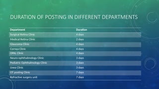
DURATION OF POSTINGIN DIFFERENT DEPARTMENTS
Department Duration
Surgical Retina Clinic 4 days
Medical Retina Clinic 2 days
Glaucoma Clinic 4 days
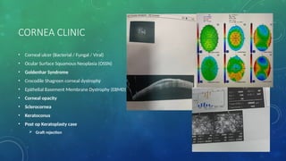
Cornea Clinic 4 days
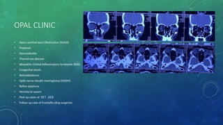
OPAL Clinic 4 days
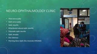
Neuro-ophthalmology Clinic 3 days
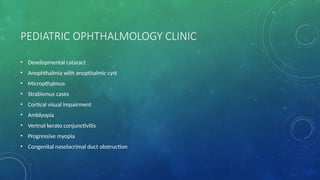
Pediatric Ophthalmology Clinic 3 days
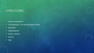
Uvea Clinic 3 days
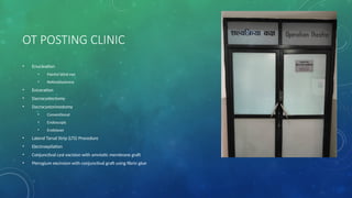
OT posting Clinic 7 days
Refractive surgery unit 7 days
5.
• Resident presenation: Monday and Wednesday
• Dr. Deep Bahadur Karki Sir class : Tuesday and Thursday
6.
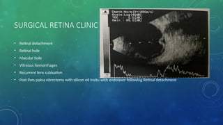
SURGICAL RETINA CLINIC
•Retinal detachment
• Retinal hole
• Macular hole
• Vitreous hemorrhages
• Recurrent lens sublxation
• Post Pars palna vitrectomy with silicon oil insitu with endolaser following Retinal detachment
7.
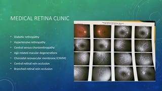
MEDICAL RETINA CLINIC
•Diabetic retinopathy
• Hypertensive retinopathy
• Central serous chorioretinopathy
• Age related macular degenerations
• Choroidal neovascular membrane (CNVM)
• Central retinal vein occlusion
• Branched retinal vein occlusion
8.
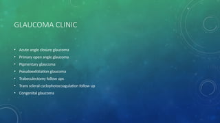
GLAUCOMA CLINIC
• Acuteangle closure glaucoma
• Primary open angle glaucoma
• Pigmentary glaucoma
• Pseudoexfoliation glaucoma
• Trabeculectomy follow ups
• Trans scleral cyclophotocoagulation follow up
• Congenital glaucoma
• SICS
• Phacoemulsificationwith FIOL
• Phacoemulsification with FIOL with pupillary stretch
• Phacoemulsification with trabeculectomy
17.
• Pars planavitrectomy with endolaser with silicon oil insitu
• Silicon oil removal
• Pars plana vitrectomy with membrane peeling with endolaser with silicon oil insitu
• Lens extraction with scleral fiixation IOL
• PPV with vitreous haemorrhage
REFRACTIVE SURGERY UNIT
•LASIK
• SMILE PRO
• SMILE +
• LASIK +
• PRK
• ICL
• C3R/ CXL
• CAIRS (Corneal Allogenic Intrastromal Ring Segments)
21.
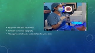
• Equipment used:Zeiss Visumax 800
• Pentacam and corneal topography
• The department follows the protocol of London Vision Clinic
22.
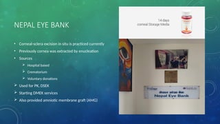
NEPAL EYE BANK
•Corneal-sclera excision in situ is practiced currently
• Previously cornea was extracted by enucleation
• Sources
Hospital based
Crematorium
Voluntary donations
Used for PK, DSEK
Starting DMEK services
Also provided amniotic membrane graft (AMG)


![CTEV [ clubfoot] DR ARUN LAL ,DR MOHAMED ASHRAF travancore medical college k...](https://cdn.slidesharecdn.com/ss_thumbnails/ctevclubfootdrarunlaldrmohamedashraftravancoremedicalcollegekollamkeralaindia-260208063247-18fc466c-thumbnail.jpg?width=640&height=640&fit=bounds)


