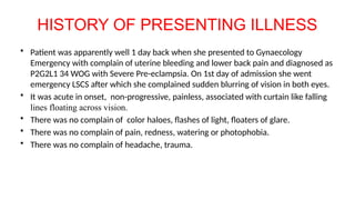
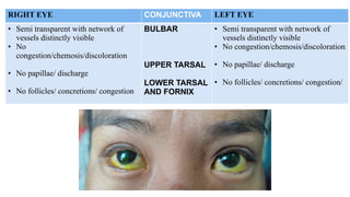
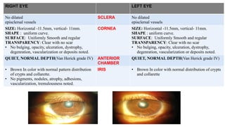
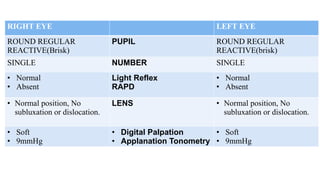
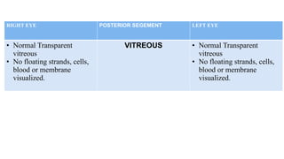
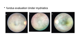
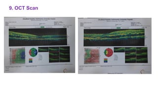
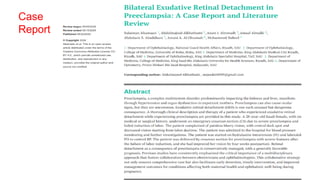
CASE PRESENTATION
Dr. Rahul Gupta
2nd year Resident
Department of ophthalmology
8th January, 2025
• H. No: 81049059
• Name: Samjhana Tamang
• Age : 30 years
• Sex : Female
• Occupation : Housewife
• Address : Melamchi-7, Sindhupalchowk
• Contact: 9703932191
• Mode of presentation: Gynaecological ER
• Date of Presentation : 2024/12/26
CHIEF COMPLAINS
1. Sudden blurring vision of both eyes since 1 day
2.Curtains lines floating across vision of both eyes since 1 day.
HISTORY OF PRESENTING ILLNESS
• Patient was apparently well 1 day back when she presented to Gynaecology
Emergency with complain of uterine bleeding and lower back pain and diagnosed as
P2G2L1 34 WOG with Severe Pre-eclampsia. On 1st day of admission she went
emergency LSCS after which she complained sudden blurring of vision in both eyes.
• It was acute in onset, non-progressive, painless, associated with curtain like falling
lines floating across vision.
• There was no complain of color haloes, flashes of light, floaters of glare.
• There was no complain of pain, redness, watering or photophobia.
• There was no complain of headache, trauma.
HISTORY OF OCULAR ILLNESS:
• No similar ocular illness in the past.
• No any ocular surgery in past.
• No history of use of glasses.
HISTORY SYSTEMIC ILLNESS:
• No any chronic illness lke Hypertension, Diabetes,
• No any surgical history.
TREATMENT HISTORY
• No any treatment or medication history known in past,
• Patient was treated with following medications during her hospital stay;
1. Injection Labetalol 100mg IV
2. Injection Magnesium Sulphate 4gm IV + 5 gm IM
then 5 gm IM 6 hourly
3. Inj. Ceftriaxone 2 gm IV BD
4. Inj. Ranitidine 50mg IV BD
5. Inj. Pethidine 50mg IM TDS
6. Inj. Phenargan 25mg IM TDS
7. Inj. Paracetamol 1gm IV TDS
8. Inj. Tranexamic Acid 1gm IV TDS
PERSONAL HISTORY
– Non Vegetarian,
– Non - Alcoholic
– Non- Smoker
– Regular bowel and bladder habit
– Good appetite
FAMILY HISTORY
-No similar history in the family
SOCIO-ECONOMIC HISTORY
-Patient comes under average middle class
DRUG AND ALLERGY HISTORY:
• No any known allergies to drugs or food.
EXAMINATION
GENERAL EXAMINATION
Physical examination
• Ill looking, concious and oriented to time, place and person
• Average built, anxious
Vitals
Temp : Afebrile
Pulse : 84/min
Respiratory rate : 22/min
Blood pressure : 160/90mm of Hg at right arm
Pallor/Icterus/Lymphadenopathy/Clubbing/Edema/
Cyanosis/dehydration : Absent
SYSTEMIC EXAMINATION
Respiratory system – B/L vesicular breath sound
Cardiovascular system - S1 & S2 audible; no murmur
Nervous system: sensory and motor system intact
Gastrointestinal system: Normal bowel sound, No
organomegaly
Ocular Examination
1. Visual acuity (VA) at bed side
2. Near vision: - Could’nt be performed at
bedside
2. Refraction
- Could’nt be performed
at bedside
Right eye Feature Left eye
Counting
Fingers at 3
meter
VA( unaided)
At bed side
Counting
Fingers at 3
meter
External Ocular Examination
HEAD POSTURE NO HEAD TILT, CHIN LIFT, OR FACE
TURN,
FACIAL SYMETRY BILATERAL SYMMETRICAL
Orbit Normal
RIGHT EYE LEFT EYE
FULL RANGE IN ALL THE
DIRECTION OF GAZE
EOM FULL RANGE IN ALL THE
DIRECTION OF GAZE
EOM- Full in all range
RIGHT EYE LEFT EYE
HIRSCHBERG TEST Reflex present at center Reflex present at center
COVER TEST NO ANY CORRECTIVE
MOVEMENT
NO ANY CORRECTIVE
MOVEMENT
UNCOVER TEST NO ANY CORRECTIVE
MOVEMENT
NO ANY CORRECTIVE
MOVEMENT
ALTRANATE COVER TEST NO ANY CORRECTIVE
MOVEMENT
NO ANY CORRECTIVE
MOVEMENT
Impression: Orthophoria
OCULAR EXAMINATION
EYEBROWS RIGHT EYE LEFT EYE
SYMMETRY Symetrically placed,
Comma- shaped.
Symetrically placed,
comma shaped.
CILIA Evenly distributed brow
hairs
Evenly distributed brow
hairs
PAF
HORIZONTAL 28mm 29mm
VERTICAL 10mm 10mm
LID CREASE HEIGHT 3mm 3mm
RIGHT EYE EYELIDS LEFT EYE
• UPPER LID-COVERS 2 MM OF
CORNEA
• LOWER LID- TOUCHES THE
LIMBUS
POSITION • UPPER LID-COVERS 2 MM OF
CORNEA
• LOWER LID- TOUCHES THE
LIMBUS
Lateral Canthi is Acute, and medial
canthus is rounded.
CANTHI Lateral Canthi is Acute, and medial
canthus is rounded.
margin flat, horizontal fold of skin
of lower lid present. no inward or
outward rolling. No swellings, no
pus point
LID MARGIN margin flat, horizontal fold of skin
of lower lid present. No inward or
outward rolling. No swellings, No
pus point
No drooping of eye lid, No loose
eyelid skin
No drooping of eye lid, No loose
eyelid skin
4 rows upper lid
around 2 rows in lower lid.
Misdirected lashes present with
eyelashes deviated inwards
toward the eye.
• No extra rows of lashes
• No loss of lashes
• No graying of lashes
EYE LASHES 4 rows upper lid
around 2 rows in lower lid.
Misdirected lashes present with
eyelashes deviated inwards
toward the eye.
• No extra rows of lashes
• No loss of lashes
• No graying of lashes
OD
OS
RIGHT EYE LEFT EYE
• Skin fold originating at upper
eyelid crease and merges into the
skin near the medial canthus.
• SWELLING,TENTDERNESS (-)
• NORMAL
• 6mm FROM INNER CANTHUS
• 6.5mm FROM INNER CNATHUS
• NO SWELLING, ERYTHEMA
INDURATION,PUS POINTS or
TENDERNESS
EPICANTHAL FOLD
LACRIMAL
APPARATUS
LACRIMAL GLAND
LACRIMAL PUNCTA
UPPER
LOWER
LACRIMAL SAC AREA
• Skin fold originating at upper eyelid
crease and merges into the skin near
the medial canthus.
• SWELLING,TENTDERNESS (-)
• NORMAL
• 6mm FROM INNER CANTHUS
• 6.5mm FROM INNER CANTHUS
• NO SWELLING, ERYTHEMA
INDURATION,PUS POINTS or
TENDERNESS
RIGHT EYE CONJUNCTIVA LEFT EYE
• Semi transparent with network of
vessels distinctly visible
• No
congestion/chemosis/discoloration
• No papillae/ discharge
• No follicles/ concretions/ congestion
BULBAR
UPPER TARSAL
LOWER TARSAL
AND FORNIX
• Semi transparent with network of
vessels distinctly visible
• No congestion/chemosis/discoloration
• No papillae/ discharge
• No follicles/ concretions/ congestion/
RIGHT EYE LEFT EYE
No dilated
episclersal vessels
SCLERA No dilated
episclersal vessels
SIZE: Horizontal -11.5mm, vertical- 11mm.
SHAPE : uniform curve.
SURFACE: Uniformly Smooth and regular
TRANSPARENCY: Clear with no scar
• No bulging, opacity, ulceration, dystrophy,
degenration, vascularization or deposits noted.
CORNEA SIZE: Horizontal -11.5mm, vertical- 11mm.
SHAPE : uniform curve.
SURFACE: Uniformly Smooth and regular
TRANSPARENCY: Clear with no scar
• No bulging, opacity, ulceration, dystrophy,
degenration, vascularization or deposits noted.
QUIET, NORMAL DEPTH(Van Herick grade IV) ANTERIOR
CHAMBER
QUIET, NORMAL DEPTH(Van Herick grade IV)
• Brown In color with normal pattern distribution
of crypts and collarette.
• No pigments, nodules, atrophy, adhesions,
vascularization, tremulousness noted.
IRIS • Brown In color with normal distribution of crypts
and collarette
RIGHT EYE LEFT EYE
ROUND REGULAR
REACTIVE(Brisk)
PUPIL ROUND REGULAR
REACTIVE(brisk)
SINGLE NUMBER SINGLE
• Normal
• Absent
Light Reflex
RAPD
• Normal
• Absent
• Normal position, No
subluxation or dislocation.
LENS • Normal position, No
subluxation or dislocation.
• Soft
• 9mmHg
• Digital Palpation
• Applanation Tonometry
• Soft
• 9mmHg
RIGHT EYE POSTERIOR SEGEMENT LEFT EYE
• Normal Transparent
vitreous
• No floating strands, cells,
blood or membrane
visualized.
VITREOUS • Normal Transparent
vitreous
• No floating strands, cells,
blood or membrane
visualized.
Fundus Examination
DIFFERENTIAL DIAGNOSIS
• Serous Retinal Detachment
• Regmatogenous Retinal Detachment
• Tractional Retinal Detachment
• Central Serous Chorioretinopathy
• Vitreous Hemorrhage
• Central Retinal Artery Occlusion
• Optic Neuritis
Reports From Previous Investigation
1. Complete blood count: Within Normal limit
2. Blood group: “B Positive”
3. Hemoglobin: 9.9 g/dl
4. Serology: Non Reactive
5. Total Protein : 5.6 low (6.5-8.2)
6. Serum Albumin : 2.6 low (3.5-5)
7. Creatinine : 1.6 High(0.4-1.1)
8. Uric Acid : 8.8 High(2.4-5.7)
9. Liver Function Test : Within Normal Limit
10. Renal Function Test : Within normal Limit
Urine RME: Within normal Limit
• INDIRECT OPHTHALMOSCOPY: ALTERED RED AND
GREYISH - WHITE REFLEX IN PUPILLARY AREA
FUNDUS EVALUATION:
• Vitreous was Transparent
• Both fundus shows balloning with fluid collection within
retina temporal to macula
• Convex elevation of retinal layer with presence of
vascular network.
• No traction band noted, no retinal foldings or tears noted.
Treatment:
1. Gtt. Acular LS 1 Drop BE TDS for 1 month.
2. control High Blood pressure
Patient Councelling done.
Follow up Advised In OPD.
Case Reviewed in OPD on 31st Dec, 2024
Chief Complain:
• Mild improved vision of both eyes
Ocular Examination
1. Visual acuity (VA) 2. Refraction
No improvement
(retinoscopy)
Right eye Feature Left eye
6/18
VA( unaided) 6/18
6/18 VA (with
pinhole)
6/18 0
0
3.Slitlamb Examination:
Eyelid : Epicanthus Tarsalis noted.
Eyelashes : Trichaisis Present = Epilation done under topical Anaesthesia.
Cornea: Superficial Puntate Keratitis noted at lower region.
Pupil : RRR, RAPD absent, Altered red and greyish glow noted.
• fundus evaluation Under mydriatics
No defect to finger confrontation
4.IOP assessment via
goldman applation
9mm Hg 9mm Hg
@10:45 AM
5. Confrontation Test
LE RE
T N
N T
6. Color Vision : Normal
7. Contrast Sensitivity : BE-1.68 logunit
8. B- SCAN
9. OCT Scan
Provisional Diagnosis
• BE Trichiasis with Epicanthus Tarsalis with Serous Retinal
Detachment Resolving (Temporal Region) with Grade 1
Hypertensive Retinopathy with Resolving seroius Pre-
eclampsia.
Treatment
• Gtt. Acular LS(Ketorolac) 1drop BE TDS for 1 month
• Other Medication continued as per Cardex
• Protective Glass advised
• Strict control of High Blood pressure advised
Note: Patient councelling done regarding visual prognosis
Follow up : 1 week / SOS
Case
Report
Summary of case reports:
• Exudative retinal detachment (ERD) is an uncommon but significant ocular complication associated
with preeclampsia, a hypertensive disorder occurring during pregnancy. ERD involves the accumulation
of fluid in the subretinal space, leading to the separation of the neurosensory retina from the retinal
pigment epithelium, which can result in visual disturbances and potential vision loss if not promptly
addressed.
• The pathophysiology of ERD in preeclampsia is thought to involve choroidal ischemia due to increased
choroidal blood flow impedance, resulting from severe hypertension during pregnancy. This disruption
may lead to fluid leakage and subsequent retinal detachment.
• Although visual symptoms are reported in 25-50% of preeclamptic patients, ERD remains a rare
manifestation, occurring in less than 1% of preeclampsia cases and about 10% of eclampsia cases.
Common visual symptoms include blurred vision, scotoma, and decreased visual acuity. Fundoscopic
examination typically reveals multiple areas of retinal detachment without signs of hypertensive
retinopathy.
• Management of ERD in preeclampsia primarily focuses on controlling blood pressure and timely
delivery of the fetus. Visual prognosis is generally favorable, with spontaneous resorption of subretinal
fluid and complete resolution of the detachment occurring within a few weeks postpartum.
• In summary, while ERD is a rare complication of preeclampsia, it necessitates prompt recognition and
management to prevent potential vision loss. Multidisciplinary care involving obstetricians and
ophthalmologists is crucial to ensure optimal outcomes for both mother and child
Retinal Detachment Ophthalmology Case.pptx

Retinal Detachment Ophthalmology Case.pptx

  • 1.
    CASE PRESENTATION Dr. RahulGupta 2nd year Resident Department of ophthalmology 8th January, 2025
  • 2.
    • H. No:81049059 • Name: Samjhana Tamang • Age : 30 years • Sex : Female • Occupation : Housewife • Address : Melamchi-7, Sindhupalchowk • Contact: 9703932191 • Mode of presentation: Gynaecological ER • Date of Presentation : 2024/12/26
  • 3.
    CHIEF COMPLAINS 1. Suddenblurring vision of both eyes since 1 day 2.Curtains lines floating across vision of both eyes since 1 day.
  • 4.
    HISTORY OF PRESENTINGILLNESS • Patient was apparently well 1 day back when she presented to Gynaecology Emergency with complain of uterine bleeding and lower back pain and diagnosed as P2G2L1 34 WOG with Severe Pre-eclampsia. On 1st day of admission she went emergency LSCS after which she complained sudden blurring of vision in both eyes. • It was acute in onset, non-progressive, painless, associated with curtain like falling lines floating across vision. • There was no complain of color haloes, flashes of light, floaters of glare. • There was no complain of pain, redness, watering or photophobia. • There was no complain of headache, trauma.
  • 5.
    HISTORY OF OCULARILLNESS: • No similar ocular illness in the past. • No any ocular surgery in past. • No history of use of glasses. HISTORY SYSTEMIC ILLNESS: • No any chronic illness lke Hypertension, Diabetes, • No any surgical history.
  • 6.
    TREATMENT HISTORY • Noany treatment or medication history known in past, • Patient was treated with following medications during her hospital stay; 1. Injection Labetalol 100mg IV 2. Injection Magnesium Sulphate 4gm IV + 5 gm IM then 5 gm IM 6 hourly 3. Inj. Ceftriaxone 2 gm IV BD 4. Inj. Ranitidine 50mg IV BD 5. Inj. Pethidine 50mg IM TDS 6. Inj. Phenargan 25mg IM TDS 7. Inj. Paracetamol 1gm IV TDS 8. Inj. Tranexamic Acid 1gm IV TDS
  • 7.
    PERSONAL HISTORY – NonVegetarian, – Non - Alcoholic – Non- Smoker – Regular bowel and bladder habit – Good appetite FAMILY HISTORY -No similar history in the family SOCIO-ECONOMIC HISTORY -Patient comes under average middle class
  • 8.
    DRUG AND ALLERGYHISTORY: • No any known allergies to drugs or food.
  • 9.
  • 10.
    GENERAL EXAMINATION Physical examination •Ill looking, concious and oriented to time, place and person • Average built, anxious Vitals Temp : Afebrile Pulse : 84/min Respiratory rate : 22/min Blood pressure : 160/90mm of Hg at right arm Pallor/Icterus/Lymphadenopathy/Clubbing/Edema/ Cyanosis/dehydration : Absent
  • 11.
    SYSTEMIC EXAMINATION Respiratory system– B/L vesicular breath sound Cardiovascular system - S1 & S2 audible; no murmur Nervous system: sensory and motor system intact Gastrointestinal system: Normal bowel sound, No organomegaly
  • 12.
    Ocular Examination 1. Visualacuity (VA) at bed side 2. Near vision: - Could’nt be performed at bedside 2. Refraction - Could’nt be performed at bedside Right eye Feature Left eye Counting Fingers at 3 meter VA( unaided) At bed side Counting Fingers at 3 meter
  • 13.
    External Ocular Examination HEADPOSTURE NO HEAD TILT, CHIN LIFT, OR FACE TURN, FACIAL SYMETRY BILATERAL SYMMETRICAL Orbit Normal
  • 14.
    RIGHT EYE LEFTEYE FULL RANGE IN ALL THE DIRECTION OF GAZE EOM FULL RANGE IN ALL THE DIRECTION OF GAZE EOM- Full in all range
  • 15.
    RIGHT EYE LEFTEYE HIRSCHBERG TEST Reflex present at center Reflex present at center COVER TEST NO ANY CORRECTIVE MOVEMENT NO ANY CORRECTIVE MOVEMENT UNCOVER TEST NO ANY CORRECTIVE MOVEMENT NO ANY CORRECTIVE MOVEMENT ALTRANATE COVER TEST NO ANY CORRECTIVE MOVEMENT NO ANY CORRECTIVE MOVEMENT Impression: Orthophoria
  • 16.
    OCULAR EXAMINATION EYEBROWS RIGHTEYE LEFT EYE SYMMETRY Symetrically placed, Comma- shaped. Symetrically placed, comma shaped. CILIA Evenly distributed brow hairs Evenly distributed brow hairs PAF HORIZONTAL 28mm 29mm VERTICAL 10mm 10mm LID CREASE HEIGHT 3mm 3mm
  • 17.
    RIGHT EYE EYELIDSLEFT EYE • UPPER LID-COVERS 2 MM OF CORNEA • LOWER LID- TOUCHES THE LIMBUS POSITION • UPPER LID-COVERS 2 MM OF CORNEA • LOWER LID- TOUCHES THE LIMBUS Lateral Canthi is Acute, and medial canthus is rounded. CANTHI Lateral Canthi is Acute, and medial canthus is rounded. margin flat, horizontal fold of skin of lower lid present. no inward or outward rolling. No swellings, no pus point LID MARGIN margin flat, horizontal fold of skin of lower lid present. No inward or outward rolling. No swellings, No pus point No drooping of eye lid, No loose eyelid skin No drooping of eye lid, No loose eyelid skin 4 rows upper lid around 2 rows in lower lid. Misdirected lashes present with eyelashes deviated inwards toward the eye. • No extra rows of lashes • No loss of lashes • No graying of lashes EYE LASHES 4 rows upper lid around 2 rows in lower lid. Misdirected lashes present with eyelashes deviated inwards toward the eye. • No extra rows of lashes • No loss of lashes • No graying of lashes OD OS
  • 18.
    RIGHT EYE LEFTEYE • Skin fold originating at upper eyelid crease and merges into the skin near the medial canthus. • SWELLING,TENTDERNESS (-) • NORMAL • 6mm FROM INNER CANTHUS • 6.5mm FROM INNER CNATHUS • NO SWELLING, ERYTHEMA INDURATION,PUS POINTS or TENDERNESS EPICANTHAL FOLD LACRIMAL APPARATUS LACRIMAL GLAND LACRIMAL PUNCTA UPPER LOWER LACRIMAL SAC AREA • Skin fold originating at upper eyelid crease and merges into the skin near the medial canthus. • SWELLING,TENTDERNESS (-) • NORMAL • 6mm FROM INNER CANTHUS • 6.5mm FROM INNER CANTHUS • NO SWELLING, ERYTHEMA INDURATION,PUS POINTS or TENDERNESS
  • 19.
    RIGHT EYE CONJUNCTIVALEFT EYE • Semi transparent with network of vessels distinctly visible • No congestion/chemosis/discoloration • No papillae/ discharge • No follicles/ concretions/ congestion BULBAR UPPER TARSAL LOWER TARSAL AND FORNIX • Semi transparent with network of vessels distinctly visible • No congestion/chemosis/discoloration • No papillae/ discharge • No follicles/ concretions/ congestion/
  • 20.
    RIGHT EYE LEFTEYE No dilated episclersal vessels SCLERA No dilated episclersal vessels SIZE: Horizontal -11.5mm, vertical- 11mm. SHAPE : uniform curve. SURFACE: Uniformly Smooth and regular TRANSPARENCY: Clear with no scar • No bulging, opacity, ulceration, dystrophy, degenration, vascularization or deposits noted. CORNEA SIZE: Horizontal -11.5mm, vertical- 11mm. SHAPE : uniform curve. SURFACE: Uniformly Smooth and regular TRANSPARENCY: Clear with no scar • No bulging, opacity, ulceration, dystrophy, degenration, vascularization or deposits noted. QUIET, NORMAL DEPTH(Van Herick grade IV) ANTERIOR CHAMBER QUIET, NORMAL DEPTH(Van Herick grade IV) • Brown In color with normal pattern distribution of crypts and collarette. • No pigments, nodules, atrophy, adhesions, vascularization, tremulousness noted. IRIS • Brown In color with normal distribution of crypts and collarette
  • 21.
    RIGHT EYE LEFTEYE ROUND REGULAR REACTIVE(Brisk) PUPIL ROUND REGULAR REACTIVE(brisk) SINGLE NUMBER SINGLE • Normal • Absent Light Reflex RAPD • Normal • Absent • Normal position, No subluxation or dislocation. LENS • Normal position, No subluxation or dislocation. • Soft • 9mmHg • Digital Palpation • Applanation Tonometry • Soft • 9mmHg
  • 22.
    RIGHT EYE POSTERIORSEGEMENT LEFT EYE • Normal Transparent vitreous • No floating strands, cells, blood or membrane visualized. VITREOUS • Normal Transparent vitreous • No floating strands, cells, blood or membrane visualized.
  • 23.
  • 24.
    DIFFERENTIAL DIAGNOSIS • SerousRetinal Detachment • Regmatogenous Retinal Detachment • Tractional Retinal Detachment • Central Serous Chorioretinopathy • Vitreous Hemorrhage • Central Retinal Artery Occlusion • Optic Neuritis
  • 25.
    Reports From PreviousInvestigation 1. Complete blood count: Within Normal limit 2. Blood group: “B Positive” 3. Hemoglobin: 9.9 g/dl 4. Serology: Non Reactive 5. Total Protein : 5.6 low (6.5-8.2) 6. Serum Albumin : 2.6 low (3.5-5) 7. Creatinine : 1.6 High(0.4-1.1) 8. Uric Acid : 8.8 High(2.4-5.7) 9. Liver Function Test : Within Normal Limit 10. Renal Function Test : Within normal Limit Urine RME: Within normal Limit
  • 26.
    • INDIRECT OPHTHALMOSCOPY:ALTERED RED AND GREYISH - WHITE REFLEX IN PUPILLARY AREA FUNDUS EVALUATION: • Vitreous was Transparent • Both fundus shows balloning with fluid collection within retina temporal to macula • Convex elevation of retinal layer with presence of vascular network. • No traction band noted, no retinal foldings or tears noted.
  • 27.
    Treatment: 1. Gtt. AcularLS 1 Drop BE TDS for 1 month. 2. control High Blood pressure Patient Councelling done. Follow up Advised In OPD.
  • 28.
    Case Reviewed inOPD on 31st Dec, 2024 Chief Complain: • Mild improved vision of both eyes
  • 29.
    Ocular Examination 1. Visualacuity (VA) 2. Refraction No improvement (retinoscopy) Right eye Feature Left eye 6/18 VA( unaided) 6/18 6/18 VA (with pinhole) 6/18 0 0
  • 30.
    3.Slitlamb Examination: Eyelid :Epicanthus Tarsalis noted. Eyelashes : Trichaisis Present = Epilation done under topical Anaesthesia. Cornea: Superficial Puntate Keratitis noted at lower region. Pupil : RRR, RAPD absent, Altered red and greyish glow noted.
  • 31.
    • fundus evaluationUnder mydriatics
  • 32.
    No defect tofinger confrontation 4.IOP assessment via goldman applation 9mm Hg 9mm Hg @10:45 AM 5. Confrontation Test LE RE T N N T
  • 33.
    6. Color Vision: Normal 7. Contrast Sensitivity : BE-1.68 logunit
  • 34.
  • 35.
  • 36.
    Provisional Diagnosis • BETrichiasis with Epicanthus Tarsalis with Serous Retinal Detachment Resolving (Temporal Region) with Grade 1 Hypertensive Retinopathy with Resolving seroius Pre- eclampsia.
  • 37.
    Treatment • Gtt. AcularLS(Ketorolac) 1drop BE TDS for 1 month • Other Medication continued as per Cardex • Protective Glass advised • Strict control of High Blood pressure advised Note: Patient councelling done regarding visual prognosis Follow up : 1 week / SOS
  • 38.
  • 39.
    Summary of casereports: • Exudative retinal detachment (ERD) is an uncommon but significant ocular complication associated with preeclampsia, a hypertensive disorder occurring during pregnancy. ERD involves the accumulation of fluid in the subretinal space, leading to the separation of the neurosensory retina from the retinal pigment epithelium, which can result in visual disturbances and potential vision loss if not promptly addressed. • The pathophysiology of ERD in preeclampsia is thought to involve choroidal ischemia due to increased choroidal blood flow impedance, resulting from severe hypertension during pregnancy. This disruption may lead to fluid leakage and subsequent retinal detachment. • Although visual symptoms are reported in 25-50% of preeclamptic patients, ERD remains a rare manifestation, occurring in less than 1% of preeclampsia cases and about 10% of eclampsia cases. Common visual symptoms include blurred vision, scotoma, and decreased visual acuity. Fundoscopic examination typically reveals multiple areas of retinal detachment without signs of hypertensive retinopathy. • Management of ERD in preeclampsia primarily focuses on controlling blood pressure and timely delivery of the fetus. Visual prognosis is generally favorable, with spontaneous resorption of subretinal fluid and complete resolution of the detachment occurring within a few weeks postpartum. • In summary, while ERD is a rare complication of preeclampsia, it necessitates prompt recognition and management to prevent potential vision loss. Multidisciplinary care involving obstetricians and ophthalmologists is crucial to ensure optimal outcomes for both mother and child

Editor's Notes

  • #3 Orbital cellulitis Orbital abscess
  • #4 Color haloes is seen in= ACG, conjunctivitis, corneal degeneration, cataract, corneal edema, Astigmatism Photopsia( flashes of light) = PVD, Retinal detachment, Vitreous hemorrhage, pressure applied on closed eye, Uvietis floaters = Vitreous degeneration,Uveitis Glare = Keratoconus, Migraine, Cataract
  • #5 No history of Tuberculosis, Diabetes.
  • #6 No any significant treatment history
  • #7 According to kuppuswami scale he belongs to average middle class family.
  • #10 96 drg fahrenheit
  • #12 On Visual acquity There was HM in right eye and on preojection of rays, it was present in all quadrants. Visual acuity charts : Snellens Chart, Landolt C chart, Allens picture chaer. etc Near Chart: jaegers chart, Romans near vision chart Snellen’s Near vision chart.
  • #13 Eye ball Examination: The two eyeballs are symmetrically placed in the orbits in such a way that line joining the central points of superior and inferior orbital margins just touches cornea. Head Posture: In paralytic squint head posture is abnormal. In complete ptosis chin is elevated to uncover pupil area. Head is turned in the direction of paralysed muscle to avoid diplopia. Forehead examination: Increased wrinkling due to overaction of frontalis muscle in patient of ptosis. Complete loss of wrinkling in one half forehead with lower motor neural palsy. Facial symetry may be affected in facial or bells palsy.
  • #14 EOM EXAMINATION: Uniocular movement: aka duction, these are: there was full range of movement in adduction, abduction, supraduction and infraduction. Adduction: medial roation along vertical axis, Abduction: lateral rotation along vertical axis, Infraduction: downward movement(depression) along horizontal axis, Supraduction: upward movement(elevation) along horizontal axis, Incycloduction(Intorsion): rotatory movement along AP axis in which superior pole of cornea moces medially, Excycloduction(Extortion): rotatory movement along AP axis in which superior pole of cornea moves laterally. Binocular movement: Version aka conjugate movement of both eyes in same direction : On binocular versions examination, there was full range of movement in Levoversion, Levoelevation, Levodepression and Dextroversion, Dextro elevation, dextro depression. Dextroversion: movement of both eyes to right, Levoversion: movement of both eyes to left. Vergence aka disjugate movement are symmetric movement of eyes: On binocular vergence evaluation, Convergence: simultaneous inward movement of eyes. Divergence: simultaneous outward movement of eyes.
  • #15 the Hirschberg test, also Hirschberg corneal reflex test, is a screening test that can be used to assess whether a person has strabismus (ocular misalignment). In normal condition corneal reflex is centrally placed. Cover and uncover is done for tropias altranate cover test done for phorias
  • #16 Note: The two eyebrows are horizontaly placed over superciliary ridge of the frontal bone seperated by glabella. Curved and with convexity upward, POSITION RE=Across superior orbital margin LE = Across superior orbital margin The Level of eyebrows may be changed in patient with ptosis due to overaction of frontalis muscle, where ptotic eye have raised eyebrow. Cilia of lateral 1/3 may be absent(madarosis) in pt. with leprosy or myxedema. In Brow ptosis there is downward pulling of eyebrows. Abnormality of PAH: 1. Ankyloblepharon is usually seen in adhesion of lids following burns, ulcerative blepharitis. 2. Blepharophimosis: congenital anamoly in which all around narrowing of Palpebral fissure.
  • #17 IN primary position if gaze the upper eyelid covers around 2 mm and lower eye lid just touches the cornea. Canthus: In Both eyes the lateral canthus forms acute angle of 60 degrees (2mm above medial canthus) and medial canthus is rounded. the lateral canthus is slightly positioned upward compared to medial canthus. LID MARGIN : In both the eyes the opposing lid margins are nearly flat 2mm in width, the lacrimal papillae with lacrimal punta present. The lacrimal portion of lid margin is rounded and devoid of lashes. The ciliary portion consist of rounded ant. border and sharp border placed against globe. distance of punta ( upper punta is 6mm and lower punta is 6.5 mm from medial canthus). with presence of horizontal linear gray line. EYE LASHES: arrenged in 2-3 rows in upperlid, directed forward upward backward. In lower lid forward downward and backward In ptosis upper lid covers more than 1/6th (2mm) cornea. Upper limbus is visible due to lid retraction seen in thyrotoxicosis and sympathetic overactivity. Movement of lids: upper lid follows eyeball in downward movement but lags behind in case of TED. blinking is decreased in trigeminal anaesthesia and absent in 7th nerve palsy.
  • #18 Epicanthus supraciliaris runs from the brow, curving downwards towards the lachrymal sac. Epicanthus palpebralis begins above the upper tarsus and extends to the inferior orbital rim. Epicanthus tarsalis originates at the upper eyelid crease and merges into the skin near the medial canthus. This is the type most often found in East Asians. Epicanthus inversus runs from the lower eyelid skin over the medial canthus and extends to the upper lid. Lacrimal Apparatus: While Examination of lacrimal apparatus, On inspection and palpation of lacrimal sac region there was no redness, swelling of fistula. While examining lacrimal punta, there was no defect such as eversion, stenosis, abscence or discharge. ROPLAS: Regurgitation on pressure over lacrimal sac. positve in Dacrosystitis and NLDO.
  • #19 Normal conjunctiva is a thin semi-transparent structure. A fine network of vessels is distinctly seen. Discoloration of conjunctiva may be brownish in melanosis and argyrosis (silver nitrate deposits), greyish due to surma deposits, pale in anaemia, bluish in cyanosis and bright red due to subconjunctival haemorrhage.
  • #20 Sclera: becomes yellowish in jaundice, bluish discoloration seen in isolated anomaly or in osteitis deformans, Marfans syndrome, Pseudoxanthoma elasticum. Inflamation of sclera: Episcleritis: Superficial localised pink or purple circumscribed flat nodule. Scleritis: deep, dusky patch associated with marked inflammation and ciliary congestion. 5. Cornea: Ant. surface elliptical with horizontal diameter of 11.7mm and vertical diameter of 11mm. SURFACE smothness of cornea can be examined by use of Placido Keratoscopic disc(disc painted with alternating black and white circles). SHEEN= Normal cornea is a bright shining structure. Sheen of corneal surface is lost in dry eye conditions. While Examining Corneal Endothelium: Specular microscopy clear morphological study of endothelial cells including cell density of endothelium is around 3000 cells/mm2 in young adults, which decreases with advancing age.
  • #21 PUPIL: Single pupil in each eye placed centrally around, circular in shape with 3 mm in size and round regular and reactive to light. On direct light reflex constriction of pupil of ipsilateral eye on both eyes. On consentual light reflex constriction of pupil on contralateral eye and on swinging light reflex RAPD was absent. There was no Synaeciae. LENS : Position of lens normally positioned in the patellar fossa. Normal thickness 3-4 mm of lens.( IOL thickness around 1mm). Shape of lens= Normal lens is a biconvex structure, which is nicely demonstrated in an optical section of the lens on slit-lamp examination. COLOR: In nuclear cataract lens may look amber, brown or black in colour
  • #23 Disc- pink colored, round regular, well defined , NRR heatly , following ISNT rule, C:D – 0.3:1:0.3, Retinal vessels originating from centre of disc to periphery forming SN, ST, IN, IT branches. Its AVR-2:3, Presence of gross arterial atenuation of supero temporal branch. Macula-is Normal situated at the posterior pole with its centre (foveola) being about 2 disc diameters lateral of disc and macula is slightly darker than the surrounding. there was abscence of foveal reflexin both eyes. There was convex elevation and tense balloning of retina with fluid collection at 7 to 11 clock hour of RE and 1 to 5 clock hour of LE around 2DD temporal side of macula of both eye. Which most probably signifies Detached retina with subretinal fluid collection.
  • #29 On Visual acquity There was HM in right eye and on preojection of rays, it was present in all quadrants. Visual acuity charts : Snellens Chart, Landolt C chart, Allens picture chaer. etc Near Chart: jaegers chart, Romans near vision chart Snellen’s Near vision chart.
  • #31 Disc- pink colored, round regular, well defined , NRR heatly , following ISNT rule, C:D – 0.3:1:0.3, Retinal vessels originating from centre of disc to periphery forming SN, ST, IN, IT branches. Its AVR-2:3, Presence of gross arterial atenuation of supero temporal branch. Macula-is Normal situated at the posterior pole with its centre (foveola) being about 2 disc diameters lateral of disc and macula is slightly darker than the surrounding. there was abscence of foveal reflex in both eyes. Gradual obsorption of fluid/ exudates within retina temporal side of macula of both eye. formation of pigments spots around resolving ares of detached retina also known as leopards spots. Which signifies resolving serous retinal detachment.
  • #34 No vitreous abnormality noted in both fundus while mild Retinal thickness noted in left retina.
  • #35 OCT shows BE Resolving Retinal Detachment with Temporal region retinal thickening LE more than RE.