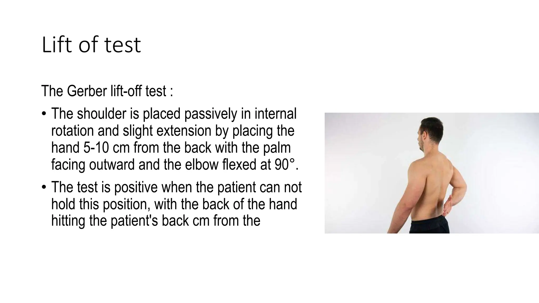
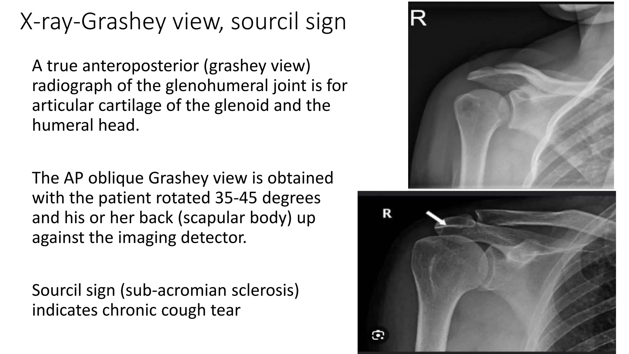
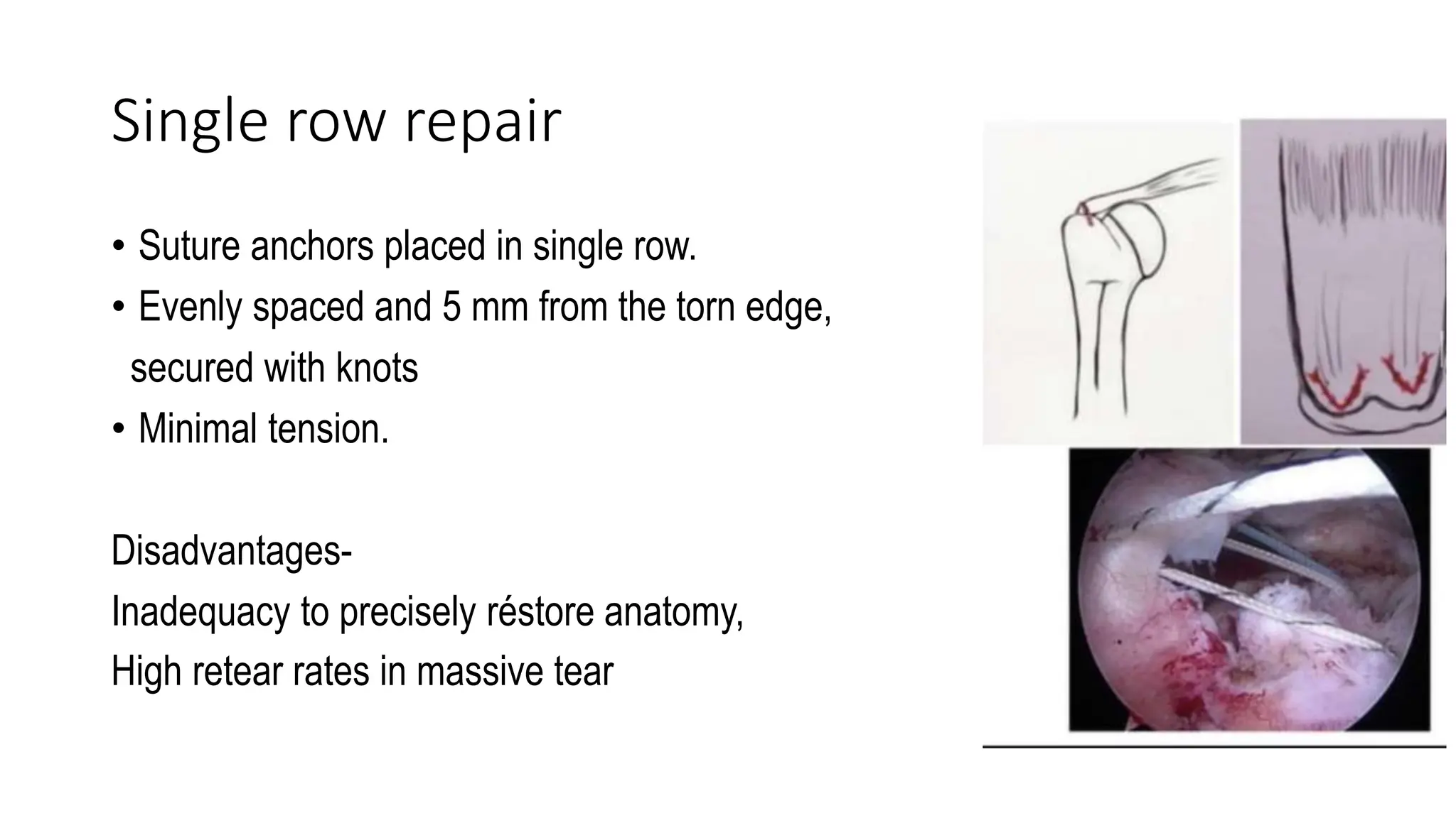
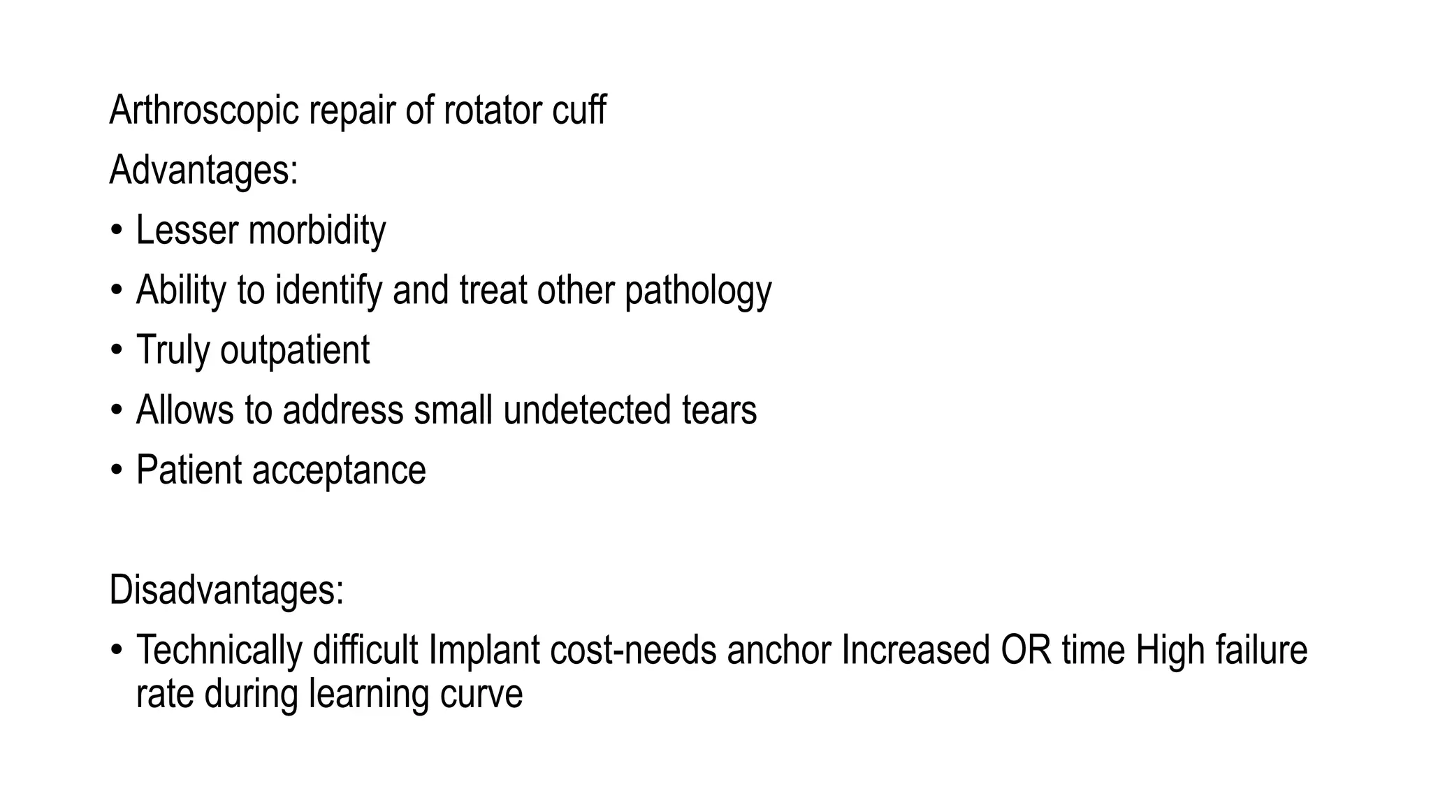
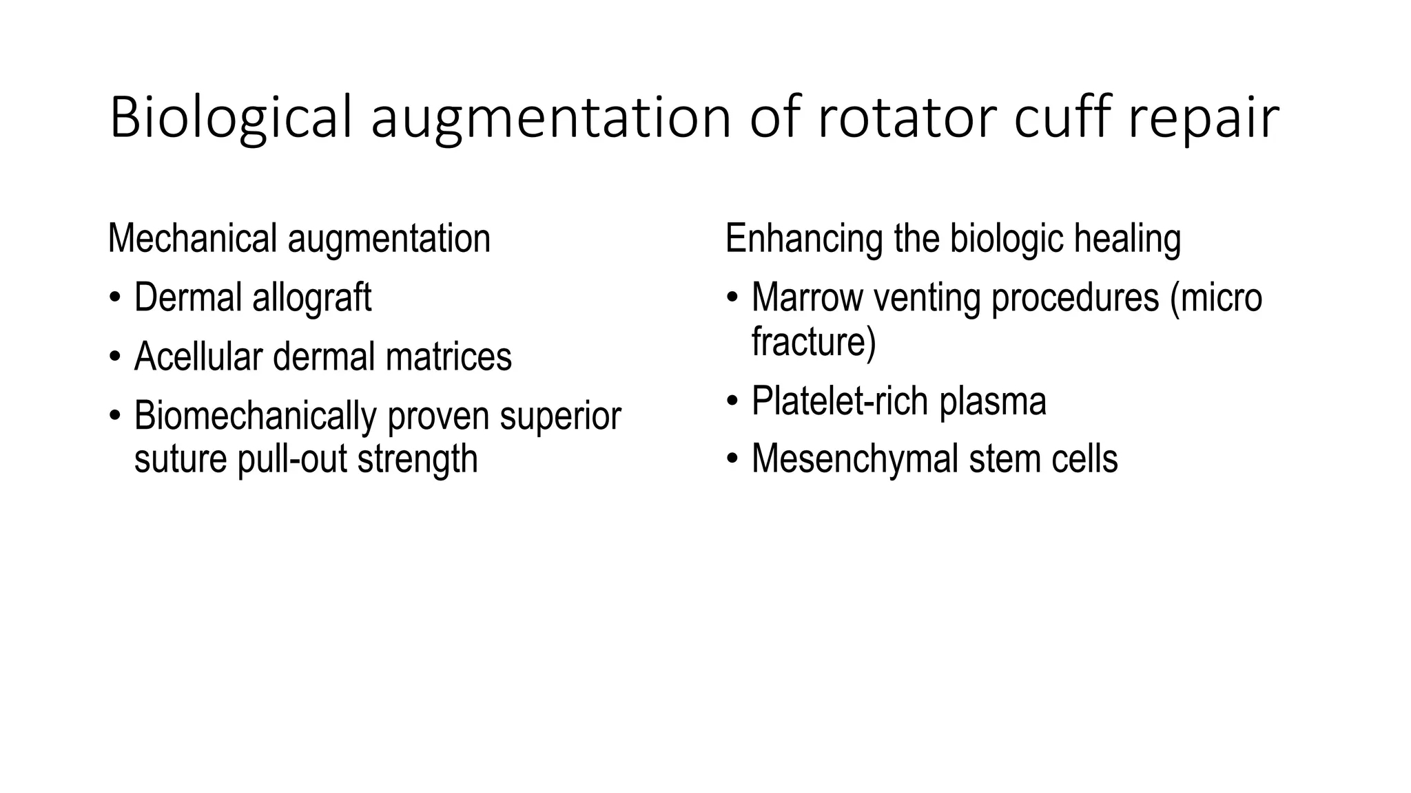
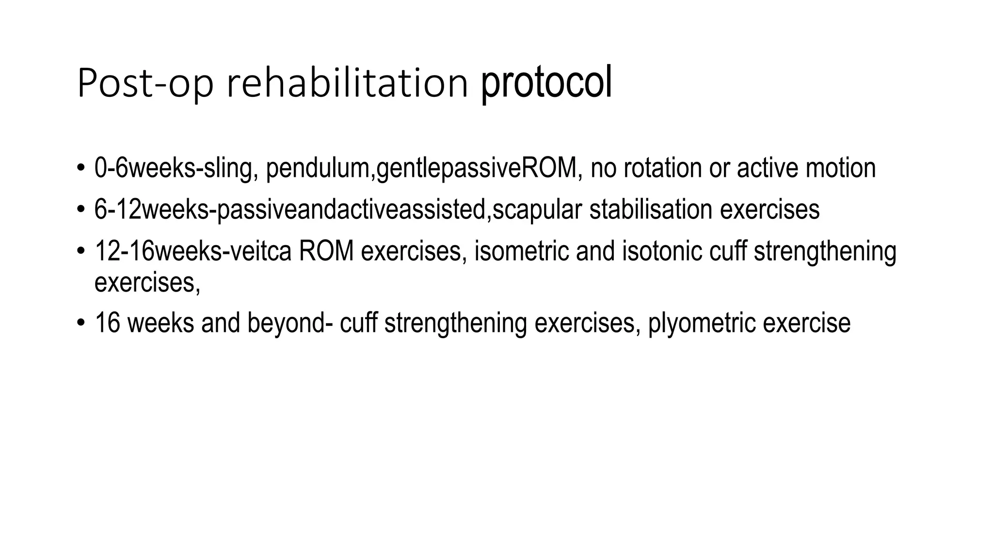
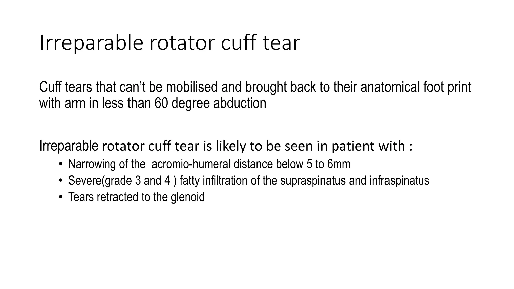
Rotator cuff injuries
Presented By:
Dr Pushpendra Singh Dhakad
Outline
Rotator cuff disorders
• Anatomy
• Aetiology of Rotator cuff tear
• Classification
• Symptoms
• Physical examinations and special tests
• Investigations
• Treatment
• Rehabilitation protocol
• Irreparable cuff tear management
• Cuff tear arthropathy
Anatomy of Rotator cuff
The rotator cuff is made up of 4 interrelated muscles arising from the scapula,
attaching to the tuberosities of the humerus.
• Supraspinatus
• Infraspinatus
• Teres minor
• Subscapularis
Expansion from these four muscles fuse with the capsule and strengthen it all
around except inferiorly
• Long head of biceps- functional part
Layers of Rotator cuff
Layer1
• Superficial fibres that overlie the cuff tendons+ coracohumeral ligament (1mm)
Layer 2
• Fibres of supraspinatus and infraspinatus tendon (3-5 mm)
Layer 3
• Adjacent infraspinatus and subscapularis tendon (3mm)
Layer 4
• Loose collagen fibre merging into deep extension of CHL
Layer 5
• True joint capsule of shoulder
Current perspective on Rotator cuff anatomy
The insertion of the rotator cuff
tendon at GT is referred to as
footprint.
Mean medial to lateral insertion widths-
• Supraspinatus- 12.7 mm
• Infraspinatus- 13.4 mm
• Teres minor-11.4 mm
• Subscapularis- 17.9 mm
Mean anteroposterior distance-
• Supraspinatus-16.3 mm
• Infraspinatus- 16.4 mm
• Teres minor- 20.7 mm
• Subscapularis- 24.3m
Rotator cable and rotator crescent
(suspension bridge)
• Rotator cable is a thick bundle
that act as suspensory support
mechanism to bear forces
applied to rotator cuff
• It offloads and protect the
rotator crescent
• Rotator cuff tear involving cable
corelates more with pain than
other
Rotator interval
It is a triangular area in the anterior and superior shoulder where no rotator cuff tendon
is present. It is bounded by
-Supraspinatus superiorly
-Subscapularis inferiorly
-Coracoid medially
-Apex is marked by transverse humeral ligament
Contents
-Coracohumeral ligament
-Biceps tendon
-Superior glenohumeral ligament
NOTE-It is Contacted in adhesive capsulitis and Expanded in shoulder instability
Classification
DURATION - Acute or chronic
ETIOLOGY- traumatic or degenerative
DEGREE OF TEAR- Partial thickness or full thicknesss.
DEGREE OF TEAR- Partial thickness or full thickness
Size of tear Degree
<1 cm Small
1- 3 cms Medium
3-5 cms Large
>5 cms Massive
Full thickness
COFIELD –
Based on size of tear
Elimans partial thickness classification
1.Articul
2.Bursal
3.Interstitial
Goutallier staging system.
On the basis of the percentage of fatty infiltration of muscle belly seen MRI
Stage Muscle description
0 Completely normal muscle
I Some fatty streaks
II Amount of muscle is greater than fatty infiltration
III Amount of muscle is equal to fatty infiltration
IV Amount of fatty infiltration is greater than muscle
Quantitative assessment of fat infiltration in
rotator cuff muscle using MRI
Pattes classification of rotator cuff tendon
retraction
Stage 1: proximal stump near the bony insertion
Stage 2: proximal stump is at the level of the humeral head
Stage 3: proximal stump at the level of glenoid or more proximal
Patte classification describes the amount of supraspinatus tendon retraction in a complete
tear of the rotator cuff
Factors contributing to pathogenesis of tear
• Traumatic factors
• Degenerative factors
• Developmental factors
• Capsuloligamentous factors
• Scapulo-thoracic neuromuscular dysfunction
• Inflammatory disease
• latrogenic
Symptoms
• Pain on the lateral aspect oft the shoulder
-may radiate to deltoid insertion
-anterior (acromion with impingement)
• Severe gnawing kind of pain preventing sleep.
• Difficulty in overhead shoulder movement (pseudo-paralysis)
• AROM<PROM
• Loss of glenohumeral rhythm
• Weakness in arm/ shoulder
Clinical examination
• Atrophy of muscles in supraspinatus and infraspinatus fossa
• ROM- passive full/almost full, but active decreased
• Atraumatic tear- impingement test +ve
• Scapular dyskinesis in atraumatic tear
Supraspinatus tear
• Commonest (most commonly used rotator cuff
muscle)
• Weakness
• Wasting in supra spinous fossa
• Spectrum of motion
• Special test- empty can, drop arm
Jobes isolation test or empty can test.
The patient is positioned sitting with
arms straight out, elbows locked,
thumbs down, and arm at 30
degrees (in scapular plane).
The patient should attempt to
abduct his arms against the
examiner's resistance
Drop arm test
Method:
• Patient abducts (or examinerpassively abducts). arm and then slowly lowers it
• May be able to lower arm slowly to 90°(deltoid function)
• Arm will then drop to side if rotator cuff tear
Positive test:
-patient unable to lower arm further with control
-If able to hold at go°, pressure on wrist will cause arm to fall
Infraspinatus tear
Associated with Volley ball and racket sports
Drop sign test
• The affected arm is held at go degrees of elevation in the scapular plane and at
almost full external rotation with the elbow flexed at 90 degrees.
• The patient is asked to maintain this position actively as the examiner releases the
wrist while supporting the elbow
Teres minor
• A/W massive tear.
• Hypertrophy->quadrilatera space
syndrome.
• Absent ER in 90 degree abduction
• Combing, hair brushing and feeding
becomes difficult.
• Drop sign, Patte/Hornblowersign-
The Patte sign is used to determine the strength of the teres minor:
With the patient standing, the examiner elevates the patient's arm to 90 degrees in the scapular
plane and flexes the elbow to 90 degrees
The patient is then asked to laterally rotate the shoulder. Weakness and/ or pain constitute a positive
test
Subscapularis
• Most commonly missed
• Largest footprint
• Biomechanically strongest muscle of rotator cuff (PCL of shoulder)
• Only weakness, no wasting due to deep location • Passive ER increased.
• Special tests
Lift off test
Bear hug test
Belly press test
Internal rotation lag test
Lift of test
The Gerber lift-off test :
• The shoulder is placed passively in internal
rotation and slight extension by placing the
hand 5-10 cm from the back with the palm
facing outward and the elbow flexed at 90°.
• The test is positive when the patient can not
hold this position, with the back of the hand
hitting the patient's back cm from the
Belly fat test
patient presses the abdomen with the flat of
the hand and attempts to keep the arm in
maximal internal rotation.the
Bear-hug test
The bear-hug test is performed with the patient's palm of the involved side placed on the
opposite shoulder with fin-gen extended and the elbow positioned anterior to the body.
'The patient is asked to hold that position (resisted internal rotation), and the examiner tries
to pull the patient's hand from the shoulder with an external rotation force applied
perpendicular to the forearm
'The test is considered positive if the patient is unable to hold the hand against the shoulder
or if he or she shows weakness of resisted internal rotation of more than 20% compared
with the opposite side.
If strength is comparable to that of the opposite side, without any pain, the test is
considered nega-tive.
A painful bear-hug test without weakness is considered negative.
Investigations
• X-ray
• USG
• CT-scan
• MRI
X-ray-Grashey view, sourcil sign
A true anteroposterior (grashey view)
radiograph of the glenohumeral joint is for
articular cartilage of the glenoid and the
humeral head.
The AP oblique Grashey view is obtained
with the patient rotated 35-45 degrees
and his or her back (scapular body) up
against the imaging detector.
Sourcil sign (sub-acromian sclerosis)
indicates chronic cough tear
Ultrasound
• Cheap and quick to perform. Good definition of rotator cuff.
• Allows dynamic examination. Operator dependant.
• Findings:
• Nonvisualization of cuff Localized absence
• Discontinuity
• Focal abnormal echogenicity
MRI
• Best diagnostic aid.
• Defines site of cuff damage.
• Demonstrates fatty changes in muscle -poor quality cuff.
• Exact size, shape and location of tear
• Non-invasive
Treatment
Non operative
• Non-traumatic or degenerative cuff tears
• Elderly patients with low demand
• Partial thickness cuff tear
• Non-dominant shoulder
Operative
• Traumatic full thickness tear
• Full thickness tear in physiologically young and
active patient
• Cuff tears not responding to adequate
conservative trial
Conservative management
• Conservative management McLaughlin in 1962 advanced reasons to avoid
early
• 25 % of cadavers had torn cuff -most of them were asymptomatic
• 50% of patients would recover comfortably • Results of early and late repair
are similar
• Repair did not always permit anatomic restoration • Early diagnosis is
difficult
• NATURAL HISTORY IS UNPREDICTABLE
NOTE : Review o f literature indicates that success rate o f nonoperative treatment
ranges from 3% to 92%
• ROM exercises, capsular stretches
• Cornerstone- scapular and rotator cuff strengthening
• Adequate oral analgesic and topical agents
• Heat therapy
• Injection therapy (Steroid and PRP)
Operative treatment
Patient selection:
Samilson & Binder :
• Patient physiologically younger than 60 yrs • Clinically or arthrographically
demonstrable full thickness cuff tear.
• Failure to improve on nonoperative management for a minimum of 6 weeks
• Need to use shoulder in overhead elevation
• Full passive range of motion
• Ability &willingness to cooperate
Poor prognostic factor
Old age group (physiological age >6o years)
Long history
No history of trauma
Smoker
Multiple steroid injection
Diffuse osteopenia
Grade 3 or less of external rotation
Upward migration of humeral head.
Operative techniques
Open repair
Arthroscopic
repair
Mini open
repair
Open rotator cuff repair
Arthroscopic repair
• Single row repair
• Double row repair
• Trans-osseus equivalent repair
Single row repair
• Suture anchors placed in single row.
• Evenly spaced and 5 mm from the torn edge,
secured with knots
• Minimal tension.
Disadvantages-
Inadequacy to precisely réstore anatomy,
High retear rates in massive tear
Double row repair
Trans-osseus equivalent repair
• Bridging and compressing the repair tendon.
• Tendon vascularity preserved
• Knotless TOE- more biological
Conclusion
• Cuff tear<1c m –SR repair
• Tear1 - 3cm-SR , DR, knotted TOE/ knotless T O E
• Tear>3 cm-TOE better outcome functionally and in terms of repair integrity
Arthroscopic repair of rotator cuff
Advantages:
• Lesser morbidity
• Ability to identify and treat other pathology
• Truly outpatient
• Allows to address small undetected tears
• Patient acceptance
Disadvantages:
• Technically difficult Implant cost-needs anchor Increased OR time High failure
rate during learning curve
Arthroscopic assisted mini open repair
• Lateral portal is expanded
• Useful for small &
moderate shape tears
• Results comparable to
open repair
Biological augmentation of rotator cuff repair
Mechanical augmentation
• Dermal allograft
• Acellular dermal matrices
• Biomechanically proven superior
suture pull-out strength
Enhancing the biologic healing
• Marrow venting procedures (micro
fracture)
• Platelet-rich plasma
• Mesenchymal stem cells
Post-op rehabilitation protocol
• 0-6weeks-sling, pendulum,gentlepassiveROM, no rotation or active motion
• 6-12weeks-passiveandactiveassisted,scapular stabilisation exercises
• 12-16weeks-veitca ROM exercises, isometric and isotonic cuff strengthening
exercises,
• 16 weeks and beyond- cuff strengthening exercises, plyometric exercise
Irreparable rotator cuff tear
Cuff tears that can’t be mobilised and brought back to their anatomical foot print
with arm in less than 60 degree abduction
Irreparable rotator cuff tear is likely to be seen in patient with :
• Narrowing of the acromio-humeral distance below 5 to 6mm
• Severe(grade 3 and 4 ) fatty infiltration of the supraspinatus and infraspinatus
• Tears retracted to the glenoid
Treatment options forirreparable rotator cuteffar
• Conservative
• Arthroscopic subacromial decompression and debridement
• Medicalisation of cuff footprint and repair
• Partial rotator cuff repair
• Tendon transfer (latissimus dorsi, trapezius, pec major, pec minor)
• Graft interposition
• Balloon spacer insertion
• Superior capsular reconstruction
• Reverse shoulder arthroplasty

Rotator Cuff Injuries present at Chirayu Medical College.pptx

  • 1.
    Rotator cuff injuries PresentedBy: Dr Pushpendra Singh Dhakad
  • 2.
    Outline Rotator cuff disorders •Anatomy • Aetiology of Rotator cuff tear • Classification • Symptoms • Physical examinations and special tests • Investigations • Treatment • Rehabilitation protocol • Irreparable cuff tear management • Cuff tear arthropathy
  • 3.
    Anatomy of Rotatorcuff The rotator cuff is made up of 4 interrelated muscles arising from the scapula, attaching to the tuberosities of the humerus. • Supraspinatus • Infraspinatus • Teres minor • Subscapularis Expansion from these four muscles fuse with the capsule and strengthen it all around except inferiorly • Long head of biceps- functional part
  • 4.
    Layers of Rotatorcuff Layer1 • Superficial fibres that overlie the cuff tendons+ coracohumeral ligament (1mm) Layer 2 • Fibres of supraspinatus and infraspinatus tendon (3-5 mm) Layer 3 • Adjacent infraspinatus and subscapularis tendon (3mm) Layer 4 • Loose collagen fibre merging into deep extension of CHL Layer 5 • True joint capsule of shoulder
  • 5.
    Current perspective onRotator cuff anatomy The insertion of the rotator cuff tendon at GT is referred to as footprint. Mean medial to lateral insertion widths- • Supraspinatus- 12.7 mm • Infraspinatus- 13.4 mm • Teres minor-11.4 mm • Subscapularis- 17.9 mm Mean anteroposterior distance- • Supraspinatus-16.3 mm • Infraspinatus- 16.4 mm • Teres minor- 20.7 mm • Subscapularis- 24.3m
  • 6.
    Rotator cable androtator crescent (suspension bridge) • Rotator cable is a thick bundle that act as suspensory support mechanism to bear forces applied to rotator cuff • It offloads and protect the rotator crescent • Rotator cuff tear involving cable corelates more with pain than other
  • 7.
    Rotator interval It isa triangular area in the anterior and superior shoulder where no rotator cuff tendon is present. It is bounded by -Supraspinatus superiorly -Subscapularis inferiorly -Coracoid medially -Apex is marked by transverse humeral ligament Contents -Coracohumeral ligament -Biceps tendon -Superior glenohumeral ligament NOTE-It is Contacted in adhesive capsulitis and Expanded in shoulder instability
  • 8.
    Classification DURATION - Acuteor chronic ETIOLOGY- traumatic or degenerative DEGREE OF TEAR- Partial thickness or full thicknesss.
  • 9.
    DEGREE OF TEAR-Partial thickness or full thickness Size of tear Degree <1 cm Small 1- 3 cms Medium 3-5 cms Large >5 cms Massive Full thickness COFIELD – Based on size of tear Elimans partial thickness classification 1.Articul 2.Bursal 3.Interstitial
  • 10.
    Goutallier staging system. Onthe basis of the percentage of fatty infiltration of muscle belly seen MRI Stage Muscle description 0 Completely normal muscle I Some fatty streaks II Amount of muscle is greater than fatty infiltration III Amount of muscle is equal to fatty infiltration IV Amount of fatty infiltration is greater than muscle
  • 11.
    Quantitative assessment offat infiltration in rotator cuff muscle using MRI
  • 12.
    Pattes classification ofrotator cuff tendon retraction Stage 1: proximal stump near the bony insertion Stage 2: proximal stump is at the level of the humeral head Stage 3: proximal stump at the level of glenoid or more proximal Patte classification describes the amount of supraspinatus tendon retraction in a complete tear of the rotator cuff
  • 13.
    Factors contributing topathogenesis of tear • Traumatic factors • Degenerative factors • Developmental factors • Capsuloligamentous factors • Scapulo-thoracic neuromuscular dysfunction • Inflammatory disease • latrogenic
  • 14.
    Symptoms • Pain onthe lateral aspect oft the shoulder -may radiate to deltoid insertion -anterior (acromion with impingement) • Severe gnawing kind of pain preventing sleep. • Difficulty in overhead shoulder movement (pseudo-paralysis) • AROM<PROM • Loss of glenohumeral rhythm • Weakness in arm/ shoulder
  • 15.
    Clinical examination • Atrophyof muscles in supraspinatus and infraspinatus fossa • ROM- passive full/almost full, but active decreased • Atraumatic tear- impingement test +ve • Scapular dyskinesis in atraumatic tear
  • 16.
    Supraspinatus tear • Commonest(most commonly used rotator cuff muscle) • Weakness • Wasting in supra spinous fossa • Spectrum of motion • Special test- empty can, drop arm
  • 17.
    Jobes isolation testor empty can test. The patient is positioned sitting with arms straight out, elbows locked, thumbs down, and arm at 30 degrees (in scapular plane). The patient should attempt to abduct his arms against the examiner's resistance
  • 18.
    Drop arm test Method: •Patient abducts (or examinerpassively abducts). arm and then slowly lowers it • May be able to lower arm slowly to 90°(deltoid function) • Arm will then drop to side if rotator cuff tear Positive test: -patient unable to lower arm further with control -If able to hold at go°, pressure on wrist will cause arm to fall
  • 19.
    Infraspinatus tear Associated withVolley ball and racket sports Drop sign test • The affected arm is held at go degrees of elevation in the scapular plane and at almost full external rotation with the elbow flexed at 90 degrees. • The patient is asked to maintain this position actively as the examiner releases the wrist while supporting the elbow
  • 20.
    Teres minor • A/Wmassive tear. • Hypertrophy->quadrilatera space syndrome. • Absent ER in 90 degree abduction • Combing, hair brushing and feeding becomes difficult. • Drop sign, Patte/Hornblowersign- The Patte sign is used to determine the strength of the teres minor: With the patient standing, the examiner elevates the patient's arm to 90 degrees in the scapular plane and flexes the elbow to 90 degrees The patient is then asked to laterally rotate the shoulder. Weakness and/ or pain constitute a positive test
  • 21.
    Subscapularis • Most commonlymissed • Largest footprint • Biomechanically strongest muscle of rotator cuff (PCL of shoulder) • Only weakness, no wasting due to deep location • Passive ER increased. • Special tests Lift off test Bear hug test Belly press test Internal rotation lag test
  • 22.
    Lift of test TheGerber lift-off test : • The shoulder is placed passively in internal rotation and slight extension by placing the hand 5-10 cm from the back with the palm facing outward and the elbow flexed at 90°. • The test is positive when the patient can not hold this position, with the back of the hand hitting the patient's back cm from the
  • 23.
    Belly fat test patientpresses the abdomen with the flat of the hand and attempts to keep the arm in maximal internal rotation.the
  • 24.
    Bear-hug test The bear-hugtest is performed with the patient's palm of the involved side placed on the opposite shoulder with fin-gen extended and the elbow positioned anterior to the body. 'The patient is asked to hold that position (resisted internal rotation), and the examiner tries to pull the patient's hand from the shoulder with an external rotation force applied perpendicular to the forearm 'The test is considered positive if the patient is unable to hold the hand against the shoulder or if he or she shows weakness of resisted internal rotation of more than 20% compared with the opposite side. If strength is comparable to that of the opposite side, without any pain, the test is considered nega-tive. A painful bear-hug test without weakness is considered negative.
  • 25.
  • 26.
    X-ray-Grashey view, sourcilsign A true anteroposterior (grashey view) radiograph of the glenohumeral joint is for articular cartilage of the glenoid and the humeral head. The AP oblique Grashey view is obtained with the patient rotated 35-45 degrees and his or her back (scapular body) up against the imaging detector. Sourcil sign (sub-acromian sclerosis) indicates chronic cough tear
  • 27.
    Ultrasound • Cheap andquick to perform. Good definition of rotator cuff. • Allows dynamic examination. Operator dependant. • Findings: • Nonvisualization of cuff Localized absence • Discontinuity • Focal abnormal echogenicity
  • 28.
    MRI • Best diagnosticaid. • Defines site of cuff damage. • Demonstrates fatty changes in muscle -poor quality cuff. • Exact size, shape and location of tear • Non-invasive
  • 29.
    Treatment Non operative • Non-traumaticor degenerative cuff tears • Elderly patients with low demand • Partial thickness cuff tear • Non-dominant shoulder Operative • Traumatic full thickness tear • Full thickness tear in physiologically young and active patient • Cuff tears not responding to adequate conservative trial
  • 30.
    Conservative management • Conservativemanagement McLaughlin in 1962 advanced reasons to avoid early • 25 % of cadavers had torn cuff -most of them were asymptomatic • 50% of patients would recover comfortably • Results of early and late repair are similar • Repair did not always permit anatomic restoration • Early diagnosis is difficult • NATURAL HISTORY IS UNPREDICTABLE NOTE : Review o f literature indicates that success rate o f nonoperative treatment ranges from 3% to 92%
  • 31.
    • ROM exercises,capsular stretches • Cornerstone- scapular and rotator cuff strengthening • Adequate oral analgesic and topical agents • Heat therapy • Injection therapy (Steroid and PRP)
  • 32.
    Operative treatment Patient selection: Samilson& Binder : • Patient physiologically younger than 60 yrs • Clinically or arthrographically demonstrable full thickness cuff tear. • Failure to improve on nonoperative management for a minimum of 6 weeks • Need to use shoulder in overhead elevation • Full passive range of motion • Ability &willingness to cooperate
  • 33.
    Poor prognostic factor Oldage group (physiological age >6o years) Long history No history of trauma Smoker Multiple steroid injection Diffuse osteopenia Grade 3 or less of external rotation Upward migration of humeral head.
  • 34.
  • 35.
  • 36.
    Arthroscopic repair • Singlerow repair • Double row repair • Trans-osseus equivalent repair
  • 37.
    Single row repair •Suture anchors placed in single row. • Evenly spaced and 5 mm from the torn edge, secured with knots • Minimal tension. Disadvantages- Inadequacy to precisely réstore anatomy, High retear rates in massive tear
  • 38.
  • 39.
    Trans-osseus equivalent repair •Bridging and compressing the repair tendon. • Tendon vascularity preserved • Knotless TOE- more biological
  • 41.
    Conclusion • Cuff tear<1cm –SR repair • Tear1 - 3cm-SR , DR, knotted TOE/ knotless T O E • Tear>3 cm-TOE better outcome functionally and in terms of repair integrity
  • 42.
    Arthroscopic repair ofrotator cuff Advantages: • Lesser morbidity • Ability to identify and treat other pathology • Truly outpatient • Allows to address small undetected tears • Patient acceptance Disadvantages: • Technically difficult Implant cost-needs anchor Increased OR time High failure rate during learning curve
  • 43.
    Arthroscopic assisted miniopen repair • Lateral portal is expanded • Useful for small & moderate shape tears • Results comparable to open repair
  • 44.
    Biological augmentation ofrotator cuff repair Mechanical augmentation • Dermal allograft • Acellular dermal matrices • Biomechanically proven superior suture pull-out strength Enhancing the biologic healing • Marrow venting procedures (micro fracture) • Platelet-rich plasma • Mesenchymal stem cells
  • 46.
    Post-op rehabilitation protocol •0-6weeks-sling, pendulum,gentlepassiveROM, no rotation or active motion • 6-12weeks-passiveandactiveassisted,scapular stabilisation exercises • 12-16weeks-veitca ROM exercises, isometric and isotonic cuff strengthening exercises, • 16 weeks and beyond- cuff strengthening exercises, plyometric exercise
  • 47.
    Irreparable rotator cufftear Cuff tears that can’t be mobilised and brought back to their anatomical foot print with arm in less than 60 degree abduction Irreparable rotator cuff tear is likely to be seen in patient with : • Narrowing of the acromio-humeral distance below 5 to 6mm • Severe(grade 3 and 4 ) fatty infiltration of the supraspinatus and infraspinatus • Tears retracted to the glenoid
  • 48.
    Treatment options forirreparablerotator cuteffar • Conservative • Arthroscopic subacromial decompression and debridement • Medicalisation of cuff footprint and repair • Partial rotator cuff repair • Tendon transfer (latissimus dorsi, trapezius, pec major, pec minor) • Graft interposition • Balloon spacer insertion • Superior capsular reconstruction • Reverse shoulder arthroplasty