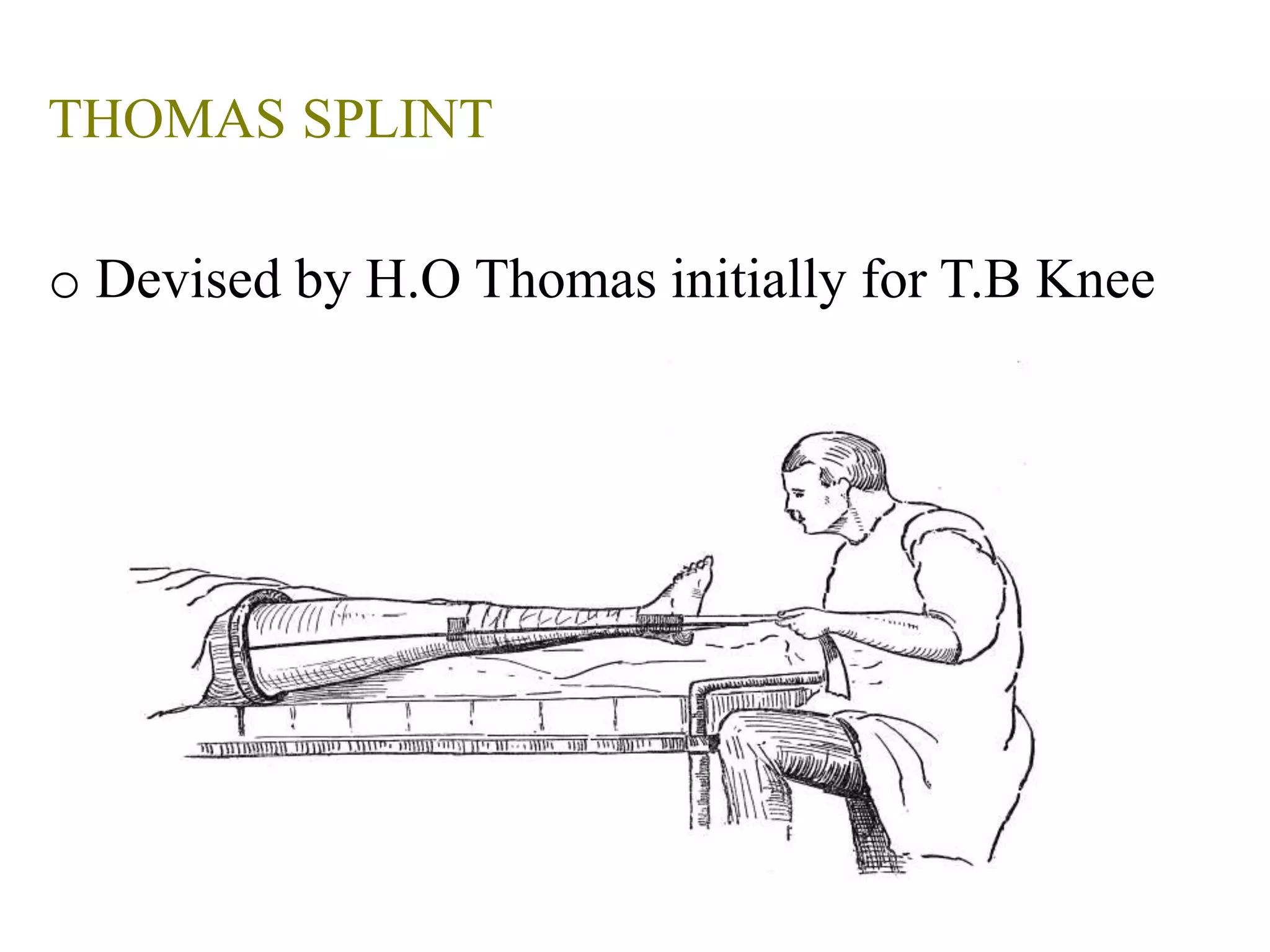
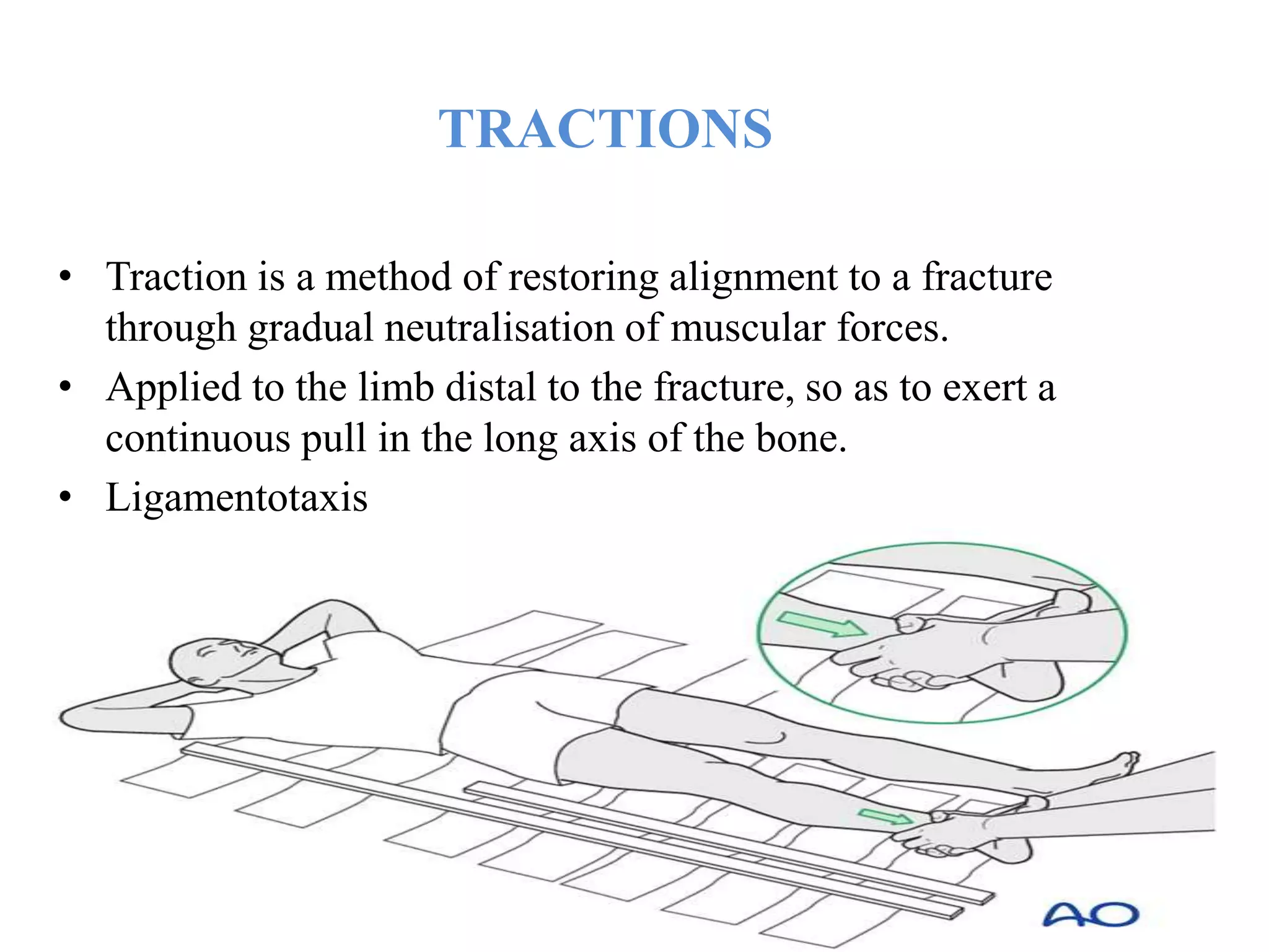
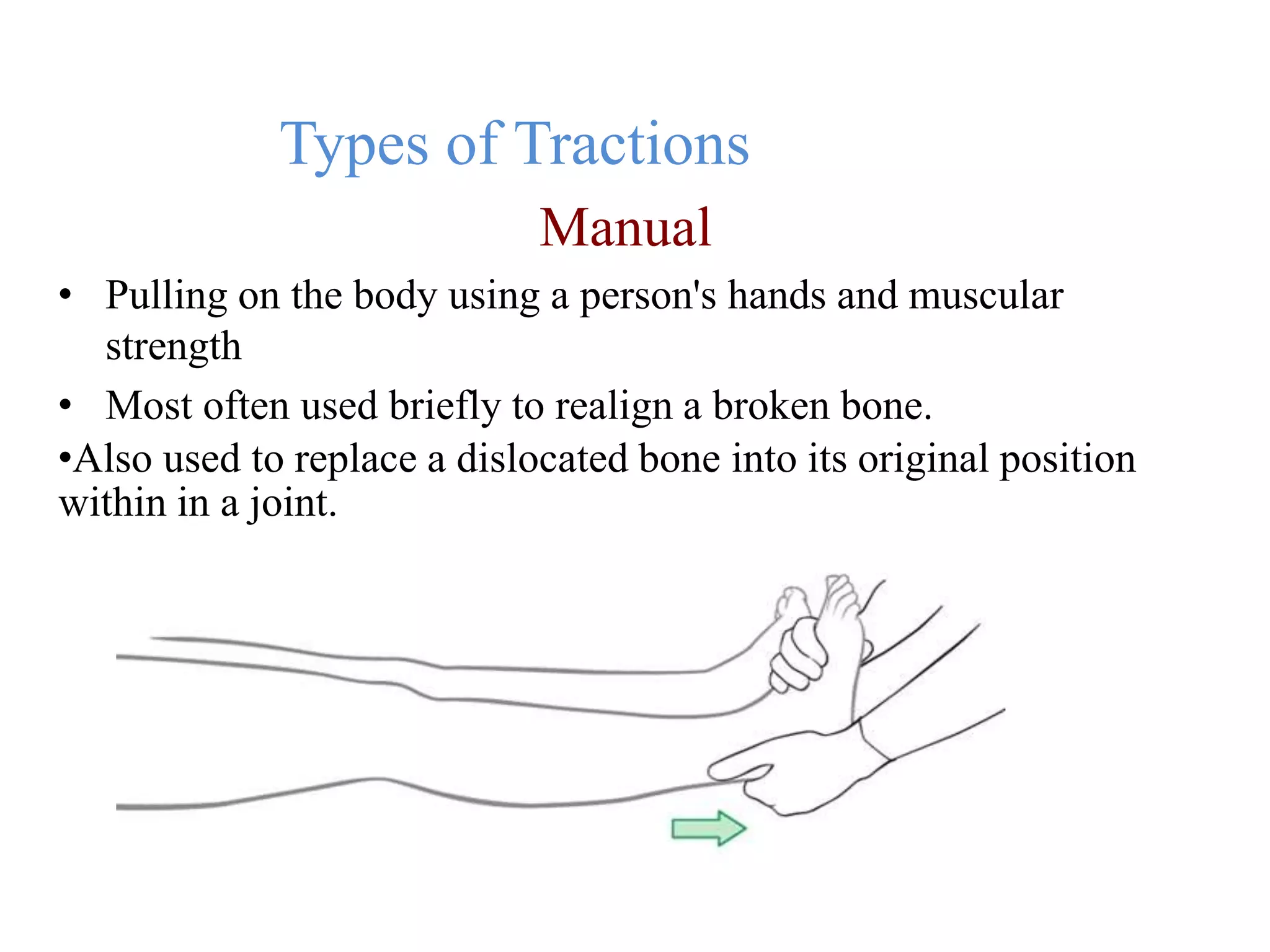
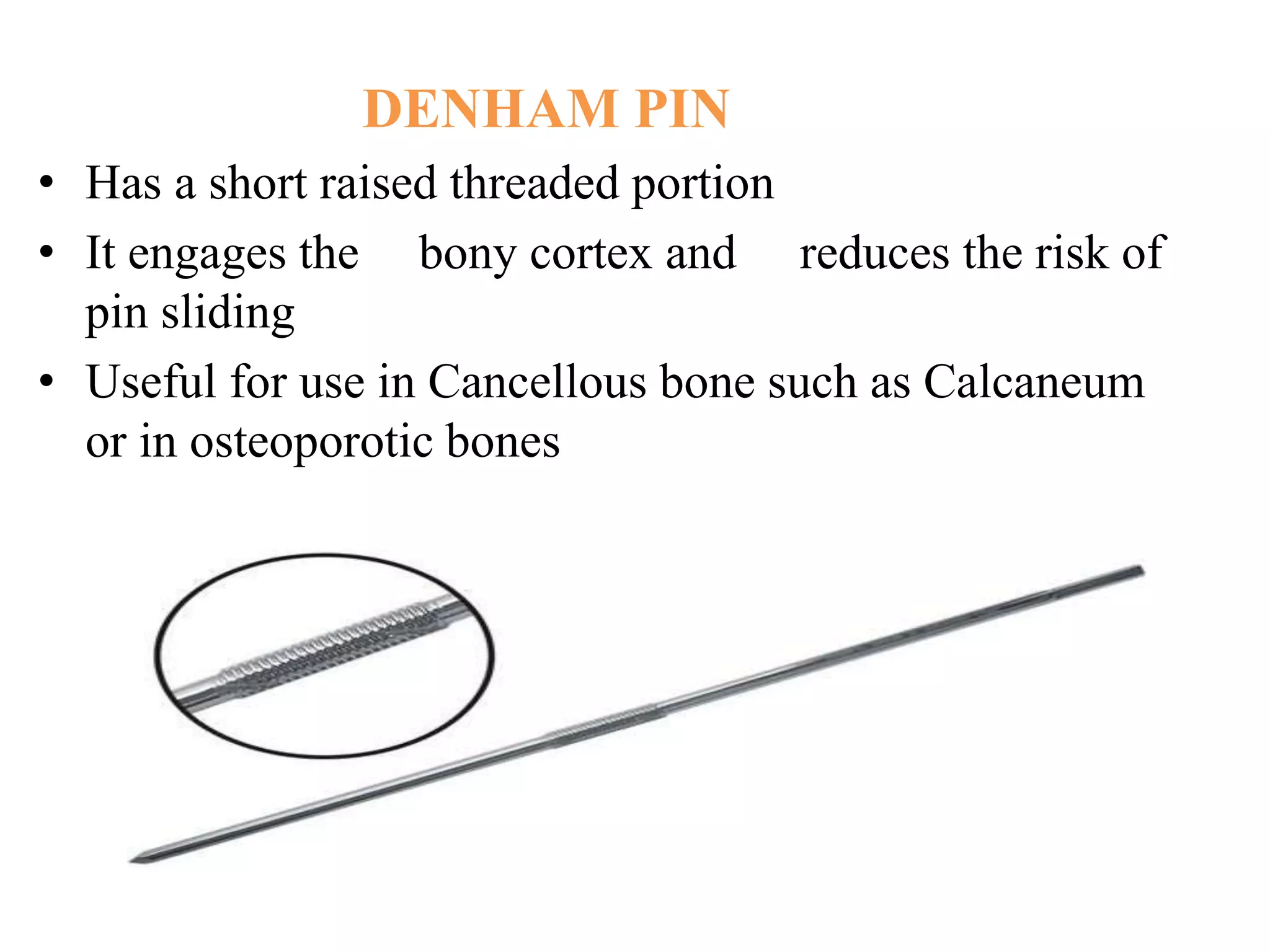
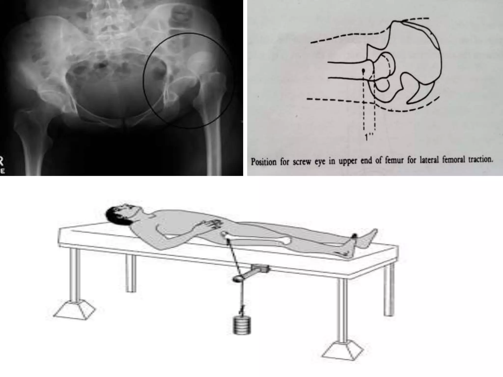
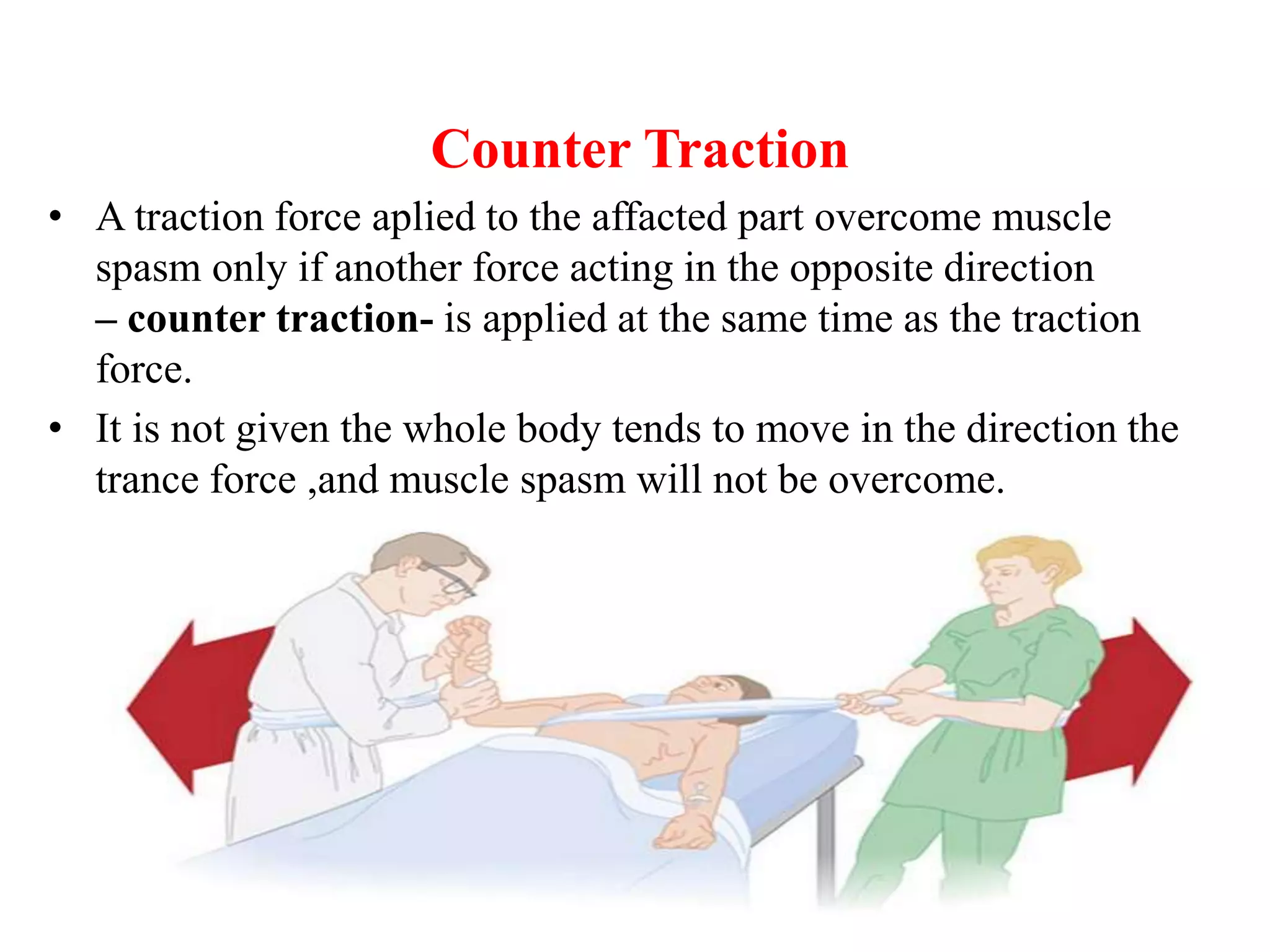
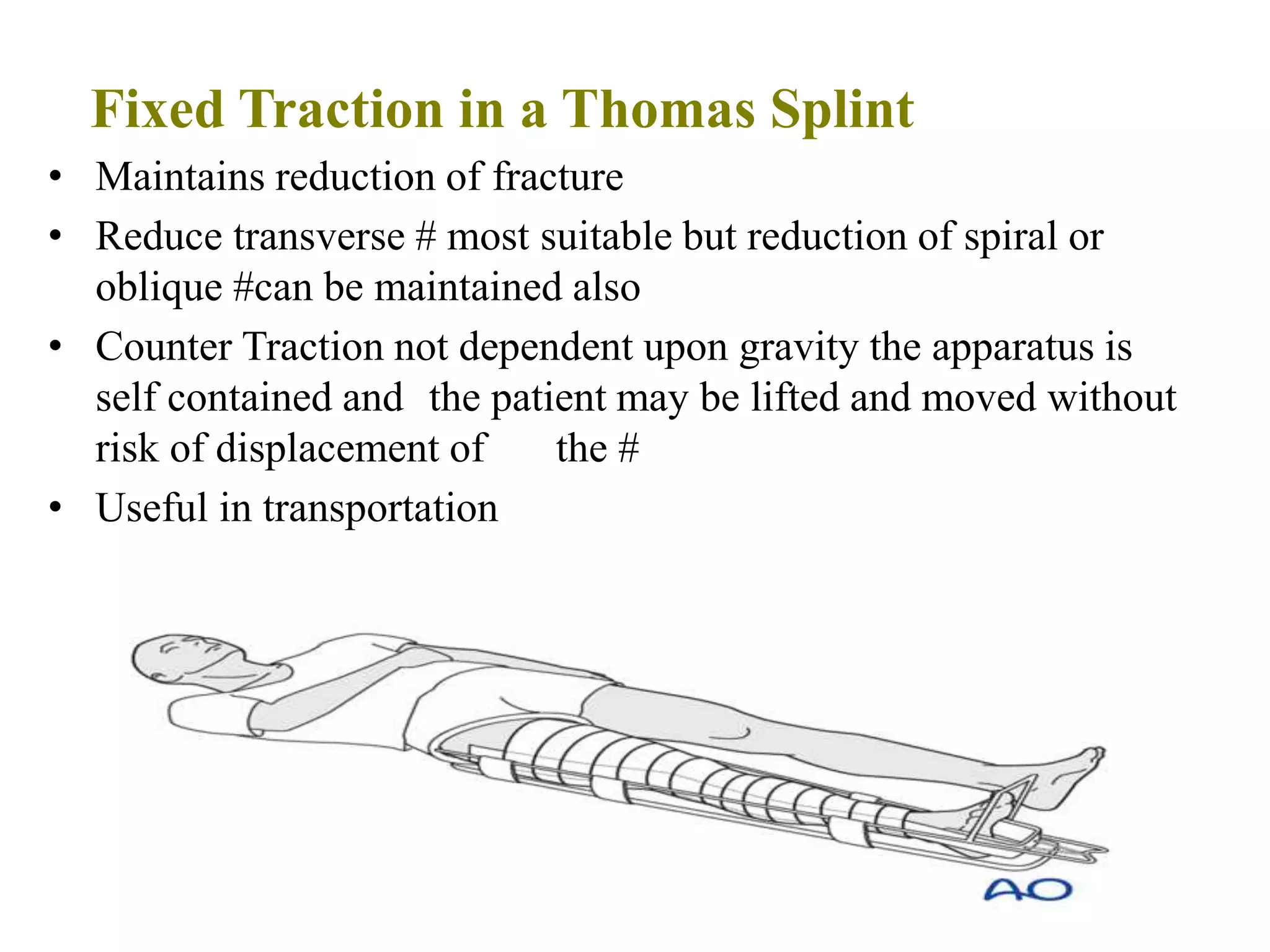
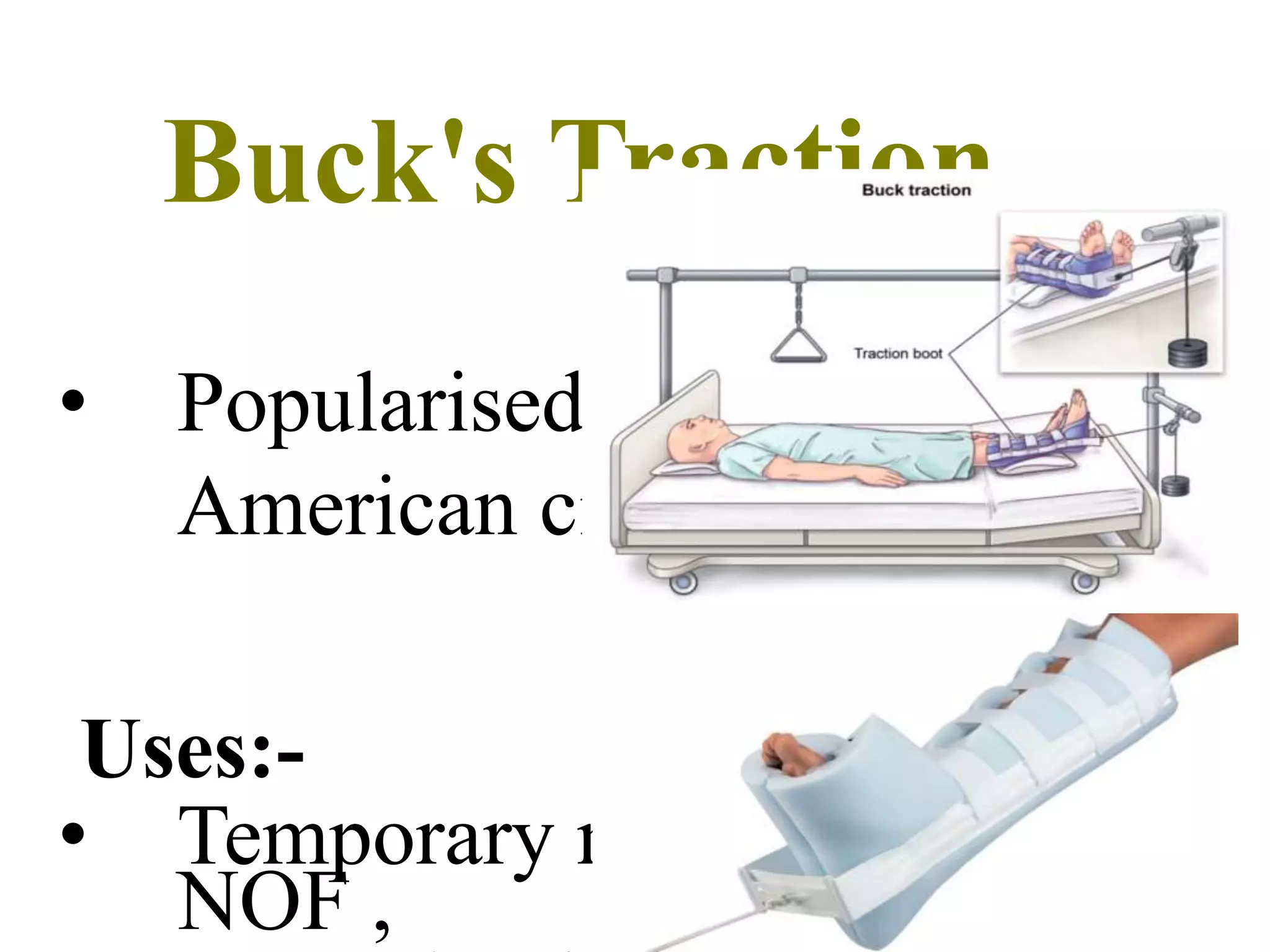
SPLINTS AND
TRACTIONS
Dr Prakhar Yadav
1st Year PG Orthopaedics
CMCH,Bhopal
SPLINT
• Is a device used for support or immobilisation of a limb or
spine.
• Any Material Used to support a fracture is a splint.
1. Conventional
2. Non Conventional
/
OUTLINES
• Functions
• Indications
• Prepartion
• Types
• Care of a patient on splinting
FUNCTIONS
• Temporary immobilization of sprains, fractures,
and reduced dislocations
• Control of pain
• Facilitates patient transportation
• Prevention of further soft tissue or
neurovascular injuries
• Decreases risk of converting a minor injury to
a major injury.
INDICATIONS
• Fractures
• -Sprains
• -Joint Infection
• -Tenosinovitis
• -Acute Arthritis/Gout
• -Lacerated Wound over joint
• -To stabilize or rest the joint in ligament injury
• -To correct deformity
• -To support and immobilize joint post op
PREPARATION
• Define injury and what splint is required
• Splint in position of function
• Document neurovascular examination before splint
application
• Anticipate ability for the patient to remove clothes
after splint is
applied
• Clean and repair skin lesions prior to splint
application
Cramer Wire Splint
• Ladder splint
• Used for temporary splintage of fractures during transportation
• Made of two thick parallel wires with interlacing wires
• Can be bend into different shapes.
THOMAS SPLINT
o Devised by H.O Thomas initially for T.B Knee
Parts
• Padded oval metal ring an angle of 120*
• To which are attached inner and outer side bars
• The two side bars joined at the distal end in the
form of “W”
• Outer bar angled out 5cm below the ring.
Bohler Braun Splint
Bohler Broun 3 pulley splint
1. Proximal pulley to privent foot drop
2. 2nd pulley – traction in line with femur
3. 3rd pulley – traction in line with the leg
PRE POST SPLINT CHECKS
• Function
• Arterial pulses
• Capillary refill
• Temperature
• Skin
TRACTIONS
• Traction is a method of restoring alignment to a fracture
through gradual neutralisation of muscular forces.
• Applied to the limb distal to the fracture, so as to exert a
continuous pull in the long axis of the bone.
• Ligamentotaxis
Functions
• Reduction of fractures and dislocations
Reduce/relieve pain
• Preventing deformities .
• Correction of soft tissue contractures
• Ensure immobilisation
• Minimize muscle spasms
Types of Tractions
Manual
• Pulling on the body using a person's hands and muscular
strength
• Most often used briefly to realign a broken bone.
•Also used to replace a dislocated bone into its original position
within in a joint.
Skin Traction
• Traction force is applied over a large area of sloan.
• Spreads the load and is Comfortable
• Generally weight used is 5lbs
• Maximum Traction weight that can be applied by sloan traction is
15lb(6.7kg)
 Two Methods of Applying
1. Adhesive Skin Traction
2. Non Adhesive Sloan Traction
Weight not exceeding 10lb or 4.5kg)
Method
• Shave the limb(Not for non adhesive)
• Protectthe malleoli(felt, foam rubber or few turns of crepe)
• Starting at the ankle/wrist,leaving a loop 'N'-N projecting 5cm
beyond the distal end of the limb and apply strapping
• Antero medially and postern laterally
-To encourage medial rotation
• Apply crepe bandage
• Check the spreader and traction cords present
• Attach required weight
Contraindications
• Abrasions of the skin
• Lacerations of the skin in the area to which
traction is applied
• Impariment of circulation- Varicose ulcers,
Impending gangrene
• Dermatitis
• Marked shortening of bony fragments
(Traction required to reduce is much more
than what can be give via skin)
Complications Cont.
• Allergic reaction to the adhesive
• Excoriations of the skin
• Pressure sores(Over the ma11eoli,
tendocalcaneus)
• Common Peroneal nerve Palsy
SKELETAL TRACTION
• Metal Pin or a wire is driven through a bone
• Traction force applied directly to the skeleton
• Frequently used in the management of Lower limb
fractures Should be reserved in cases in which
Skin traction is Contraindicated
• Serious complication is 'Osteomyelitis
Steinmann Pin
• Rigid Stainless Steel Pins of varying lengths,4-6 mm in dia.
• After insertion Bohler Stirrup is attached to the pin
• Bohler stirrup allows the direction of the traction to be varied
without tunning the pin in the bone
DENHAM PIN
• Has a short raised threaded portion
• It engages the bony cortex and reduces the risk of
pin sliding
• Useful for use in Cancellous bone such as Calcaneum
or in osteoporotic bones
Kirschner(K) Wire
• Small in diameter.
• Insufficiently rigid until pulled taut in a special stirrup
• Rotation of the stirrup is imparted to the wire.
• Wire easily cuts out of bone if a heavy weight is applied
• Mostly used in the upper limb
Olecranon Traction
• #Supracondylar Humerus
• Comminuted # of lower end of Humerus
• Unstable # of the Shaft of Humerus
METHOD
• Deep to the s/c border of the upper end of the
ulna, 1 .25 inches(3cm) distal to the tip of
olecranon
• This avoids the elbow joint
•Medial to Lateral
2NDAND 3RDMETACARPALS TRACTION
• Comminuted # of the bones of the forearm (particularly
comminuted # of the Lower end of radius)
Method
• K wire inserted 0.75-l inch(2-2.5cm) proximal to the distal end
of 2nd MC
• Wire traverses 2nd and 3rd MC transversely to lie at right
angles to the longitudinal axis of the radius.
Complication:
•Fibrosis of the Interosseus muscles and can lead to stiffness
of fingers.
Upper end of femur- Greater
trochanter
• Central # Dislocations of the hip
• To restore the relationship of the weight
bearing part of the femoral head to the dome of the
acetabulum.
Method
• Lateral surface of the femur,l inch(2.5 cm) below
the most prominent part of the greater
trochanter,mid way between the anterior and
posterior surfaces of femur.
• 10-20 lbs
• Active hip and knee movements started
Lower end of femur
• Method of choice for Acetabular and Proximal femur
fractures
• If there is a knee ligament injury usually use distal femur
instead of proximal tibial traction
• 3 cm proximal to the articluation b/w lateral femoral Condyle
and lateral Tibial plateau
Place pin from
Medial to
Lateral at the
adductor
tubercle –
slightly
proximal to
epicondyle
Vastus lateralis muscle -
-
Vastus intermedius muscle
l
I l
II
Biceps Femoris muscle -
Common peroneol nerve
Tibial nerve "
I
I
Vastus medialis
I
'
Gracilis muscle
Semimembranosis
Semitendinosus muscle
Popliteal artery and vein
Upper end of Tibia
• 0.75 inch(2.0cm) behind the crest, just below the level of the
tubercle of the tibia
• Pin driven from Lateral to Medial side of the limb to avoid
damage to the CPN
Lower end of Tibia
• 2 inches(5 cm) above the level of the ankle joint, mid
way between the anterior and posterior borders of the
tibia
Calcaneus
• 0.75 inch(2 cm) below and behind the lateral mallelous or 1.25
inch(3cm) below and behind the medial malleolus
•Care must be taken to avoid entering the sub taler joint
• Most commonly used with a spanning ex fix for "travelling
traction" or a Bohlen Braun frame
• Stiffness of the subtalar joint
Complications of Skeletal Traction
• Introduction of Infection
• Incorrect placement
Cut out of the bone causing pain and failure
Control of rotation difficult
Uneven pull being applied.
• Distraction
• Ligamentous Damage
•Damage to episphyseal growth plates(Genu recurvatum in the treatment of # SOF by
UTST
• Depressed scars
Counter Traction
• A traction force aplied to the affacted part overcome muscle
spasm only if another force acting in the opposite direction
– counter traction- is applied at the same time as the traction
force.
• It is not given the whole body tends to move in the direction the
trance force ,and muscle spasm will not be overcome.
Fixed Traction
• Counter traction by applying a force against a fixed point on the body,
proximal to the attachments of the muscles in spasm
• To apply a force on the fixed point on the body an appliance such as the
Thomas splint is used
• Traction force balances the pull of the muscles and as the muscular pull and
hematoma decrease, the traction force decrease
• Distraction at the # site and accompanying danger of delayed union or non
union is less likely to occur
Fixed Traction in a Thomas Splint
• Maintains reduction of fracture
• Reduce transverse # most suitable but reduction of spiral or
oblique #can be maintained also
• Counter Traction not dependent upon gravity the apparatus is
self contained and the patient may be lifted and moved without
risk of displacement of the #
• Useful in transportation
Sliding Traction
• Gravity utilized to provide Counter traction by tilting the bed so that patient tends to slide in Opposite
direction to traction force.
• Traction force continues to act as long as the weight remains clear of the
floor
• When used to reduce a # the initial wt used is greater than the wt required
to maintain (Great care taken to avoid distraction)
• Femoral shaft # an initial wt of 10% of the weight of the patient is usually
sufficient
• Counter traction 1 inch (2.5cm) for each of 1b(0.46kg) of traction wt.
Buck's Traction
• Popularised during the
American civil war
Uses:-
• Temporary management of#
NOF ,
Method
• Adhesive strapping or Ventfoam skin traction Bandage
• Support the leg on a soft pillow to keep the heel clear of the bed
• Pass the cord from the spreader over a pulley attached to the
end of the bed
• 5-7lb(2.3-3.2k8)
• Elevate the foot end of the bed
• Lateral rotation of the limb is not controlled by this method
Perkins Traction
• Fracture tibia
• # femur from subtrochanteric region distally in all ages racture #Trochanter
in <50 yrs
• Principle is use of Skeletal traction without any external splintage coupled
with active movements of injured limb
• Perkins showed that by encouraging early stiffness of joint was prevented
by extensibility of muscles by reciprocal innervation.
Method
• Under GA and aseptic precautions
• Insert Denham pin through upper Tibia
• Attach Simonis swivel to each end of pin
• Connect 2 traction cords to each swivel
• Pass each cordover separate pulleys
• Attach wt(4.6kgs) to each traction cord making total
traction wt of 9.2kgs.
• Elevate foot of bed by l inch for every 0.46 kg.
• Place pillows under thigh to maintain normal bowing of
thigh.
• CHECK LIMB LENGTH WITH TAPE & INCREASE
OR DECREASE THE TRACTION WEIGHT
• Start active quadriceps exercises immediately.
Hamilton Russel Traction
• # Shaft of Femur
• After Arthroplasty operations of the hip
Method :-
• Adults- 8lb(3.6kg)
• Infants and older children- 0.5 - 41b(0.28 - 1.8 kg)
Tulloch Brown Traction
• Tulloch Brown or U loop tibial pin traction and suspension with Nissen foot plate
and Stirrup
Uses:-
• After Arthroplasty of the Hip
• Pseudoarthrosis operation on the hip
• # Shaft of Femur
• Not used in children
Ninety/Ninety Traction
• Obletz(1946) as an aid to the operative and early post op
management of compound fractures of the femur with wounds
on the post aspect of thigh
• Subtrochanteric #
• Proximal l/3rd of Femur
• Children as well as adults
• Three ways of applying it
• l.Using a Tulloch Brown U loop
2. Using a second Steinmann Pin
3.Using a B/K Plaster cast
Complications of Ninety/Ninety Traction
• Those of Skeletal Traction
• Stiffness and loss of extension of the Knee
• Flexion contracture of the Hip
• Injury to the Lower femoral or upper tibial epiphyseal
growth plates
• Neurovascular damage
Sliding Traction with a Thomas Splint and
Knee Flexion piece (Pearson Modification)
• Used to Obtain the reduction ofan oblique or spiral # of the shaft of the femur and then to
retain it until union occurs
• Suspend the knee flexion piece to maintain 20-30 degrees of flexion at knee joint (extended
position is zero)
Knee _flexion piece
• Helps in easier mobilization of the knee
• Controls Rotation
• prevents stretching of the post capsule and
cruciate ligaments of the knee.
Bryant's (Gallows Traction)
• #SOF in children Less than 2 years
• Weigh less than 35-40lb(15.9-18.2 kg)
• Fractures in children unite rapidly
• Seldom applied for more than 4 week
Complications
• Vascular complication of Bryants traction may occur in either
the injured or normal limb.
• A careful check must be done in both limbs during first 24-72
hrs. -By checking color and temp of limbs. -Dorsiflexion of
both ankle passively.
• Bryants traction in children under 2 yrs – SAFE
• 2-4 yrs vascular complications more (can be prevented by using
posterior splint).
• Over 4yrs -- absolutely contraindicated.
Modified Bryant's Traction
• Sometimes used in the initial management of CDH when
diagnosed over the age of 1 year
• Abduction begun after 5 days
• 10degrees on alternate days
• By 3 weeks fully abducted
Agnes Hunt Traction
4.2! A¢no HonmcIiun.TheulrIiEIrs:cdiou¢rIhnb umuunednnndngkb be
Pelvic Traction
• Conservative management of prolapsed Intervertebral disc
•Special Canvas harness is buckled around the patient's pelvis
•Long cords or straps attach the harness to the foot of the bed
•Foot of the bed raised amount of which depends upon patients weight
• Function is to ensure that Me patient lies quickly in bed rather
than to attempt to distract the vertebral bodies .
Alternative
• Buck's traction
Pelvic traction is superior but however because it leaves patient's
legs free and therefore able to move freely
References
Traction and Orthopaedic Appliances 2
nd
Edition
John D.M Stewart
Jefferey P Hallet

Tractions in Orthopaedics.ppt

  • 1.
    SPLINTS AND TRACTIONS Dr PrakharYadav 1st Year PG Orthopaedics CMCH,Bhopal
  • 2.
    SPLINT • Is adevice used for support or immobilisation of a limb or spine. • Any Material Used to support a fracture is a splint. 1. Conventional 2. Non Conventional
  • 3.
  • 4.
    OUTLINES • Functions • Indications •Prepartion • Types • Care of a patient on splinting
  • 5.
    FUNCTIONS • Temporary immobilizationof sprains, fractures, and reduced dislocations • Control of pain • Facilitates patient transportation • Prevention of further soft tissue or neurovascular injuries • Decreases risk of converting a minor injury to a major injury.
  • 6.
    INDICATIONS • Fractures • -Sprains •-Joint Infection • -Tenosinovitis • -Acute Arthritis/Gout • -Lacerated Wound over joint • -To stabilize or rest the joint in ligament injury • -To correct deformity • -To support and immobilize joint post op
  • 7.
    PREPARATION • Define injuryand what splint is required • Splint in position of function • Document neurovascular examination before splint application • Anticipate ability for the patient to remove clothes after splint is applied • Clean and repair skin lesions prior to splint application
  • 8.
    Cramer Wire Splint •Ladder splint • Used for temporary splintage of fractures during transportation • Made of two thick parallel wires with interlacing wires • Can be bend into different shapes.
  • 9.
    THOMAS SPLINT o Devisedby H.O Thomas initially for T.B Knee
  • 10.
    Parts • Padded ovalmetal ring an angle of 120* • To which are attached inner and outer side bars • The two side bars joined at the distal end in the form of “W” • Outer bar angled out 5cm below the ring.
  • 11.
    Bohler Braun Splint BohlerBroun 3 pulley splint 1. Proximal pulley to privent foot drop 2. 2nd pulley – traction in line with femur 3. 3rd pulley – traction in line with the leg
  • 12.
    PRE POST SPLINTCHECKS • Function • Arterial pulses • Capillary refill • Temperature • Skin
  • 13.
    TRACTIONS • Traction isa method of restoring alignment to a fracture through gradual neutralisation of muscular forces. • Applied to the limb distal to the fracture, so as to exert a continuous pull in the long axis of the bone. • Ligamentotaxis
  • 14.
    Functions • Reduction offractures and dislocations Reduce/relieve pain • Preventing deformities . • Correction of soft tissue contractures • Ensure immobilisation • Minimize muscle spasms
  • 15.
    Types of Tractions Manual •Pulling on the body using a person's hands and muscular strength • Most often used briefly to realign a broken bone. •Also used to replace a dislocated bone into its original position within in a joint.
  • 16.
    Skin Traction • Tractionforce is applied over a large area of sloan. • Spreads the load and is Comfortable • Generally weight used is 5lbs • Maximum Traction weight that can be applied by sloan traction is 15lb(6.7kg)  Two Methods of Applying 1. Adhesive Skin Traction 2. Non Adhesive Sloan Traction Weight not exceeding 10lb or 4.5kg)
  • 17.
    Method • Shave thelimb(Not for non adhesive) • Protectthe malleoli(felt, foam rubber or few turns of crepe) • Starting at the ankle/wrist,leaving a loop 'N'-N projecting 5cm beyond the distal end of the limb and apply strapping • Antero medially and postern laterally -To encourage medial rotation • Apply crepe bandage • Check the spreader and traction cords present • Attach required weight
  • 19.
    Contraindications • Abrasions ofthe skin • Lacerations of the skin in the area to which traction is applied • Impariment of circulation- Varicose ulcers, Impending gangrene • Dermatitis • Marked shortening of bony fragments (Traction required to reduce is much more than what can be give via skin)
  • 20.
    Complications Cont. • Allergicreaction to the adhesive • Excoriations of the skin • Pressure sores(Over the ma11eoli, tendocalcaneus) • Common Peroneal nerve Palsy
  • 21.
    SKELETAL TRACTION • MetalPin or a wire is driven through a bone • Traction force applied directly to the skeleton • Frequently used in the management of Lower limb fractures Should be reserved in cases in which Skin traction is Contraindicated • Serious complication is 'Osteomyelitis
  • 22.
    Steinmann Pin • RigidStainless Steel Pins of varying lengths,4-6 mm in dia. • After insertion Bohler Stirrup is attached to the pin • Bohler stirrup allows the direction of the traction to be varied without tunning the pin in the bone
  • 23.
    DENHAM PIN • Hasa short raised threaded portion • It engages the bony cortex and reduces the risk of pin sliding • Useful for use in Cancellous bone such as Calcaneum or in osteoporotic bones
  • 24.
    Kirschner(K) Wire • Smallin diameter. • Insufficiently rigid until pulled taut in a special stirrup • Rotation of the stirrup is imparted to the wire. • Wire easily cuts out of bone if a heavy weight is applied • Mostly used in the upper limb
  • 26.
    Olecranon Traction • #SupracondylarHumerus • Comminuted # of lower end of Humerus • Unstable # of the Shaft of Humerus METHOD • Deep to the s/c border of the upper end of the ulna, 1 .25 inches(3cm) distal to the tip of olecranon • This avoids the elbow joint •Medial to Lateral
  • 28.
    2NDAND 3RDMETACARPALS TRACTION •Comminuted # of the bones of the forearm (particularly comminuted # of the Lower end of radius) Method • K wire inserted 0.75-l inch(2-2.5cm) proximal to the distal end of 2nd MC • Wire traverses 2nd and 3rd MC transversely to lie at right angles to the longitudinal axis of the radius. Complication: •Fibrosis of the Interosseus muscles and can lead to stiffness of fingers.
  • 30.
    Upper end offemur- Greater trochanter • Central # Dislocations of the hip • To restore the relationship of the weight bearing part of the femoral head to the dome of the acetabulum. Method • Lateral surface of the femur,l inch(2.5 cm) below the most prominent part of the greater trochanter,mid way between the anterior and posterior surfaces of femur. • 10-20 lbs • Active hip and knee movements started
  • 32.
    Lower end offemur • Method of choice for Acetabular and Proximal femur fractures • If there is a knee ligament injury usually use distal femur instead of proximal tibial traction • 3 cm proximal to the articluation b/w lateral femoral Condyle and lateral Tibial plateau
  • 33.
    Place pin from Medialto Lateral at the adductor tubercle – slightly proximal to epicondyle Vastus lateralis muscle - - Vastus intermedius muscle l I l II Biceps Femoris muscle - Common peroneol nerve Tibial nerve " I I Vastus medialis I ' Gracilis muscle Semimembranosis Semitendinosus muscle Popliteal artery and vein
  • 34.
    Upper end ofTibia • 0.75 inch(2.0cm) behind the crest, just below the level of the tubercle of the tibia • Pin driven from Lateral to Medial side of the limb to avoid damage to the CPN
  • 35.
    Lower end ofTibia • 2 inches(5 cm) above the level of the ankle joint, mid way between the anterior and posterior borders of the tibia
  • 36.
    Calcaneus • 0.75 inch(2cm) below and behind the lateral mallelous or 1.25 inch(3cm) below and behind the medial malleolus •Care must be taken to avoid entering the sub taler joint • Most commonly used with a spanning ex fix for "travelling traction" or a Bohlen Braun frame • Stiffness of the subtalar joint
  • 37.
    Complications of SkeletalTraction • Introduction of Infection • Incorrect placement Cut out of the bone causing pain and failure Control of rotation difficult Uneven pull being applied. • Distraction • Ligamentous Damage •Damage to episphyseal growth plates(Genu recurvatum in the treatment of # SOF by UTST • Depressed scars
  • 38.
    Counter Traction • Atraction force aplied to the affacted part overcome muscle spasm only if another force acting in the opposite direction – counter traction- is applied at the same time as the traction force. • It is not given the whole body tends to move in the direction the trance force ,and muscle spasm will not be overcome.
  • 39.
    Fixed Traction • Countertraction by applying a force against a fixed point on the body, proximal to the attachments of the muscles in spasm • To apply a force on the fixed point on the body an appliance such as the Thomas splint is used • Traction force balances the pull of the muscles and as the muscular pull and hematoma decrease, the traction force decrease • Distraction at the # site and accompanying danger of delayed union or non union is less likely to occur
  • 40.
    Fixed Traction ina Thomas Splint • Maintains reduction of fracture • Reduce transverse # most suitable but reduction of spiral or oblique #can be maintained also • Counter Traction not dependent upon gravity the apparatus is self contained and the patient may be lifted and moved without risk of displacement of the # • Useful in transportation
  • 41.
    Sliding Traction • Gravityutilized to provide Counter traction by tilting the bed so that patient tends to slide in Opposite direction to traction force. • Traction force continues to act as long as the weight remains clear of the floor • When used to reduce a # the initial wt used is greater than the wt required to maintain (Great care taken to avoid distraction) • Femoral shaft # an initial wt of 10% of the weight of the patient is usually sufficient • Counter traction 1 inch (2.5cm) for each of 1b(0.46kg) of traction wt.
  • 42.
    Buck's Traction • Popularisedduring the American civil war Uses:- • Temporary management of# NOF ,
  • 43.
    Method • Adhesive strappingor Ventfoam skin traction Bandage • Support the leg on a soft pillow to keep the heel clear of the bed • Pass the cord from the spreader over a pulley attached to the end of the bed • 5-7lb(2.3-3.2k8) • Elevate the foot end of the bed • Lateral rotation of the limb is not controlled by this method
  • 44.
    Perkins Traction • Fracturetibia • # femur from subtrochanteric region distally in all ages racture #Trochanter in <50 yrs • Principle is use of Skeletal traction without any external splintage coupled with active movements of injured limb • Perkins showed that by encouraging early stiffness of joint was prevented by extensibility of muscles by reciprocal innervation.
  • 45.
    Method • Under GAand aseptic precautions • Insert Denham pin through upper Tibia • Attach Simonis swivel to each end of pin • Connect 2 traction cords to each swivel • Pass each cordover separate pulleys • Attach wt(4.6kgs) to each traction cord making total traction wt of 9.2kgs. • Elevate foot of bed by l inch for every 0.46 kg. • Place pillows under thigh to maintain normal bowing of thigh. • CHECK LIMB LENGTH WITH TAPE & INCREASE OR DECREASE THE TRACTION WEIGHT • Start active quadriceps exercises immediately.
  • 46.
    Hamilton Russel Traction •# Shaft of Femur • After Arthroplasty operations of the hip Method :- • Adults- 8lb(3.6kg) • Infants and older children- 0.5 - 41b(0.28 - 1.8 kg)
  • 47.
    Tulloch Brown Traction •Tulloch Brown or U loop tibial pin traction and suspension with Nissen foot plate and Stirrup Uses:- • After Arthroplasty of the Hip • Pseudoarthrosis operation on the hip • # Shaft of Femur • Not used in children
  • 48.
    Ninety/Ninety Traction • Obletz(1946)as an aid to the operative and early post op management of compound fractures of the femur with wounds on the post aspect of thigh • Subtrochanteric # • Proximal l/3rd of Femur • Children as well as adults • Three ways of applying it • l.Using a Tulloch Brown U loop
  • 49.
    2. Using asecond Steinmann Pin 3.Using a B/K Plaster cast
  • 50.
    Complications of Ninety/NinetyTraction • Those of Skeletal Traction • Stiffness and loss of extension of the Knee • Flexion contracture of the Hip • Injury to the Lower femoral or upper tibial epiphyseal growth plates • Neurovascular damage
  • 51.
    Sliding Traction witha Thomas Splint and Knee Flexion piece (Pearson Modification) • Used to Obtain the reduction ofan oblique or spiral # of the shaft of the femur and then to retain it until union occurs • Suspend the knee flexion piece to maintain 20-30 degrees of flexion at knee joint (extended position is zero) Knee _flexion piece • Helps in easier mobilization of the knee • Controls Rotation • prevents stretching of the post capsule and cruciate ligaments of the knee.
  • 52.
    Bryant's (Gallows Traction) •#SOF in children Less than 2 years • Weigh less than 35-40lb(15.9-18.2 kg) • Fractures in children unite rapidly • Seldom applied for more than 4 week
  • 53.
    Complications • Vascular complicationof Bryants traction may occur in either the injured or normal limb. • A careful check must be done in both limbs during first 24-72 hrs. -By checking color and temp of limbs. -Dorsiflexion of both ankle passively. • Bryants traction in children under 2 yrs – SAFE • 2-4 yrs vascular complications more (can be prevented by using posterior splint). • Over 4yrs -- absolutely contraindicated.
  • 54.
    Modified Bryant's Traction •Sometimes used in the initial management of CDH when diagnosed over the age of 1 year • Abduction begun after 5 days • 10degrees on alternate days • By 3 weeks fully abducted
  • 55.
    Agnes Hunt Traction 4.2!A¢no HonmcIiun.TheulrIiEIrs:cdiou¢rIhnb umuunednnndngkb be
  • 56.
    Pelvic Traction • Conservativemanagement of prolapsed Intervertebral disc •Special Canvas harness is buckled around the patient's pelvis •Long cords or straps attach the harness to the foot of the bed •Foot of the bed raised amount of which depends upon patients weight • Function is to ensure that Me patient lies quickly in bed rather than to attempt to distract the vertebral bodies . Alternative • Buck's traction Pelvic traction is superior but however because it leaves patient's legs free and therefore able to move freely
  • 58.
    References Traction and OrthopaedicAppliances 2 nd Edition John D.M Stewart Jefferey P Hallet