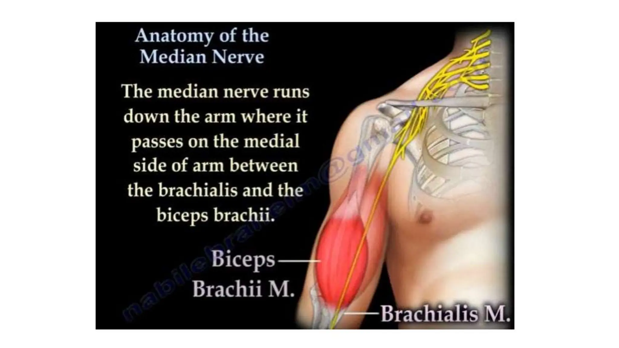
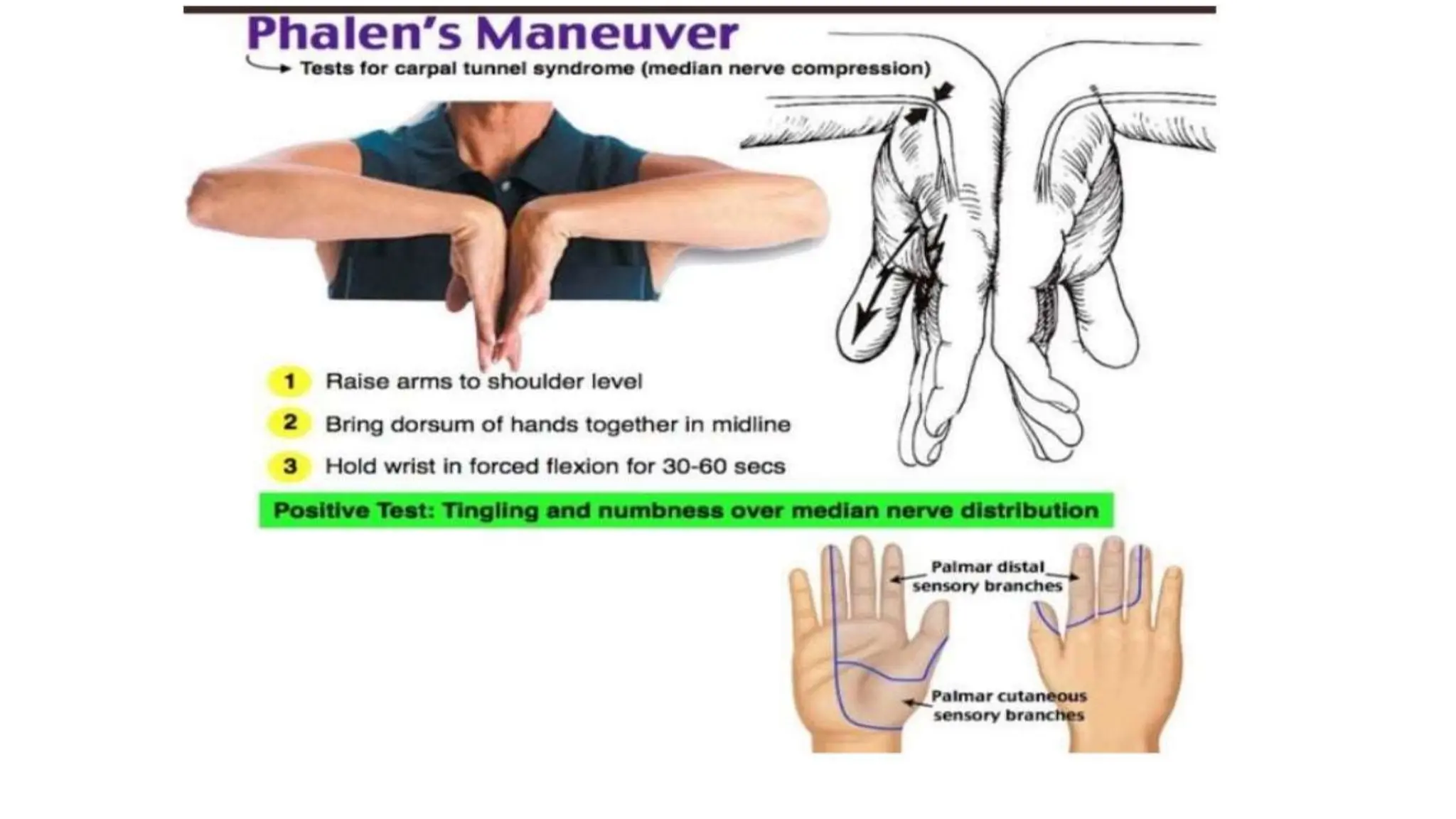
💎VVIP Kolkata Call Girls Parganas🩱7001035870🩱Independent Girl ( Ac Rooms Avai...
median nerve power point presentation.pptx
1. Presented by - Dr AVINASH KHARE,
PG RESIDENT
DEPARTMENT OF ORTHOPAEDICS
2.
3.
4.
5.
6. • Anatomy:Axilla
• After arising from lateral and medial cord of brachial plexus
• It runs on lateral aspect of axillary artery.
• Anatomy:Arm
• Continues to run lateral to the brachial artery till the mid-arm.
• Crosses the artery anteriorly and passes anterior to the elbow joint into
forearm.
27. • Motor functions-The flexors and pronators in the forearm are paralysed,with
the exception of the flexor carpi ulnaris and medial half of FDP.
• The forearm constantly supinated,and flexion is weak(often accompanied by
adduction,because of the pull of the flexor carpi ulnaris).
• Flexion at thumb is also prevented,as both the longus and brevis muscles are
paralysed.
• The lateral two lumbrical muscles are paralysed,and the patient will not be
able to flex at MCP joints or extend at IP joints of the index and middle
fingers.
28. • Sensory functions: lack of sensation over the areas that the median
nerve innervates.
• The thenar eminence is wasted,due to atrophy of thenar muscles.
29.
30. Examination
• Pronator teres assessment:The patients forearm is extended and fully
pronated.The patient is then instructed to resist supination of forearm by
the examiner.
31. • Flexor carpi radialis assessment-The patient flexes the wrist along
trajectory of forearm-wrist deviates ulnarly.
32.
33.
34.
35.
36. • Pronator quadratus assessment-Have the
patient resist supination of a fully flexed and
pronated forearm.
• With full forearm flexion ,pronation by usually
dominant pronator teres is minimised.
37. • When AIN injury is present,the patient will be unable to bring together the
tips of distal phalanx of the thumb and the index finger.
• Will be unable to make the OK SIGN.
OK SIGN / KLEIN NIOH SIGN
38. APE THUMB DEFORMITY
• The thumb is adducted and laterally rotated so that the thumb lies in the
same plane as the other fingers.It is due to over action of adductor
pollicis(supplied by ulnar nerve).
56. KAPLAN’S CARDINAL LINE
• It is a more predictable landmark for
the superficial palmer arch.
• In referencing this landmark as the
distal most extent of an open or
endoscopic carpal tunnel release,the
superficial palmar arch should be free
of transection.
57. Complications
• Injury to palmar cutaneous/recurrent motor branch of the median nerve.
• Hypertrophic scarring.
• Hematoma/Arterial imjury.
58. Pronator teres syndrome
• High compression neuropathy
• It is rare compared to CTS and AIS
• It is caused by compression of median nerve by PT muscle in proximal
forearm.
59. Symptoms and signs
• Symptoms are similar to those of carpal tunnel syndrome.
• Sensory disturbances.
• Night pain is unusual and forearm pain is more common.
• Hand numbness on gripping.
• Phalen’s test negative.
• Symptoms provoked by -
• resisted elbow flexion with forearm supinated (tightening of bicipital aponeurosis).
• resisted forearm pronation with the elbow extended (pronator tension).
60.
61.
62.
63. General indications of surgery
• In sharp injury exploration for diagnostic as well as therapeutic
purpose.Neurorrhaphy (end to end suturing of nerve) can be done at time
of exploration.
• In avulsion or blast injury-to identify and suture nerve ends for delayed
repair.
• No improvements since last 12 weeks following close injury.
64. Time of surgery
• Primary repair within 6-8 hours gives best results.
• Delayed primary repair-between 7-18 days.
• Secondary repair- 3-6 weeks later.preferable in
crushed,avulsed,contaminated wounds where patients life is seriously
endangered.
65. Critical limit of delay of suture
• Motor recovery in the intrinsic muscles of the hand does not occur if suture
is delayed 9 months in high lesions or 12 months in low ones.
• Useful sensory recovery only rarely occurs after 9 months in high lesions
or 12 months in low ones.