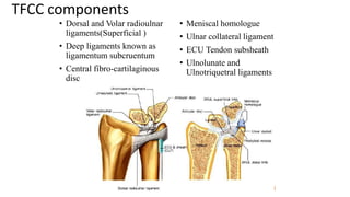
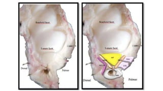
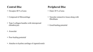
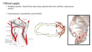
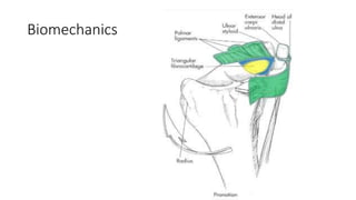
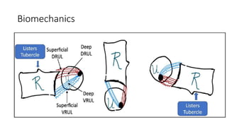
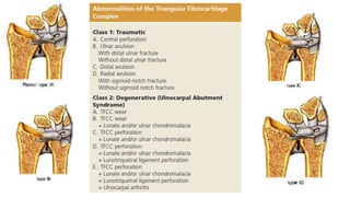
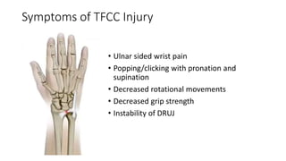
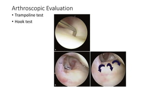
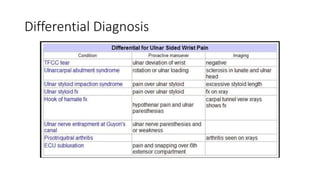
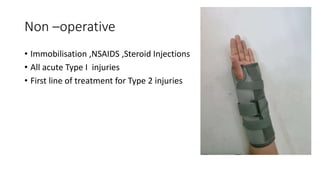
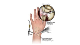
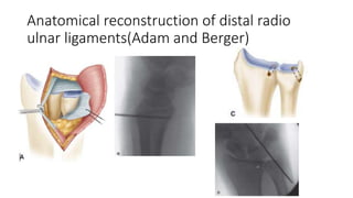
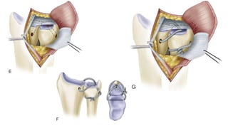
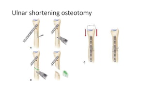
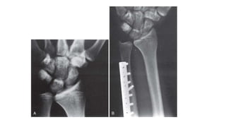
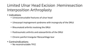
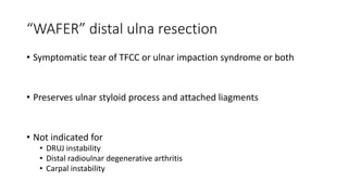
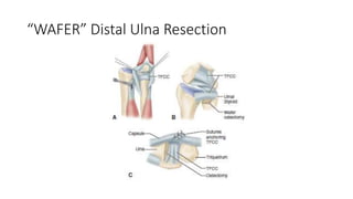
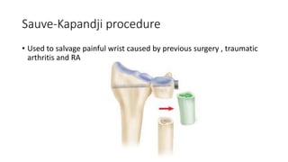
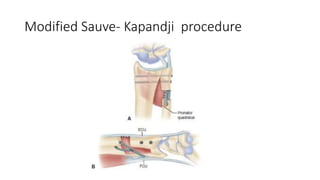
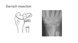
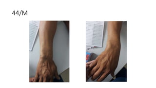
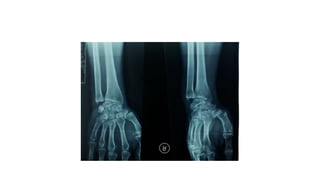
2. Importance
• TFCC Injury – m/c ligament injury
• >40 % of displaced distal radial fractures associated with instability of DRUJ
• In 1981, Palmer and Werner introduced term TFCC
4. • Origin
• Concave sigmoid notch medial to the distal end of radius from its
hyaline cartilage
• Insertion
• Fovea
• Base of ulnar styloid
• ECU subsheath
• Ulnocarpal ligaments
5. TFCC components
• Dorsal and Volar radioulnar
ligaments(Superficial )
• Deep ligaments known as
ligamentum subcruentum
• Central fibro-cartilaginous
disc
• Meniscal homologue
• Ulnar collateral ligament
• ECU Tendon subsheath
• Ulnolunate and
Ulnotriquetral ligaments
6.
7. Central Disc
• Occupies 80 % of area
• Composed of fibrocartilage
• Type I collagen bundles with interspersed
chondrocytes
• Avascular
• Poor healing potential
• Attaches to hyaline cartilage of sigmoid notch
Peripheral Disc
• Outer 20 % of area
• Vascular connective tissue along with
fibroblasts
• Good healing potential
8. • Blood supply
• Periphery portion : branch from ulnar artery and also from Ant. and Post. interosseous
arteries
• Central portion :nourished by synovial fluid
12. Symptoms of TFCC Injury
• Ulnar sided wrist pain
• Popping/clicking with pronation and
supination
• Decreased rotational movements
• Decreased grip strength
• Instability of DRUJ
13. Diagnosis of TFCC Injuries
• Physical examination of the TFCC
• Palpable tenderness over the TFCC
• Combined ulnar deviation and pronation /supination may cause pain and
popping/clicking sounds
• Ulnar impingement sign
• TFCC Stress Test
• “Press Test”: has been shown to have 100 % sensitivity for TFCC tears
14. Physical Examination
• Ulnar impaction test
• Wrist hyperextension and ulnar deviation with
axial compression
• Press Test
• Seated patient is asked to lift himself/herself out of
chair while bearing weight on extended wrists
15. • Piano key test
• Shuck test
• Radial aspect of wrist
stabilized
• Anteroposterior stress is
applied to the ulnar side of
wrist
• Fovea sign
16. Radiology
• X-ray
• Ulnar variance is calculated in neutral position
• (+) is associated with TFCC Tears
• MRI
• Radioulnar ligaments ,ulnocarpal ligaments and the TFCC with its foveal
attachment to ulna can be visualized
• Sensitivity and specificity for TFCC tears (100%)
19. Non –operative
• Immobilisation ,NSAIDS ,Steroid Injections
• All acute Type I injuries
• First line of treatment for Type 2 injuries
20. Surgical repair
• For patients with isolated or associated injuries yielding significant
DRUJ instability
• Often does not itself restore stability of the joint
• Careful patient selection
21. Traumatic Lesion of TFCC (Palmer Class 1A):
• Traumatic central tears of the TFCC with no instability
• Initial treatment non-operative for 4 weeks.
• Persistent pain relieved by
• Arthroscopic debridement of the flap portion of tear
• No more than two thirds of the central disc should be excised
• 2mm of the TFCC peripheral rim should be preserved
22.
23. Lesions of TFCC (Palmer Class 1B):
• Traumatic detachment of TFCC from ulna with or without ulnar
styloid fracture
• Initially conservative
• A/w ulnar styloid fracture
• ORIF or
• Excision of small fragment –usual T/T
• Open repair of TFCC
• A/w injury to ECU sheath
• Arthroscopic repair of TFCC
• Open ECU sheath reconstruction
24. Class 1C Lesions
• Disruptions of ulnocarpal ligaments
• A/w injuries include lunotriquetral and class 1B tears
• Difficult to diagnose
• Usually conservative management
• Operative treatment
• Late open or arthroscopic repair
25. Class 1D Lesions
• Tear of TFCC from the radius at the distal end of sigmoid notch
• Oriented in AP direction
• May involve radioulnar ligaments
• Frequently a/w distal radius fractures
• Satisfactory reduction of radial fracture-healing with stable DRUJ
• If instability after fracture reduction
• Arthrocopic /Open repair
28. Chronic Instability of DRUJ
• After unsuccessful attempts to repair the TFCC
• Failed primary TFCC repair
• Other causes
• After isolated trauma to the DRUJ
• After fracture of distal radius and ulna
• Inflammatory arthritis
29. Procedures to stabilize DRUJ
• Soft tissue procedures
• TFCC Repair
• TFCC Reconstruction
• Malunited distal radius fracture
• Distal radial osteotomy and bone grafting
• Ununited displaced ulnar styloid fracture
• Open reduction and internal fixation
35. Limited Ulnar Head Excision :Hemiresection
Interposition Arthroplasty
• Indications
• Unreconstructable fractures of ulnar head
• Ulnocarpal impingement syndrome with incongruity of the DRUJ
• Rheumatoid arthritis involving the DRUJ
• Posttraumatic arthritis and osteoarthritis of the DRUJ
• Chronic painful triangular fibrocartilage tear
• Contraindications
• No reconstructable TFCC
37. “WAFER” distal ulna resection
• Symptomatic tear of TFCC or ulnar impaction syndrome or both
• Preserves ulnar styloid process and attached liagments
• Not indicated for
• DRUJ instability
• Distal radioulnar degenerative arthritis
• Carpal instability
42. • Procedure to stabilize unstable proximal ulnar segment after distal
ulnar excision
• Volar, distally based capsular flap attached to the ulna proximally
• Slip of the ECU tendon, based proximally or distally, passed through drill
holes in the ulna or wrapped around ulna
• PQ as a combination interposition-stabilizer
• Slip of FCU tendon, usually distally based and passed through drill holes in
ulna
• Combination of ECU tenodesis and dorsal transfer of PQ
48. Take Home Message
• TFCC injury is most common
• Multiple causes of ulnar sided wrist pain
• Conservative treatment is mainstay of treatment
• Persistent pain and instability only requires operative treatment
49. References
• Campbell’s Operative Orthopaedics ,14th Edition
• Hand and Wrist Surgery ,Operative Techniques ,13th edition
• IFSSH Scientific Committee on Anatomy and Biomechanics ,2013
• Anatomy of the Triangular Fibrocartilage Complex (TFCC)
,ISSHACADEMICS
TFCC begins on the ulnar side of the lunate fossa of the radius, Articular surface contact in the shallow sigmoid notch accounts for
about 20% of DRUJ stability
During forearm rotation, the ulnar head at its articulation with the sigmoid notch appears to move from dorsal and distal in full pronation to proximal and palmar in full supination.
The anterior interosseous artery is one of the two branches of the short common interosseous artery (from the ulnar artery)
The volar portion of the TFCC is innervated by a branch of the ulnar nerve and the dorsal sensory branch of the ulnar nerve.
Keep 1 mri photo
If positive ulnar variance- arthroscopic wafer resection or ulnar shortening osteotomy
ORIF for ulnar styloid- perform open TFCC repair
- ULNAR STYLOID FRACTURE- ORIF -Excision of small fragment
Relieves pain and instability
Open reapir- if displaced fracture of sigmoid notch
Arthroscopic repair of class 1D injury of triangular fibrocartilage complex. A, Edge of sigmoid notch is abraded with
motorized burr. B, Holes are drilled through radius with Kirschner wire. C, Sutures are placed into triangular fibrocartilage complex
through drill holes with long meniscal repair needles. D, Sutures are tied on surface of radius.
. A, Approach to TFCC, reflection of dorsal radioulnar ligament and periosteum over lunate fossa.
B, Suture placement into TFCC through holes drilled in dorsoulnar aspect of distal radius; sutures are directed in palmar and ulnar
direction to exit at edge of lunate fossa and sigmoid notch. Horizontal mattress sutures are placed in TFCC
Angulation of distal radius fractures with volar convexity of more than 20 or 30 degree
More than 5 mm of radial shortening
DRUJ unstable,
Restored stability, preserved motion, and relieved pain
Type 2 with pos UV more than 2mm, App- plate- mark- osteotomy- interfrag-plate fixation
Ulnar shortening osteotomy. A, Wrist of 16-year-old male wrestler with bilateral
ulnar wrist pain and positive ulnar variance of 2 mm. B, Four weeks after surgery, wrist is in ulnar
neutral variance and osteotomy is healed.
Bowers technique of hemiresection arthroplasty.A, Because ulna is too long, it impinges on stylocarpal ligament.
B and C, This problem can be corrected by interposition (B) or shortening
Type 2 with pos UV less than 2mm pain relief, preserves motion, , partial wafer excision at distal ulna- 2-4mm ulnar head including articular surface and subchondral bone excised, preserving ulnar styloid and lig attachments- capsule closed with sutures anchoring TFCC
Salvage of painful wrist – in cases of persistant pain after surgery/ traumatic arthritis/ RA- dru ARTHRODESIS with distal ulna pseudoarthrosis-10mm
Potential for untable prox ulna stump/painful pseudoarthrosis
Distal based FCU- tenodesis with distal end of prox ulna, placement of P.Q in osteotomy site and suturing it with ECU
SALVAGE elderly pt with limited activity- distal ulnar head excision (1.5-2cm )
Loss of grip strength, ulnar translation of carpus, ulnar impingement, ecu subluxation and pain, distal jn unstable
Tenodesis of ECU and pronator Q transfer
Combination tenodesis of ECU & FCU
Ulnar sided right wrist pain – 2 months, painful clicking on wrist motion, no h/o trauma- tailor, ulnar impaction test positive- provisional diagnosis of RT TFCC injury- under pain managemet with NSAID and wrist splint