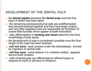
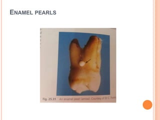
Ecosystem Interactions Class Discussion Presentation in Blue Green Lined Styl...
Root formation and periodontal ligament
1. DEVELOPMENT OF THE DENTAL PULP
ROOT FORMATION
reference :Oral anatomy , histology and embryology by
berkovitz
2. DEVELOPMENT OF THE DENTAL PULP
starts at the bud stage as condensation of
mesenchymal cells
some of these cells will become odontoblasts ,
those come from the neural crest
(ectomesenchyme)
the others are of mesenchymal origin
all cells look the same under the microscope
-these cells rapidly divide and are seperated by little
extracellular fluid.
3. DEVELOPMENT OF THE DENTAL PULP
in the cap stage the cells forming this mass are
known as the dental papilla
dental follicle : mesenchymal cells surrounding
the developing enamel organ and later on they will
form the periodontal ligement and the supporting
tooth structures.
during the bell stage cytodiffrentiation of cells by
signals from internal enamel epithelium take place
into :
1.central mass of fibroblasts
2. peripheral layer of odontoblasts.
4.
5.
6.
7. DEVELOPMENT OF THE DENTAL PULP
the dental papilla becomes the dental pulp once the first
layer of dentin has been layed.
the remaining ectomesenchymal cells are undiffrentiaited ,
they become packed together and they have large nucleus
and very little cytoplasm and are responsible for formation of
coarse fiber bundles which appear at tooth maturation.
cells differentiation in hertwig root sheet determins the final
morphology of pulp space .
the development of pulp is considered complete once the final
length of the roots has been reached.
cell rich zone : layer present under the odontoblasts , formed
by migration of central cells.
cell free zone : some believe it is a fixation artifact , appears
around tooth eruption.
cells of dental pulp can differentiate to different types to
respond to insult or stimulus or whatever.
8. BLOOD SUPPLY
during the bell stage branches of the jaws blood supply
enter the base of the papilla.
some of these branches enlarge and become the basic
arteriols of the dental papilla
they form a complete network of arteriols and venules
in the odontoblast and the subodontoblast layer.
-the vascularity of odontoblast increase with increasing
dentin thickness as a result of cells invasion into the
vascular bed.
exact lymphatic pattern and timing of pulp is not clear
yet.
mature pulp cells that enter with the blood vessels are :
pericytes , lymphoid cells and macrophages.
9.
10. NERVE SUPPLY
nerve fibers enter the dental papilla at a late stage.
first fibers to enter the papilla are from the trigiminal
nerve close to the blood vessels
those fibers are sensory autonomic fibers and they
control blood flow , this might help them to control tooth
development as well.
Neurotrophin brain derived growth factor is seen at the
periphery of developing pulp and this indicates
that this region is innervated by trigiminal nerve.
nerve fibers enter the pulp before final root formation but
the formation of plexus such as plexus of rashkowtakes
place after root formation is complete.
11. ROOT DEVELOPMENT
it starts with axial tooth eruption.
interaction between the dental follicle , the dental
papilla and hertwig epithelial root sheet ( derived from
cervical loop region of enamel ) .
at the late bell stage root formation starts when
dentinogenesis and amelogenesis are advanced ,
internal and external epethelial layers form and
proliferate apically to determine root formation.
number of roots is determined by number of epithelial
shelves ( these also proliferate to form multiple
secondary apical foramane ).
12.
13. ROOT DEVELOPMENT
root eruption takes place when two thirds of root is
complete , in this case we have open wide apex
apically.
and it closes 3 years later.
during eruption epethelial root sheath encloses the
enamel organ except for the apical foramen.
between the 2 epithelial layers surrounding the
dental papilla theres NO statum intermedium or
stellate reticulum , these are seen only in region of
enamel pearls..
dental follicle is external to root sheath and forms
the cementum alvoelar bone and periodontal
ligement.
14.
15. Adjacent to the epithelial root sheath is the inner layer of the
dental follicle which is ectomesenchyme ( derived from neural
crest cells).
adjacent to the alveolar bone is the outer layer of the dental
follicle which is separated from the inner layer by an intermediate
layer.
Both the middle and intermediate layers are mesodermal in origin
and their cells contain few organelles and appear relatively
structureless.
Cells of inner layer of dental follicle differentiate into
cementoblasts , and form a cuboidal layer on surface of root
dentin .
In primary acellular cementum , where collagen is of the extrinsic
type , cementoblasts from a little bit of the extracellular material ,
later with the formation of intrincic cellular cementum ,
cementoblasts will asllo secrete collagen.
16.
17. Once cementogenesis has begun , cells of dental
follicle become obliquely oriented to root surface
and their organelles increase and become the
fibroblasts of PL , and they secrete collagen in the
extracellular fluid and become embedded in
cementum on the root surface and in bone on
alveolar surface.
18. ROOT DEVELOPMENT
the cells of the internal layer of root epithelium
stimulate the cells of the dental papilla to become
odontoblasts.
after odontigenesis start , epithelial root sheat cells
loose their continuity and become rest cells of
malassez.
and the mesenchymal cells of the dental follicle
start differentiating into cementoblasts and
cementogenesis start.
19. CUSHION HAMMOCK LIGAMENT
Connective tissue lying immediately below the developing
root apex
Was described as fibrous network with fluid filled
compartments , connected to the alveolus on either side
Was thought to provide a strong base to prevent bone
resorption by heavy forces of eruption underneath the
developing root, this view is no longer held.
It was proven that this fibrous membrane is not attached
to bone but merges with fibers of PL , so more correctly
was called the pulp limiting membrane
Its surgical removal does not affect tooth eruption.
20.
21. It was proposed that changes in vascular
permeability around developing roots can affect
eruption behavior.
Dense accumulation of fluid ( effusion) was seen
beneath root apices , when they used radioactive
fibrinogen to study this , it was quickly incorporated
into the effusion .
Vascular hypotheses of eruption
Effusion forces the root and the bone apart on
eruption , and this contributes to eruption and
further root growth.
22. ROOT DEVELOPMENT
FORMATION OF COLLAGEN FIBERS WITHIN THE
PERIODONTAL LIGAMENT
stage one : before eruption the dentogingival and oblique
fibers are formed in permanent molars.
in premolars only the dentogingival are formed.
stage 2 as the tooth erupts in the oral cavity , PL of
permenant molars is well organized.
in premolars PL fibers are organised coronally only (
near the alveolus ) apically they are present yet they are not
organized.
stage 3 in molars cervical fibers become organised in
occlusion
in premolars cervical fibers become organized and
apically are not organized.
stage 4 after a while in function , fibers of all teeth become
organized.
23. FORMATION OF COLLAGEN FIBERS WITHIN
THE PERIODONTAL LIGAMENT
bulk of PL fibers is not fully organized during eruption
and this was proposed to help dissipate the forces
during tooth eruption.
the more time in function , the inclination of fibers
decrease and the thickness increase , however this
differ between species.
during tooth eruption , active resorption takes place at
the base of the socket. , some species have the
opposite e.g the dogs premolars ! this depends on the
distance of eruption , if the distance more than the
root length bone deposition is a must.
25. PRIMARY ACELLULAR CEMENTUM
cementogenesis begins at the cervical margin and progress downward.
epethelial root sheet cells do not enlarge in this phase in contrats to
enamel organ cells during amelogenesis.
the epethelial root sheet is seperated from both the dental follicle and
the dental papilla by basal lamina.
enteraction between epethelial root sheath and cells of dental papilla
induces them to differentiate into odontoblasts.
first formed layer of dentine is called predentin : structureless organic
glass like matrix under the polarised light. called hayaline once it is fully
mineralized and is about 10 microns.
cells of the epithelial root sheath are close to the predentine cells for a
short distance only , this allows the fibroblast like cells of dental follicle -
represent cementoblasts to be close and interact with the unminiralised
hayalyne layer.
at their deep surface they unite with the hayaline layer cells , and on
their superficial surface they form fibrous fringe extending
perpendicular in PL space for 10-20 microns.
26. at the beginning of cementogenesis epithelial root sheet cells secrete proteins
such as ameloblastin +- amelogenin into the united interface because they have
golgi complex in their cells . their function is unclear
might relate to epithelial mesenchymal interactions to induce cementoblasts or
odontoblasts.
during subsequent mineralization they are lost , may remain some of them in
dentin granular layer
as the odontoblasts move to the pulp , they leave their process behind forming
the granular layer of tomes and then circumpulal dentine ( basic tubular
structure of dentin ).
miniralisation starts few microns from the hayaline layer , thus the innermost layer
of it is continous with the periphery of the pulp .
similar staining pattern is observed after administration of antibiotic tetracyclin.
mineralization of cellular cementum speards outwaord , and this layer is firmly
attached to root dentin.
at this stage PL fibers are more parallel t root surface and not attached to root
fringe.
27. in early stages of cementogenesis , cementoblasts secrete many
proteins such as bone sialoprotein and osteopontin.
and they play a role in cell attraction and differentiation.
they might be related to a type of osteopetrosis in which osteocytes are
dificient.
subsequent development of acellular cementum includes
1. slow increase in thickness,
2.continouty between PL fibers and fibrous fringe at the surface of root
dentine,) this is the only way a tooth can be supported in the socket and
timing is different between permanent and primary teeth
for permanent teeth this attachment happens once the tooth erupts and
Acellular cementum thickness is 10 micron
acellular cementum lining the tooth before this time is known as intrinsic
acellular cementum
3. slow miniralisation of collagen.
28. once complete union has occured between PL
fibers and surface of cenentum this cementum is
classified as
acellular extrinsic fibre cementum , increases in
thikness about 2-2.5 microns throughout life , the
cells cannot be readily recognised as a layer from
the surrounding , they rather mingle .
29. cementoblasts contain organelles responsible for protein synthesis ,
and they secrete the surrounding ground material around the collagen
bundels.
miniralisation of cementum is initiated by the hydroxyappatite crystals
in the adjacent dentin not by cells.
PL fibroblasts also play a role in mineralization.
mineralization is a long slow process , this explains the absence of a
precementum layer.
mineralization of cementum is linked to the initial root dentine formation
, thats why drugs which inhibit this processsuch as bisphosphonates
inhibit also cementogenesis.
cementogenesis is rythemetic process with periods of increased and
decreased activity , this explains the presence of incremental lines ,
incremental lines are associated with decreased activity , they contain
high content of ground materialand water and less amount of collagen.
acellular cementum forms more slowly , than cellular cementum , the
incremental lines are packed more together.
30. ACELLULAR AFIBRILLAR CEMENTUM
thin layer overlying enamel in cervical tooth area.
reduced enamel epethelium overlying tooth
becomes damaged , cells of connective tissues
adjacent to enamel organ diffrintiate into
cementoblasts which secrete afibrillar matrix and
then calcifies .
31. SECONDARY CELLULAR CEMENTUM
forms in the apical region and furcation at time of tooth
eruption.
formation is similar to primary cementum and is related to
dentin formation.
following loss of continuity of epithelial root sheath , large
basophiles of dental follicle differentiate into cementoblasts
near root surface.
under light microscope , they have endoplasmic reticulum
and secrete the matrix and the collagen fibers of the
seconday cellular cementum.
fibers are parallel to root surface , with increased rate
formation , unmeneralized layer of pre cementum forms
around 5 microns thickness.
overall secondary cementum is less mineralized than
primary cementum , miniralization takes place in linear
matter.
32. cells become gradually incorporated within the matrix
they secrete , then the become osteoyte , this
necissates the new stem cells from PL differentiate
into cementoblasts.
cellular cementum has no sharpeys fibers and is
present only in apical and furcation areas , it alternates
with acellular extrinsic fiber cementum to form mixed
stratified cementum.
when cellular intrinsic fiber cementum is covered by
acellular extrinsic fiber cementum sharpeys fibers are
attached to root surface , in the opposite situation ,
sharpeys fibers are detached from root surface.
33. CELLULAR MIXED FIBER CEMENTUM :
cellular intrinsic cementum gives attachment to
extrinsic fibers from PL which are aligned perpindicular
to root surface incontrast to intrinsic fibers secreted by
cementoblasts themselves which are secreted parallel
to root surface.
as for acellular cementum , exact origin of cells
responsible for formation of secondary cementum still
needs clarification , its believed they come from
different cell populations.
due to similarity between cementoblasts and
osteoblasts it has been suggested that proginertaor
cells responsible for formation osteoblast could
migrate to PL and provide new source for
cementoblasts.
34. ENAMEL PEARLS
small spherical of enamel located on tooth surface
near cervical end popular at the bifurcation,
in region affected : stallate reticulum and stratum
intermedium develop between external and internal
enamel sheath