Downloaded 11 times


The document summarizes the process of dentinogenesis or dentin formation. It involves differentiation of odontoblasts from dental papilla cells, secretion of an organic matrix, and mineralization of the matrix. Odontoblasts secrete collagen fibers and matrix vesicles that initiate mineralization. Dentin is formed in mantle dentin near enamel and circumpulpal dentin further inside via continuous mineralization. Root dentin formation begins after crown completion, guided by Hertwig's epithelial root sheath.
Presentation by Dr. Sonal Aggarwal on Dentinogenesis, focusing on dentin formation processes.
Formation of dentin involves cell differentiation, organic matrix synthesis, and mineralization, starting at the bell stage.
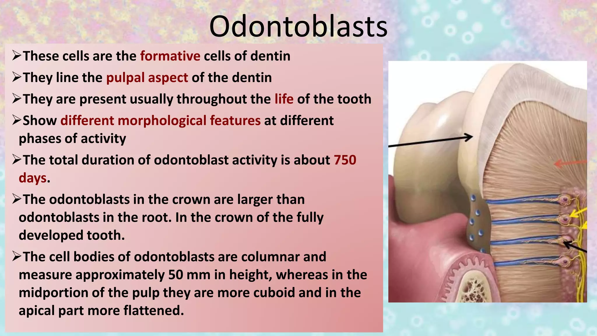
Odontoblasts form dentin, vary in size and shape during activity, and have a life span of about 750 days.
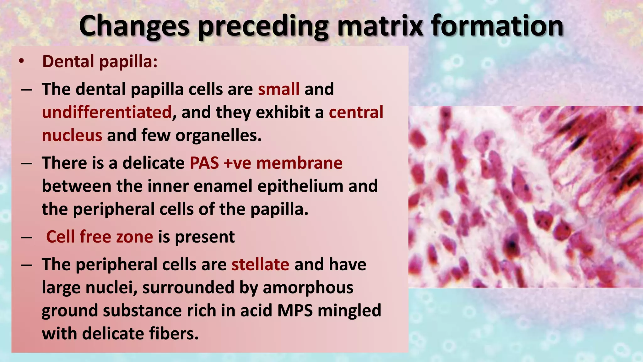
Dental papilla cells are undifferentiated and lead to matrix formation, surrounded by various cell types.
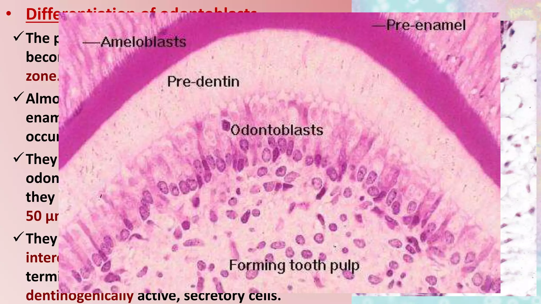
Differentiation involves pre-odontoblasts becoming odontoblasts with structural changes, leading to active secretion.
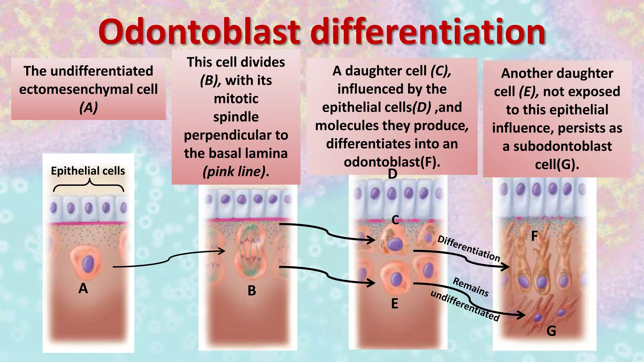
Detailing the differentiation pathway of ectomesenchymal cells into odontoblasts influenced by epithelial cells.
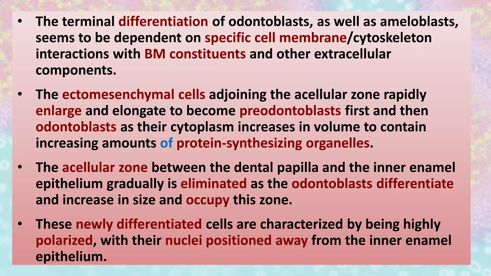
Odontoblast differentiation relies on cytoskeleton interactions and cell membrane activities as they polarize.
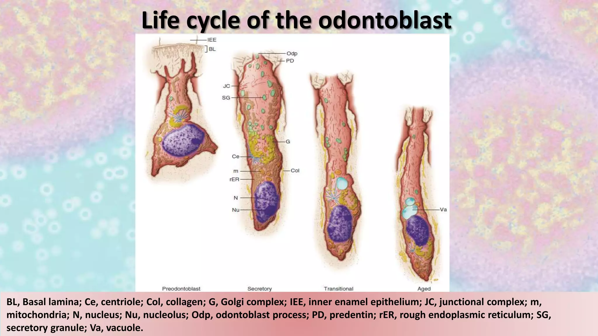
Visual representation of odontoblast life cycle and associated cellular structures necessary for formation.
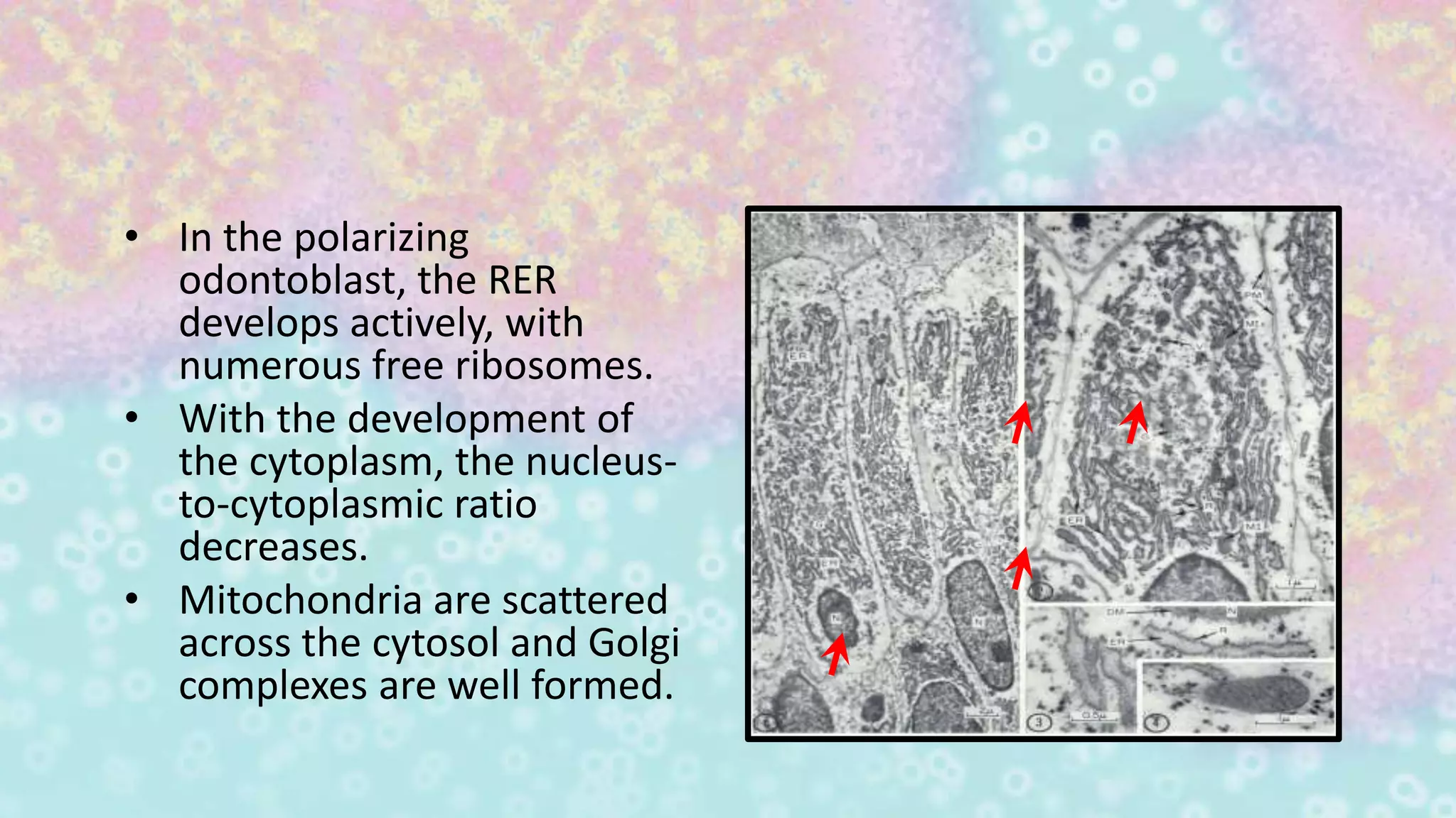
Development of odontoblasts involves active rough endoplasmic reticulum and nuclear-to-cytoplasmic ratio changes.
Describes the presence and role of junctional complexes between odontoblast cell bodies and processes.
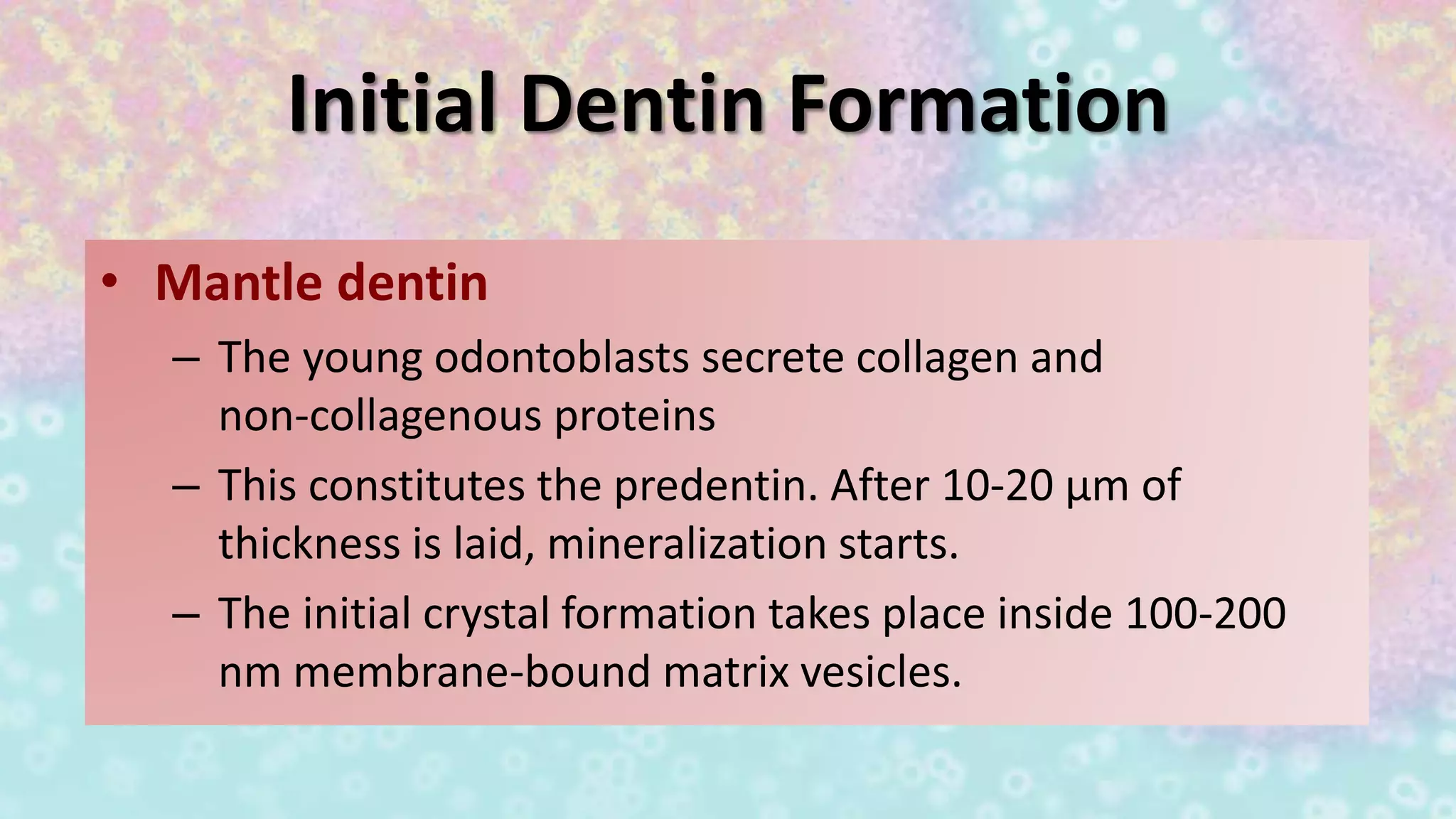
Young odontoblasts secrete collagen to form predentin which mineralizes after reaching 10-20 μm thickness.
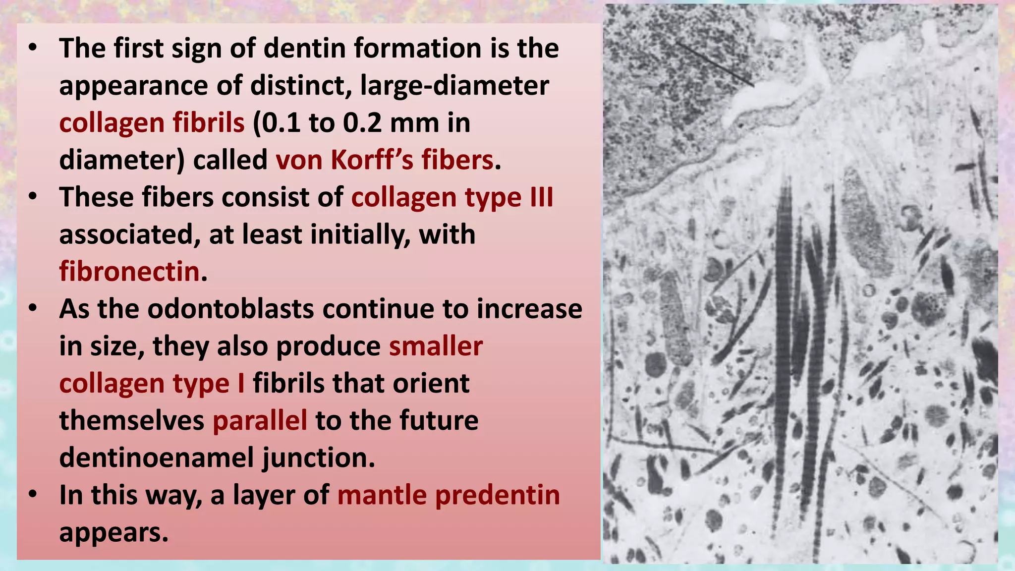
Collagen type III fibers, von Korff's fibers, mark the beginning of distinct dentin formation processes.
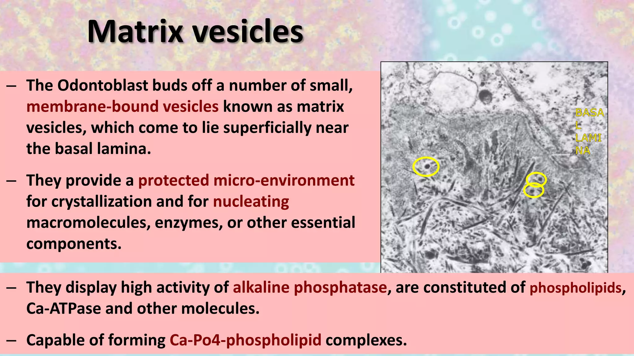
Matrix vesicles from odontoblasts create a micro-environment enabling crystallization and mineralization.
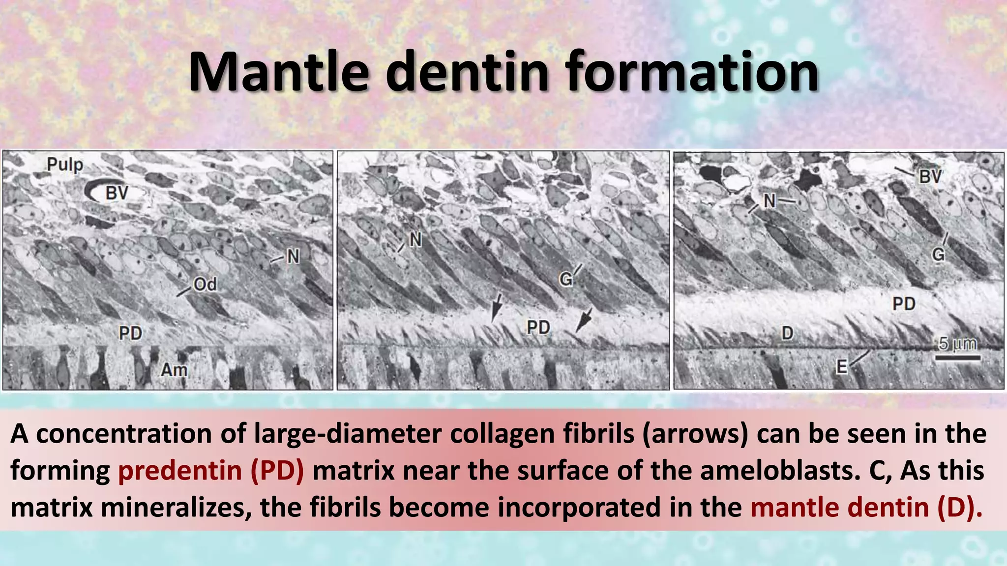
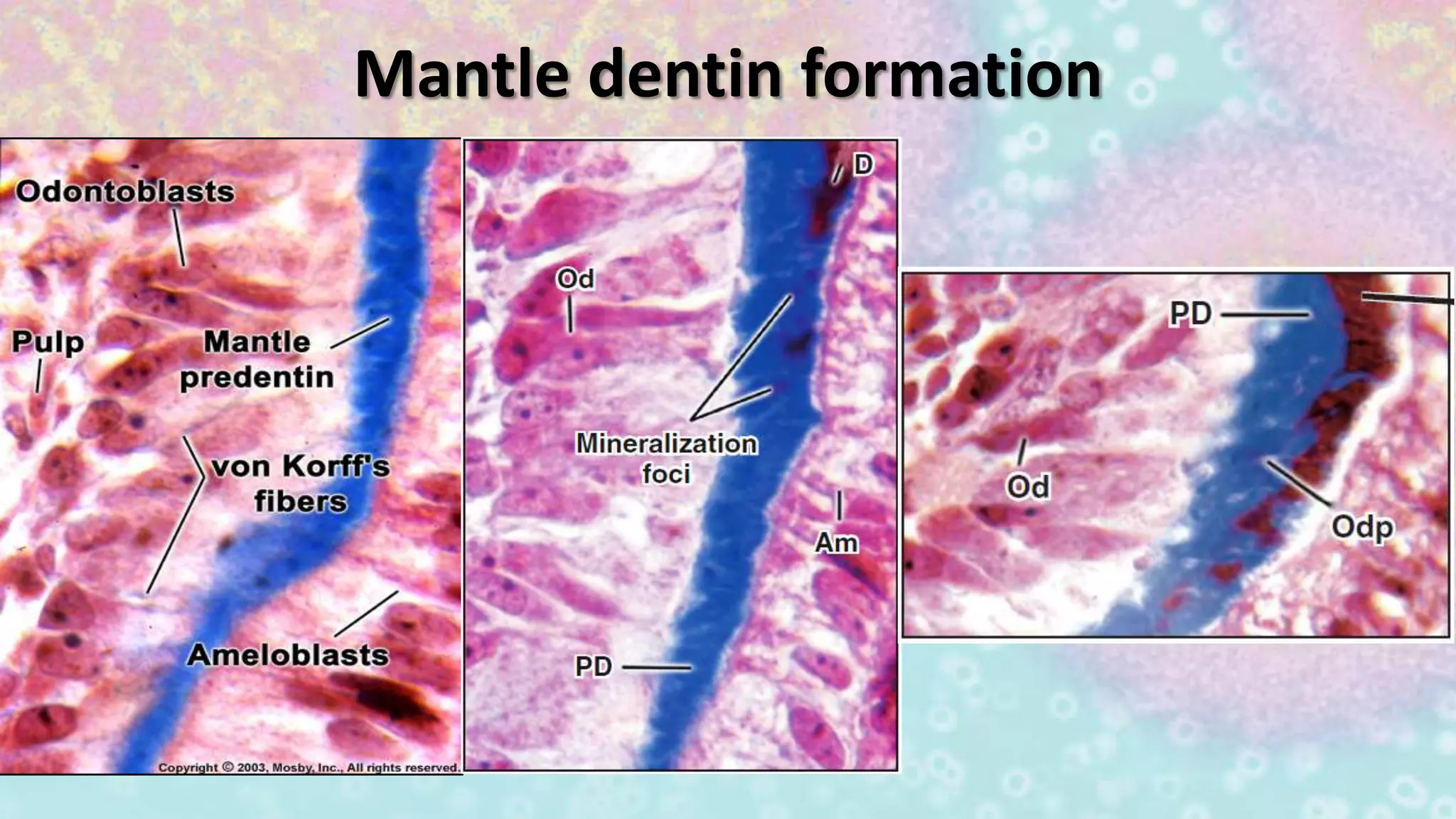
Formation of mantle dentin involves alignment of collagen fibrils within the emerging predentin matrix.
Continued discussion on mantle dentin formation mechanisms previously introduced.
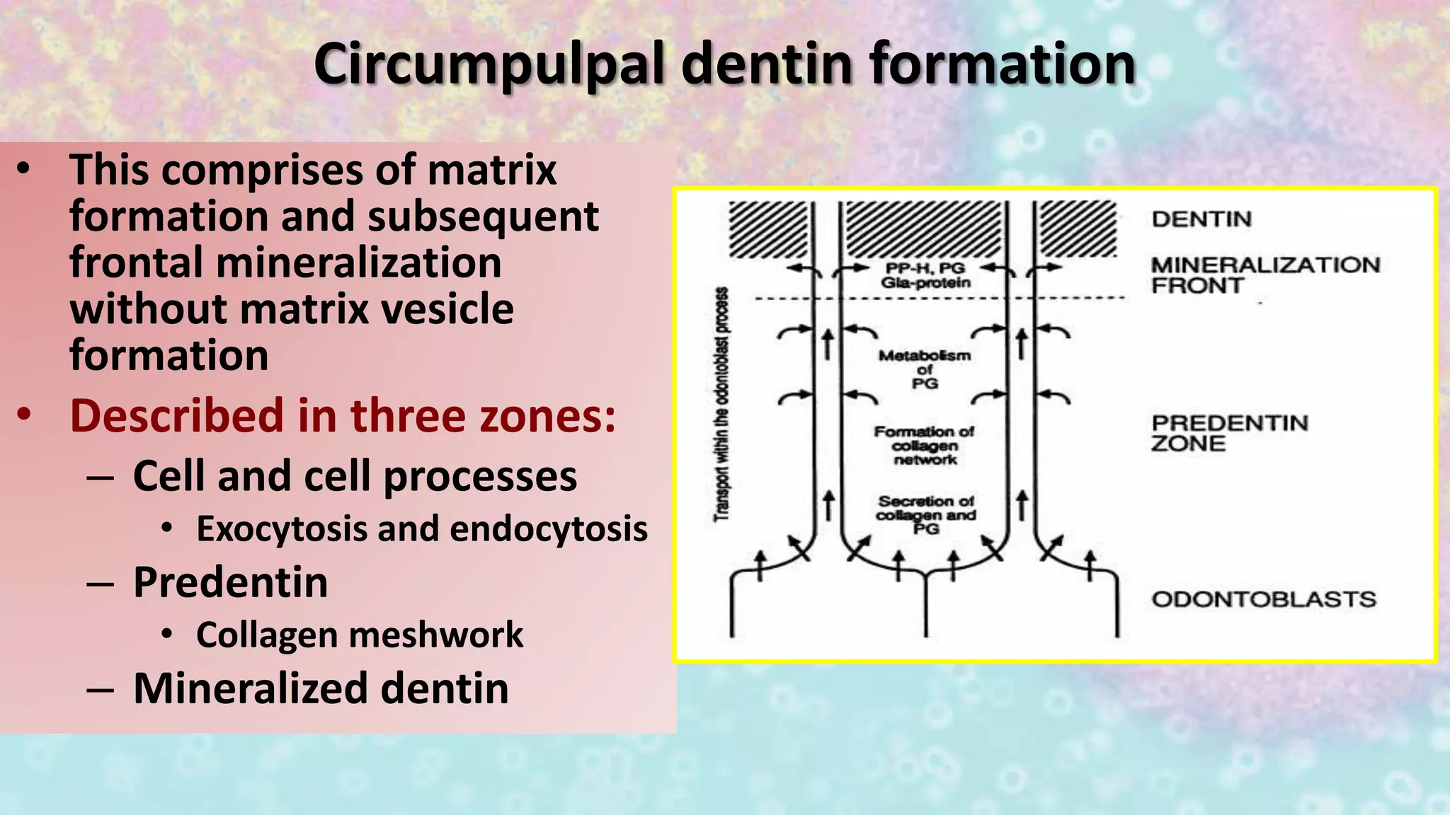
Describes circumpulpal dentin formation in three zones, emphasizing matrix formation and mineralization.
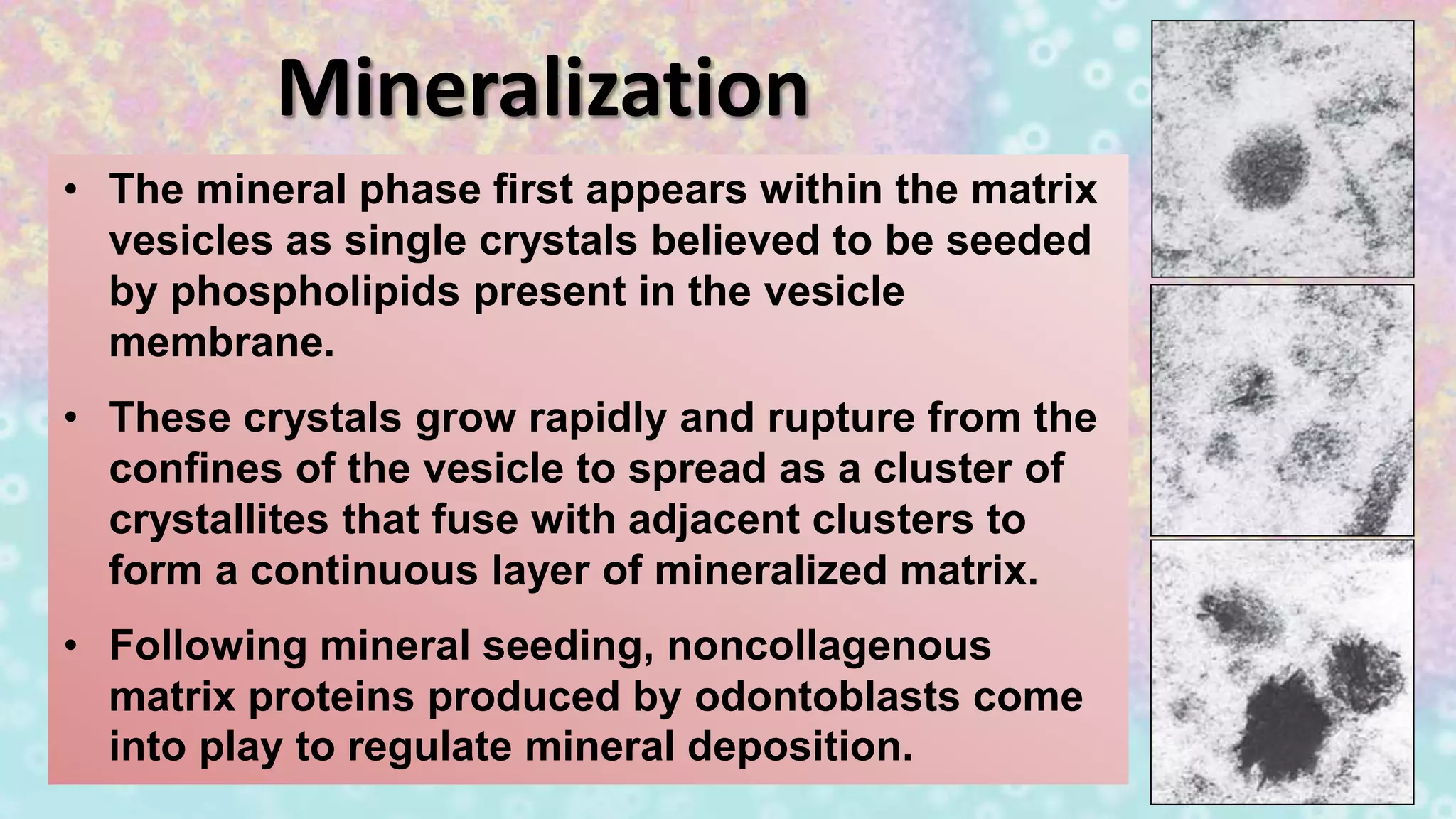
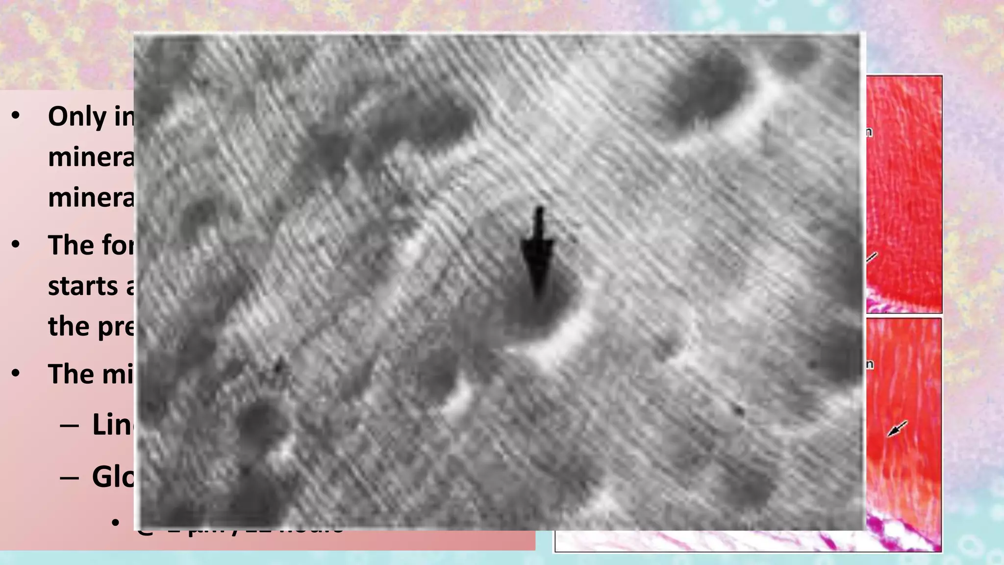
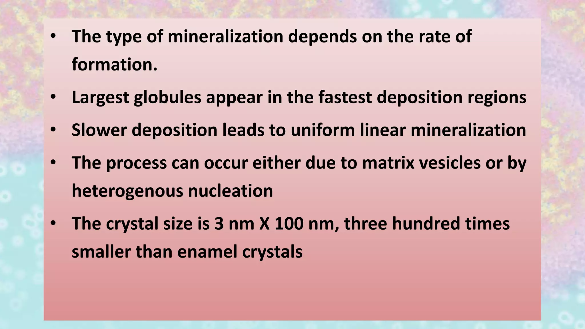
Mineralization occurs within matrix vesicles, leading to the creation of a mineralized matrix in dentin.
Differentiates between intertubular and intratubular dentin mineralization with specifics on rates of deposition.
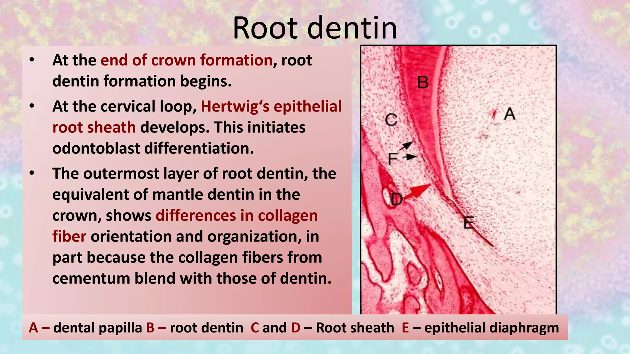
Root dentin formation initiates at the cervical loop with unique collagen organization compared to crown dentin.


