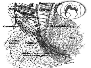
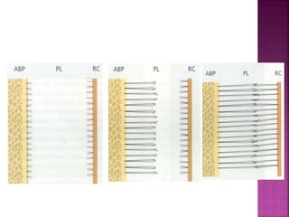
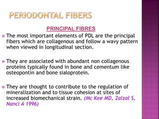
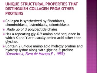
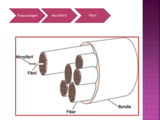
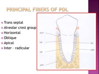
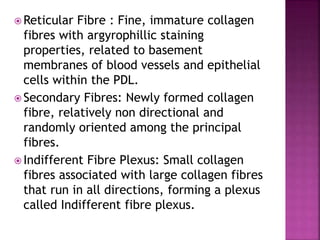
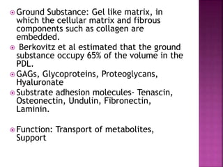
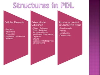
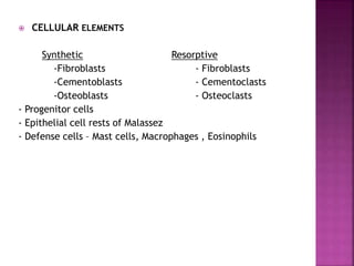
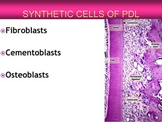
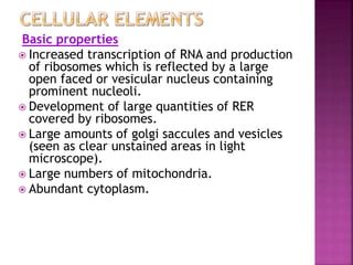
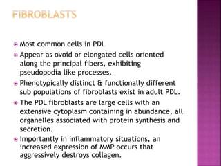
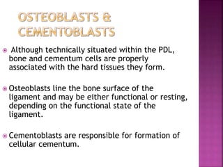
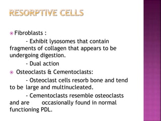
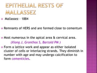
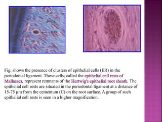
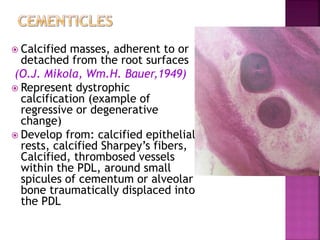
The periodontium is the connective tissue that surrounds and supports the teeth. It consists of gingiva, periodontal ligament, cementum, and alveolar bone. The periodontal ligament is a soft, vascular connective tissue that joins the cementum and alveolar bone. It contains collagen fibers that provide support and flexibility to the teeth. The ligament is populated by fibroblasts that synthesize collagen fibers, as well as other cells like cementoblasts and osteoblasts that maintain the hard tissues.
This document summarizes a presentation on the periodontal ligament given by Dr. Abhishek Gakhar. It discusses the structure, development, constituents including cells, fibers and blood/nerve supply of the periodontal ligament. The functions of the periodontal ligament and its clinical correlations are also reviewed. Diagrams illustrate the fiber groups and cells found in the periodontal ligament.
The periodontal ligament is a fibrous connective tissue between the tooth root and jaw bone. It contains fibroblasts, cementoblasts, osteoclasts, blood vessels, nerves, and lymphatics. The principal fibers of the ligament are collagen and they are arranged in groups to withstand forces from chewing. The ligament receives blood supply from gingival, intra-alveolar, and apical vessels. It functions to support teeth, sense touch and pressure, provide nutrients, form new fibers, and protect teeth by distributing forces. In aging, the periodontal ligament shows reduced vascularity, cellularity and thickness and may contain calcified cementicles.
This document provides information on cementum, including its definition, physical characteristics, chemical composition, formation (cementogenesis), classification, functions, anomalies, and clinical considerations. Cementum is the mineralized tissue covering tooth roots. It is softer than dentin and lacks enamel's luster. Cementum formation involves acellular and cellular stages. Cementum attaches the periodontal ligament fibers to the tooth root and allows for tooth repair. Abnormalities include hypercementosis, ankylosis, and cementomas. Cementum is an important part of the periodontium that aids in tooth attachment and repair.
The document discusses the periodontal ligament (PDL), which is the soft connective tissue that surrounds tooth roots and attaches cementum to alveolar bone. It defines PDL and describes its extent, average width, development from the dental follicle, orientation of collagen fibers, cellular elements including fibroblasts, cementoblasts, osteoblasts, and epithelial rests of Mallassez. The document also covers the biochemical composition and ground substance of PDL, as well as its blood supply, nerve supply, age-related changes, and role in healing after periodontal surgery.
The periodontium refers to the tissues that surround and support teeth. The periodontal ligament is a specialized connective tissue that connects the tooth root to the inner surface of the alveolar bone. It is made up of collagen fibers, fibroblasts, and contains blood vessels. The periodontal ligament develops from cells of the dental follicle that differentiate into cementoblasts, fibroblasts, and other cells after the root forms and erupts. It contains principal fibers that connect the cementum to bone and resist various forces on the teeth. Other components include cementoblasts, osteoblasts, epithelial cell rests, and defense cells that maintain the periodontium.
The periodontal ligament (PDL) is a soft connective tissue located between the cementum on the root of a tooth and the alveolar bone. It consists of collagen fibers, cells like fibroblasts and cementoblasts, blood vessels, and nerves. The principal fibers of the PDL are arranged in groups to help support the tooth, resist movement, and absorb forces during chewing. The PDL transmits occlusal forces to the bone, attaches the tooth, and maintains the gingiva.
The junctional epithelium is a non-keratinized stratified squamous epithelium that forms an attachment to the tooth surface. It develops from the reduced enamel epithelium during tooth eruption. The junctional epithelium acts as a barrier against oral pathogens and allows for host defense mechanisms to reach the gingival sulcus. It has a rapid turnover rate of 4-6 days and can quickly regenerate after injury. The attachment to enamel is mediated by hemidesmosomes in the epithelial cells that are connected to the internal basal lamina on the tooth surface. Disruption of this attachment can initiate periodontal pocket formation and disease.
Bone is a living tissue that provides structure and support. It can be classified based on shape, development, histology, and composition. The alveolar process forms with tooth development and eruption to support teeth in the jaw. It consists of cortical and cancellous bone layers surrounded by osteoblasts and osteoclasts, which build and resorb bone through various signaling pathways and enzymes.
Alveolar bone is the specialized bone that forms the sockets for teeth in the maxilla and mandible. It consists of alveolar bone proper surrounding the tooth root, supporting alveolar bone made of cortical plates and spongy bone, and bundle bone where periodontal ligament fibers insert. Osteoblasts build bone matrix while osteoclasts resorb it, allowing remodeling. With age, alveolar bone thins with wider marrow spaces and more fragile trabeculae, leading the alveolar crest to slope down distally as teeth tilt mesially.
The document discusses the periodontium and its components, with a focus on cementum. It defines cementum as the hard calcified tissue covering tooth roots that attaches the tooth to the surrounding alveolar bone via periodontal ligament fibers. The document describes the physical properties, chemical composition, classification, and histological structure of cementum. It also discusses acellular cementum, cellular cementum, afibrillar cementum, cementoblast cells, cementocyte cells, the cemento-enamel junction, incremental lines of Salter, and age-related changes such as hypercementosis.
The document discusses the periodontal ligament (PDL), which is a specialized connective tissue that surrounds tooth roots and attaches them to the alveolar bone. It provides definitions of the PDL, describes its development, structure, cellular components, principal fibers, blood supply, and functions. The PDL maintains the space between the root and bone despite forces on the teeth and is composed of fibroblasts, extracellular fibers and ground substance that connect the cementum and alveolar bone. It summarizes the key anatomical and functional aspects of the PDL.
This document provides an overview of bone histology and development. It discusses that bone is a specialized mineralized connective tissue that can be divided into compact and cancellous bone microscopically. Bone develops through either endochondral or intramembranous ossification. The key cells involved are osteoblasts, which form new bone, and osteoclasts, which resorb bone. Bone is remodeled throughout life by the balanced actions of these cells.
Cementum is the mineralized connective tissue covering tooth roots. It has several functions including anchoring collagen fibers from the periodontal ligament to provide attachment between the tooth and bone. Cementum can be classified based on its location, cellularity, fiber content, and other characteristics. It plays roles in adaptation, repair, and maintaining the periodontium. The cemento-enamel junction describes the interface between cementum and enamel at the cervical portion of the tooth root.
This document provides an overview of cementum, the calcified tissue that forms the outer covering of tooth roots. It discusses the development, composition, histology, classification, and functions of cementum. Cementum begins forming at the cementoenamel junction and extends to the root apex. It is made up of inorganic hydroxyapatite and organic collagen fibers. Cementum provides a medium for periodontal ligament attachment and protects underlying dentin, helping to maintain tooth integrity under forces. It is capable of continuous deposition to repair damage or resorption on root surfaces.
The document discusses the peridontium and its components, which include the gingiva, periodontal ligament, cementum, and alveolar bone. It focuses on cementum, describing it as a hard connective tissue that covers tooth roots and provides attachment for collagen fibers. Cementum begins forming at the cementoenamel junction and continues to the root apex. It contains cementoblasts and cementocytes that aid in its formation and structure. Cementum comes in cellular and acellular varieties and demonstrates incremental lines from its continuous deposition over time.
The document discusses alveolar bone, which forms the primary support structure for teeth. It defines alveolar bone and discusses its classification, composition, function, histology, cells, development, remodeling, and age-related changes. Alveolar bone holds teeth firmly in position, supplies vessels to periodontal ligaments and cementum, and houses developing permanent teeth. It is a specialized part of the maxilla and mandible composed of lamellar and bundle bone that surrounds tooth roots and provides attachment for periodontal ligament fibers. Alveolar bone is constantly remodeled through formation and resorption to adapt to functional forces.
The periodontium consists of cementum, the periodontal ligament (PDL), alveolar bone, and gingiva surrounding the tooth. The PDL is a specialized connective tissue between cementum and alveolar bone that functions to support the teeth, absorb chewing forces, provide sensory feedback, and enable nutrient transport. It contains collagen fiber bundles arranged to withstand forces. Cementum is a hard, avascular tissue covering roots that provides attachment for collagen fibers. The alveolar bone forms the tooth sockets and contains the PDL. Molecular factors regulate tissue development and regeneration.
The periodontal ligament (PDL) is a soft connective tissue that surrounds tooth roots and attaches them to the alveolar bone in the jaw. It ranges from 0.15-0.38mm in width and is narrowest at the mid-root level. The PDL contains principal collagen fibers, blood vessels, nerves and cells that allow it to absorb forces and remodel throughout life. Diseases can widen the PDL space and disrupt its fibers. The document discusses the development, structure, functions and clinical implications of the PDL.
Cementum is the mineralized tissue covering the roots of teeth that provides attachment for collagen fibers linking the tooth to surrounding bone. It begins at the cementoenamel junction and continues along the root to the apex. Cementum is avascular and less hard than dentin. It contains both inorganic minerals and organic materials including collagen. Cementoblast cells synthesize cementum by laying down an organic matrix that subsequently mineralizes. Cementum thickness varies along the root and increases with age. It provides for functional adaptation and resistance to resorption during orthodontic tooth movement.
The document discusses the dentogingival junction and junctional epithelium. It begins with an introduction and overview of the three zones of gingival epithelium. It then covers the historical aspects and development of the junctional epithelium. The structure of the junctional epithelium is described including its anatomical features, epithelial attachment apparatus involving hemidesmosomes, and dynamic aspects like rapid turnover. The permeability and various functions of the junctional epithelium are also summarized.
Alvelor bone has several important functions including supporting tissues, providing muscle attachments, and storing ions like calcium. It has the ability to remodel according to functional demands. Alveolar bone development depends on the presence of teeth. Bone is classified as either endochondral or intramembranous bone developmentally, and as compact or cancellous bone histologically. The main cell types in bone are osteoblasts, osteocytes, bone lining cells, osteoprogenitor cells, and osteoclasts. Bone undergoes remodeling through the stages of resorption, reversal, formation, and resting. Microdamage signals bone remodeling through resorption and calcified matrix filling cracks. Clinical considerations for bone include resorption
Coronal and radicular pulp
Apical foramen
Accessory canal
Functions of dental pulp
Components of dental pulp
Functions of pulpal extracellular matrix
Organization of cells in the pulp
The principle cells of the pulp
The pathways of collagen synthesis
Matrix and ground substances
Vasculature and lymphatic supply
Innervation of Dentin- pulp complex
Disorders of the dental pulp
Advances in pulp vitality testing
The periodontal ligament is a dense connective tissue that occupies the space between the root of a tooth and the alveolar bone. It contains collagen fibers, cells, blood vessels, and nerves. The fibers are arranged in different groups to support the tooth and allow movement during mastication while protecting the underlying bone. The periodontal ligament provides nutrition to cementum and bone, senses pressure, and facilitates tooth movement during orthodontic treatment by stimulating bone remodeling through fiber tension and compression. It also protects the tooth and bone by distributing forces and containing mechanoreceptors.
5. alveolar bone in health part a dr-ibrahim_shaikhDrIbrahim Shaikh
The document provides an overview of alveolar bone structure and function. It begins with definitions of key terms like the periodontium and alveolar process. It then discusses the classification, composition, development and typical morphology of alveolar bone. Specific structures are described like the alveolar bone proper, cortical and spongy bone, bundle bone, and lamina dura. The final section outlines some common radiographic features seen in alveolar bone like the lamina dura, alveolar crest, and trabecular patterns. In summary, the document serves as a comprehensive review of alveolar bone anatomy and histology.
The gingiva is a masticatory mucosa that covers the alveolar process of the jaw and surrounds the neck of the teeth. It is made up of epithelium and connective tissue. The gingiva can be divided into three types - free gingiva, gingival sulcus, and attached gingiva. Microscopically, the gingival epithelium consists of outer oral epithelium, sulcular epithelium, and junctional epithelium. The gingiva also contains dense collagen fibers called the gingival ligament. Blood supply to the gingiva is provided by the alveolar artery and it receives nerve innervation from various nerves depending on location.
The periodontal ligament is a dense connective tissue that connects the tooth to the alveolar bone. It consists of collagen fibers arranged in bundles, cells, blood vessels and nerves. The fibers are organized into groups that resist various forces on the tooth. The periodontal ligament provides support, sensory function, nutrition and protects the tooth from excessive forces. It undergoes changes with age, becoming less cellular and vascular with possible calcifications forming.
The periodontal ligament is a specialized connective tissue that connects the cementum of teeth to the alveolar bone. It develops from the dental follicle during root formation and tooth eruption. The periodontal ligament is composed of collagen fibers, fibroblasts, blood vessels and nerves. The principal collagen fibers are arranged in bundles and attach to the cementum and bone. The periodontal ligament helps maintain homeostasis between the teeth and surrounding tissues and allows for tooth mobility.
The periodontal ligament is a complex connective tissue that connects the tooth to the alveolar bone. It contains principal collagen fiber bundles arranged in six groups, cellular elements like fibroblasts and immune cells, and ground substance. The periodontal ligament functions include transmitting forces, remodeling bone in response to forces, supplying nutrients to surrounding structures, and sensing tactile sensations.
The periodontal ligament is a connective tissue that connects the cementum of teeth to the alveolar bone. It contains principal collagen fibers, fibroblasts that produce the fibers, undifferentiated cells, and a ground substance of proteoglycans and glycoproteins. The principal fibers develop in stages from the cementum to bone and resist various forces on teeth. The periodontal ligament plays an important role in tooth support and is vital for tooth function.
The document summarizes the key components and functions of the periodontium, which provides support to teeth. It consists of the periodontal ligament, cementum, and alveolar bone. The periodontal ligament is a complex connective tissue that surrounds the tooth root and connects it to the alveolar bone. It contains principal fiber groups that help distribute forces. The periodontium provides physical support, plays a role in remodeling, and provides sensory and nutritional functions to maintain teeth. Age changes and diseases can impact the periodontium and tooth support. Regenerative therapies are being explored to repair periodontal ligament destruction.
Indian Dental Academy: will be one of the most relevant and exciting training center with best faculty and flexible training programs for dental professionals who wish to advance in their dental practice,Offers certified courses in Dental implants,Orthodontics,Endodontics,Cosmetic Dentistry, Prosthetic Dentistry, Periodontics and General Dentistry.
The periodontium consists of the investing and supporting tissues of the tooth, including the gingiva, periodontal ligament, cementum, and alveolar bone. The gingiva is divided into marginal, attached, and interdental areas. The periodontal ligament connects the cementum and alveolar bone and contains collagen fibers that provide attachment, transmit forces, and allow for remodeling. Cementum is the calcified tissue covering the tooth root. The alveolar bone surrounds and provides support to the tooth sockets. Together, these tissues support and protect the teeth.
The periodontal ligament is a soft, vascular connective tissue that connects tooth roots to the alveolar bone socket. It develops from the dental follicle during root formation and tooth eruption. The periodontal ligament contains principal collagen fiber bundles oriented in different directions, as well as fibroblasts, cementoblasts, osteoblasts, and progenitor cells. It maintains homeostasis through a balance of synthetic and resorptive cells and extracellular substances. The unique structure and cellular composition of the periodontal ligament allow it to function in tooth attachment and as a sensory organ.
The document provides information about the periodontal ligament (PDL). It defines the PDL as the specialized connective tissue that surrounds tooth roots and attaches them to the alveolar bone. The summary discusses that the PDL is composed of collagen fibers, fibroblasts and other cellular elements embedded in a ground substance. It has an average width of 0.2mm and develops along with root formation. The principal collagen fibers of the PDL, including alveolar crest fibers, horizontal fibers, oblique fibers and apical fibers, develop from the cementoblast surface and resist various forces on the teeth.
The periodontal ligament is a complex connective tissue that surrounds the tooth root and connects it to the alveolar bone. It is composed of collagen fibers, cells like fibroblasts and cementoblasts, blood vessels and nerves. The PDL develops from the dental follicle during root formation and ranges in width from 0.15-0.38mm. It contains principal fibers that extend obliquely from cementum to bone and adapt to functional changes in teeth. The PDL maintains homeostasis between the hard tissues of cementum and bone through regulatory molecules and cells.
The periodontal ligament connects the tooth to the alveolar bone and is made of specialized connective tissue. It develops as cells from the dental follicle migrate into the newly formed root cementum. The principal fibers of the ligament develop with tooth eruption, first entering the alveolar bone and later organizing into bundles. Microscopically, the ligament contains fibroblasts, progenitor cells, and extracellular fibers and ground substance. The fibroblasts are responsible for collagen synthesis and turnover and have a higher rate of protein production compared to gingiva or bone fibroblasts.
The periodontal ligament is a complex connective tissue that surrounds the tooth root and attaches the tooth to the alveolar bone. It is composed of collagen fibers, ground substance, blood vessels, nerves and cells. The principal collagen fibers resist various forces on the tooth and help transmit these forces to the bone. The periodontal ligament provides nutrition to surrounding structures, allows for remodeling and repair, and has proprioceptive functions through its nerve endings.
The document discusses the composition and structure of the periodontal ligament. It notes that the periodontal ligament consists of cells such as fibroblasts and an extracellular matrix containing collagen fibers, ground substance, and other proteins. It connects tooth cementum to alveolar bone and functions to support teeth and withstand forces. The fibroblasts are responsible for maintaining and remodeling the ligament in response to forces through synthesis and degradation of the extracellular matrix.
The periodontium is composed of gingiva, periodontal ligament, cementum, and alveolar bone. The gingiva is divided into marginal, attached, and interdental regions. It surrounds the neck of the tooth and is lined by sulcular and junctional epithelium. The gingiva contains collagen fibers that attach it to the tooth and alveolar bone. It receives blood supply from the periodontal ligament and alveolar bone and drains into local lymph nodes.
The periodontal ligament is a complex connective tissue that connects teeth to the alveolar bone. It contains fibroblasts, cementoblasts, osteoblasts, osteoclasts and epithelial rests of Malassez. The fibroblasts are the predominant cells and produce collagen fibers that develop into principal fiber bundles. The periodontal ligament allows for adaptation during function by remodeling its collagen fibers and allows movement within the periodontal space. It develops from the dental follicle prior to tooth eruption and contains stem cells that can differentiate into cementoblasts, osteoblasts or fibroblasts.
This document discusses the biology of tooth movement. It begins by classifying tooth movement into physiological, pathological, and orthodontic categories. It then discusses the historical studies on tooth movement dating back to the early 1900s. The bulk of the document describes the relevant biological structures - cementum, periodontal ligament, alveolar bone, and their cells and composition. It explains the fiber groups within the periodontal ligament. Finally, it discusses the biological events and tissue reactions that occur during orthodontic tooth movement.
The alveolar process develops during tooth eruption from bony septa that form as tooth germs develop in the mandible and maxilla. It contains alveolar bone proper that lines the tooth socket, as well as bundle bone that is penetrated by Sharpey's fibers connecting it to the cementum. The periodontal ligament situated between cementum and alveolar bone contains principal fiber bundles, fibroblasts, and a ground substance that allows it to withstand masticatory forces. It receives its blood supply from the alveolar arteries and innervation from the inferior and superior alveolar nerves.
The periodontal ligament is a fibrous connective tissue that attaches the tooth to the alveolar bone. It is composed of fibers, cells, blood vessels and ground substance. The fibers are arranged in bundles that provide support, resist displacement, and allow movement. The principal fiber groups include gingival fibers around the neck of the tooth and dento-alveolar fibers along the root. Blood supply comes from the superior and inferior alveolar arteries.
Dental hygienists need to understand nutrition because diet is closely related to oral health. They should assess patients' nutritional status and provide dietary advice, such as recommending soft foods after procedures to aid healing. Key nutrients support oral health, while sugars increase caries risk and fiber aids cleaning. Malnutrition can suppress immunity and impair healing. The document then defines various nutrition terms and classifications of foods and nutrients, describing the functions and recommendations for carbohydrates, proteins, and fats.
Post operative complications of periodontal surgeryRitam Kundu
This document discusses various factors that can cause bleeding during oral surgical procedures and how to manage it. It notes that the extent of tissue reflection, inflammation, presence of unhealthy tissues, perforation of blood vessels, time to complete treatment, vasoconstrictors used, and patient medications all impact bleeding. It provides examples of average blood loss from different procedures and defines hypovolemic shock. Techniques to reduce bleeding and hematoma formation during flap surgery are outlined.
This document provides an overview of dental implants. It discusses the history of implant dentistry from ancient civilizations to modern developments like osseointegration. It defines a dental implant and describes the phases of osseointegration. The document outlines different implant components, surfaces, and classifications. It notes the benefits of implants compared to other options as well as potential complications. Examples of implant cases from the Department of Periodontics are also mentioned.
This document outlines the phases of periodontal therapy, including:
1) Preliminary phase focusing on emergencies and extractions.
2) Nonsurgical phase involving plaque control, non-surgical treatments like scaling and root planing.
3) Surgical phase using various periodontal surgeries and other treatments like implants and endodontics.
4) Restorative phase for final restorations and prosthodontics.
5) Maintenance phase for long-term supportive periodontal therapy.
Surgical periodontal therapy aims to eliminate pathologic changes, create a stable periodontium, and promote regeneration through techniques like pocket reduction surgeries and correction of anatomic defects
The document summarizes the key phases and techniques involved in nonsurgical periodontal therapy (NSPT). It discusses the goals of NSPT to eliminate pathogens and halt disease progression. Techniques include scaling and root planing to remove calculus, contaminated cementum, and bacterial toxins. Studies found that aggressive root planing is not needed and that clinical improvements result from scaling alone or with root planing. The effects of NSPT on subgingival microflora and selection of instrumentation techniques are also summarized.
This document discusses guidelines for smoking cessation interventions and treatment. It outlines recommendations from the WHO and US Public Health Service on promoting non-smoking as the norm, prohibiting tobacco promotion, and reducing tobacco industries. The goals of smoking cessation treatment are achieving long-term abstinence, offering treatment to all tobacco users, and consistently identifying and treating tobacco use. Dental professionals can play a key role in educating patients and the community about the harms of tobacco and helping to enroll them in cessation programs. Assessment tools like the Fagerstrom Test and stages of change model are also discussed to guide treatment and counseling approaches. A variety of nicotine replacement therapies and non-nicotine medications are described for treating nic
Necrotising periodontal diseases, Necrotising periodontal diseases as a manifestation of systemic diseases.
By Dr. Ritam Kundu, MDS PGT, Dr. R. Ahmed Dental College & Hospital, Kolkata, India.
The document discusses acute periodontal infections including abscesses, pericoronitis, and herpetic gingivostomatitis. It describes the clinical features, causes, microbiology, diagnosis and treatment of each condition. Abscesses are classified as gingival, periodontal or pericoronal depending on their location. Pericoronitis is inflammation around an unerupted tooth, usually due to food debris trapped under the gums. Herpetic gingivostomatitis is caused by the herpes simplex virus and presents as diffuse gingival swelling and vesicles that rupture, leaving painful ulcers. Prompt diagnosis and treatment including drainage, debridement and antibiotics are important to resolve the infections
share - Lions, tigers, AI and health misinformation, oh my!.pptxTina Purnat
• Pitfalls and pivots needed to use AI effectively in public health
• Evidence-based strategies to address health misinformation effectively
• Building trust with communities online and offline
• Equipping health professionals to address questions, concerns and health misinformation
• Assessing risk and mitigating harm from adverse health narratives in communities, health workforce and health system
Local Advanced Lung Cancer: Artificial Intelligence, Synergetics, Complex Sys...Oleg Kshivets
Overall life span (LS) was 1671.7±1721.6 days and cumulative 5YS reached 62.4%, 10 years – 50.4%, 20 years – 44.6%. 94 LCP lived more than 5 years without cancer (LS=2958.6±1723.6 days), 22 – more than 10 years (LS=5571±1841.8 days). 67 LCP died because of LC (LS=471.9±344 days). AT significantly improved 5YS (68% vs. 53.7%) (P=0.028 by log-rank test). Cox modeling displayed that 5YS of LCP significantly depended on: N0-N12, T3-4, blood cell circuit, cell ratio factors (ratio between cancer cells-CC and blood cells subpopulations), LC cell dynamics, recalcification time, heparin tolerance, prothrombin index, protein, AT, procedure type (P=0.000-0.031). Neural networks, genetic algorithm selection and bootstrap simulation revealed relationships between 5YS and N0-12 (rank=1), thrombocytes/CC (rank=2), segmented neutrophils/CC (3), eosinophils/CC (4), erythrocytes/CC (5), healthy cells/CC (6), lymphocytes/CC (7), stick neutrophils/CC (8), leucocytes/CC (9), monocytes/CC (10). Correct prediction of 5YS was 100% by neural networks computing (error=0.000; area under ROC curve=1.0).
Promoting Wellbeing - Applied Social Psychology - Psychology SuperNotesPsychoTech Services
A proprietary approach developed by bringing together the best of learning theories from Psychology, design principles from the world of visualization, and pedagogical methods from over a decade of training experience, that enables you to: Learn better, faster!
Here is the updated list of Top Best Ayurvedic medicine for Gas and Indigestion and those are Gas-O-Go Syp for Dyspepsia | Lavizyme Syrup for Acidity | Yumzyme Hepatoprotective Capsules etc
These lecture slides, by Dr Sidra Arshad, offer a quick overview of the physiological basis of a normal electrocardiogram.
Learning objectives:
1. Define an electrocardiogram (ECG) and electrocardiography
2. Describe how dipoles generated by the heart produce the waveforms of the ECG
3. Describe the components of a normal electrocardiogram of a typical bipolar lead (limb II)
4. Differentiate between intervals and segments
5. Enlist some common indications for obtaining an ECG
6. Describe the flow of current around the heart during the cardiac cycle
7. Discuss the placement and polarity of the leads of electrocardiograph
8. Describe the normal electrocardiograms recorded from the limb leads and explain the physiological basis of the different records that are obtained
9. Define mean electrical vector (axis) of the heart and give the normal range
10. Define the mean QRS vector
11. Describe the axes of leads (hexagonal reference system)
12. Comprehend the vectorial analysis of the normal ECG
13. Determine the mean electrical axis of the ventricular QRS and appreciate the mean axis deviation
14. Explain the concepts of current of injury, J point, and their significance
Study Resources:
1. Chapter 11, Guyton and Hall Textbook of Medical Physiology, 14th edition
2. Chapter 9, Human Physiology - From Cells to Systems, Lauralee Sherwood, 9th edition
3. Chapter 29, Ganong’s Review of Medical Physiology, 26th edition
4. Electrocardiogram, StatPearls - https://www.ncbi.nlm.nih.gov/books/NBK549803/
5. ECG in Medical Practice by ABM Abdullah, 4th edition
6. Chapter 3, Cardiology Explained, https://www.ncbi.nlm.nih.gov/books/NBK2214/
7. ECG Basics, http://www.nataliescasebook.com/tag/e-c-g-basics
TEST BANK For Basic and Clinical Pharmacology, 14th Edition by Bertram G. Kat...rightmanforbloodline
TEST BANK For Basic and Clinical Pharmacology, 14th Edition by Bertram G. Katzung, Verified Chapters 1 - 66, Complete Newest Version.
Does Over-Masturbation Contribute to Chronic Prostatitis.pptxwalterHu5
In some case, your chronic prostatitis may be related to over-masturbation. Generally, natural medicine Diuretic and Anti-inflammatory Pill can help mee get a cure.
Basavarajeeyam is a Sreshta Sangraha grantha (Compiled book ), written by Neelkanta kotturu Basavaraja Virachita. It contains 25 Prakaranas, First 24 Chapters related to Rogas& 25th to Rasadravyas.
2. Periodontium : “peri” - “odont”
In greek, Peri means Around & Odont means
tooth
The PERIODONTIUM is a connective
tissue organ, covered by epithelium that
attaches the teeth to the bone of the
jaws and provides a continually adapting
apparatus for support of the teeth.
4. It is that soft, specialized CT situated between the cementum covering
the root of the tooth and bone forming the socket wall.
(A.R.Tencate 1971)
Periodontal ligament is the soft, richly vascular and cellular connective
tissue which surrounds the roots of the teeth and joins the root
cementum with the socket wall. (Jan Lindhe Clinical
Periodontology and Implant Dentistry , 6th ed)
The periodontal ligament occupies the periodontal space, which is located between the
cementum and the periodontal surface of alveolar bone and extends coronally to the
most apical part of the lamina propria of the gingiva. (Orban’s 12th Edition)
Periodontal membrane, Alveolo-dental ligament, Desmodont ,
Pericementum,Dental periosteum Gomphosis
Different names of PDL :
The periodontal ligament is composed of a complex vascular and highly cellular
connective tissue that surrounds the tooth root and connects it to the inner wall of
the socket. (Carranza’s Clinical Periodontology 12th Edition)
5. In the coronal direction it is continuous with
lamina propria of gingiva & is demarcated
by the alveolar crest fibers.
At the root apex it merges with the dental pulp.
PDL has the shape of an hour glass and is
narrowest at the mid root level.
It ranges in width from 0.15-0.38mm.
Depending on age
11-16 yrs - 0.21mm
32-52 yrs - 0.18mm
51-67 yrs - 0.15mm
According to functional state of the tissues
Time of eruption - 0.1- 0.5mm
At function - 0.2-0.35mm
Hypo function - 0.1-0.15mm
6. Thin radiolucent line interposed between the
root & lamina dura.
Occlusal Trauma → widened PDL space or
funneling of coronal aspect of PDL space.
It can also widened in case of vertical
fractures & progressive systemic sclerosis
(Scleroderma).
7. Begins with root formation
(Tencate et al.,1972)
& prior to tooth eruption.
The periodontal tissues are derived from dental
follicle which in turn originates from dental papilla.
8.
9. As the root formation continues, cells in
the peri follicular mesenchyme gain their
polarity, cellular volume &become
widely separated
Actively synthesize & deposit collagen
fibrils in developing PDL
(Grant’s 1989; Ten Cate’s 1971)
Type I collagen is secreted
Assembles as collagen bundles on
the bone and cementum surface
Establish continuity across the
ligament space
10.
11. Terminal portions of principal fibers that insert into
cementum and bone are termed as Sharpey’s fibers.
The principal fibers embedded in the cementum
have a small diameter but are more numerous than
those embedded in the alveolar bone proper.
In addition to these fiber types, small collagen
fibers associated with larger principal fibers have
been called as “Indifferent fiber plexus of
Shefforfold”
13. PRINCIPAL FIBRES
The most important elements of PDL are the principal
fibers which are collagenous and follow a wavy pattern
when viewed in longitudinal section.
They are associated with abundant non collagenous
proteins typically found in bone and cementum like
osteopontin and bone sialoprotein.
They are thought to contribute to the regulation of
mineralization and to tissue cohesion at sites of
increased biomechanical strain. (Mc Kee MD, Zalzal S,
Nanci A 1996)
14. Collagen is synthesized by fibroblasts,
chondroblasts, osteoblasts, odontoblasts.
Made up of 3 polypeptide chains
Has a repeating gly-X-Y amino acid sequence in
which X and Y are usually amino acid other than
glycine.
Contain 2 unique amino acid hydroxy proline and
hydroxy lysine along with glycine & proline
(Carneiro J, Fava de Moraes F , 1955)
16. The ligament fibers and Sharpey’s fibers are
composed of interstitial collagen type III and I
Type IV is found in the Basal Lamina
Collagen V : found in the spaces between the
fiber bundles.
Type VI - microfibrillar component associated
with oxytalan fiber system.
Type XII contribute to the construction of 3-
dimensional fibril arrangement- hence closely
associated with regeneration of PDL and also seen
during tooth development
17. Trans septal
Alveolar crest group
Horizontal
Oblique
Apical
Inter – radicular
18. Extend Inter proximally over the alveolar
bone crest and are embedded in the
cementum of adjacent tooth.
Are a remarkably constant finding and are
reconstructed even after destruction of the
alveolar bone has occurred in periodontal
disease.
Considered as belonging to the gingiva
because they do not have osseous
attachment.
19. Extend obliquely from the cementum just
beneath the junctional epithelium to the
alveolar crest.
Functions –
1. Prevent extrusion of tooth (Carranza, 1956)
2. Resist lateral tooth movement
20. HORIZONTAL GROUP (10-15%)
Extend at right angles to the long axis of tooth
from cementum to the alveolar bone.
OBLIQUE GROUP (80-85%)
Largest group in the PDL. Extend from the
cementum in a coronal directing obliquely to the
bone.
Function - Bear the brunt of vertical masticatory
stresses and transform them into tension on the
alveolar bone.
21. APICAL GROUP
Radiate in a rather irregular fashion from the
cementum to the bone at the apical region of
the socket .
Do not occur on incompletely formed roots.
Resist forces of luxation, may prevent tooth
tipping and protect the delicate blood vessels,
nerves and lymphs at the apex.
INTER RADICULAR FIBERS
Fan out from cementum to the tooth in the
furcation areas of multi rooted teeth.
Other well formed fiber bundles inter digitate
at right angles or splay around.
22. The terminal portion of principal fibers of
periodontal ligament, that are inserted into
cementum, on one side and alveolar bone, on the
other are called Sharpey’s fibers.
The number and size of sharpey’s fibers varies
with functional status of the tooth.
Calcification of Sharpey’s Fibre are related to the
non collagenous protein: Osteopontin & Bone
Sialoprotein
23. • A distinctly different zone
previously thought to be the junction
of PDL fibres from alveolar bone and
cementum.
• Previous Concept: Zone with
increased metabolic activity
• Current Concept: Fibres branching
and inter joining with each other
•Entire fibre metabolically active,
not just the mid portion
•Observed only in Longitudinal
Sections, not in cross sections
24. Three histochemically and structurally
different fibres
Elastin – Mature Fiber. High percentage of
Glycine, Proline & Hydroxyproline residues.
Observed in the walls of the afferent blood
vessels.
Elaunin – Immanture Fibre. Found within the
fibres of gingival ligament
Oxytalan- Immature elastic fibre. Oriented in
axial direction.
Function : Load bearing, supporting blood vessels
25. Reticular Fibre : Fine, immature collagen
fibres with argyrophillic staining
properties, related to basement
membranes of blood vessels and epithelial
cells within the PDL.
Secondary Fibres: Newly formed collagen
fibre, relatively non directional and
randomly oriented among the principal
fibres.
Indifferent Fibre Plexus: Small collagen
fibres associated with large collagen fibres
that run in all directions, forming a plexus
called Indifferent fibre plexus.
26. Ground Substance: Gel like matrix, in
which the cellular matrix and fibrous
components such as collagen are
embedded.
Berkovitz et al estimated that the ground
substance occupy 65% of the volume in the
PDL.
GAGs, Glycoproteins, Proteoglycans,
Hyaluronate
Substrate adhesion molecules- Tenascin,
Osteonectin, Undulin, Fibronectin,
Laminin.
Function: Transport of metabolites,
Support
30. Basic properties
Increased transcription of RNA and production
of ribosomes which is reflected by a large
open faced or vesicular nucleus containing
prominent nucleoli.
Development of large quantities of RER
covered by ribosomes.
Large amounts of golgi saccules and vesicles
(seen as clear unstained areas in light
microscope).
Large numbers of mitochondria.
Abundant cytoplasm.
31. Most common cells in PDL
Appear as ovoid or elongated cells oriented
along the principal fibers, exhibiting
pseudopodia like processes.
Phenotypically distinct & functionally different
sub populations of fibroblasts exist in adult PDL.
The PDL fibroblasts are large cells with an
extensive cytoplasm containing in abundance, all
organelles associated with protein synthesis and
secretion.
Importantly in inflammatory situations, an
increased expression of MMP occurs that
aggressively destroys collagen.
32. Although technically situated within the PDL,
bone and cementum cells are properly
associated with the hard tissues they form.
Osteoblasts line the bone surface of the
ligament and may be either functional or resting,
depending on the functional state of the
ligament.
Cementoblasts are responsible for formation of
cellular cementum.
33. Fibroblasts :
- Exhibit lysosomes that contain
fragments of collagen that appears to be
undergoing digestion.
- Dual action
Osteoclasts & Cementoclasts:
- Osteoclast cells resorb bone and tend
to be large and multinucleated.
- Cementoclasts resemble osteoclasts
and are occasionally found in normal
functioning PDL.
34. Mallassez – 1884
Remnants of HERS and are formed close to cementum
Most numerous in the apical area & cervical area.
(Xiong J, Gronthos S, Bartold PM )
Form a lattice work and appear as either isolated
cluster of cells or interlacing strands. They diminish in
number with age and may undergo calcification to
form cementicles.
35. Fig. shows the presence of clusters of epithelial cells (ER) in the
periodontal ligament. These cells, called the epithelial cell rests of
Mallassez, represent remnants of the Hertwig's epithelial root sheath. The
epithelial cell rests are situated in the periodontal ligament at a distance of
15-75 μm from the cementum (C) on the root surface. A group of such
epithelial cell rests is seen in a higher magnification.
36. Calcified masses, adherent to or
detached from the root surfaces
(O.J. Mikola, Wm.H. Bauer,1949)
Represent dystrophic
calcification (example of
regressive or degenerative
change)
Develop from: calcified epithelial
rests, calcified Sharpey’s fibers,
Calcified, thrombosed vessels
within the PDL, around small
spicules of cementum or alveolar
bone traumatically displaced into
the PDL
37. Periodontal ligament stem cells were first
isolated in 2004. They are capable of developing
into adipocytes, osteoblasts & cementoblasts
like cells in vitro. As well as producing
cementum like and periodontal ligament like
tissues in vivo.
Recent studies have also shown their ability to
differentiate into neuronal precursors
PDL stem cells expresses an array of
cementoblasts and osteoblast markers as well as
STRO 1, CD146 Antigens, which are found in
Dental Pulp Stem Cells and Bone marrow
mesenchymal stem cells.
38. 1. Provision for a soft tissue ‘CASING’ to
protect the vessels and nerves from injury
by mechanical forces .
2. Transmission of occlusal forces to the bone
3. Attachment of teeth to bone.
4. Maintenance of gingival tissues in their
proper relationship to the teeth.
5. Resistance to impact of occlusal forces
SHOCK ABSORPTION : Tensional theory &
Viscoelastic theory
39. Ascribes the principal fibers of PDL the major
responsibility in supporting the tooth and transmitting
the forces to the bone.
Force is applied to the tooth
Principal fibers first unfold and straighten
Transmit force to the alveolar bone causing elastic
deformation of the socket.
When alveolar bone has reached its limit, the force is
transferred to underlying basal bone
Many investigators find this theory insufficient to
explain available experimental evidence.
40. According to this, the displacement of tooth is largely
controlled by fluid movement, with fibers having only
secondary role (Bien SM, 1966 and Birn H, 1966)
Force applied on tooth
Extra-cellular fluid from PDL escapes to marrow spaces
Depletion of fluid, Fibers absorb slack and tighten
Blood vessels stenosis
Arterial back pressure created
Ballooning of vessels
Passage of blood ultra filtrates
Lost fluid replenished
41. Arrangement is like suspension bridge or
hammock.
The oblique fibers alter their wavy pattern
and sustain the major part of the axial force
AXIS OF ROTATION
Single rooted tooth at junction of middle and
apical 3rd of the root.
Multirooted tooth in the bone between the
roots below furcation.
42. Distribution of faciolingual forces (arrow) around the axis of rotation (black
circle on root) in a mandibular premolar. The periodontal ligament fibers are
compressed in areas of pressure
43. The apical portion of the root moves in a
direction opposite to the coronal portion.
Areas of tension→ Principal fiber bundles are
taut rather than wavy.
Areas of pressure→ fibers are compressed,
the tooth is displaced & a corresponding
distortion of bone exists in a direction of
root movement (Picton DC, 1967)
44. Cells have the capacity to resorb & synthesize
the extracellular substance of the CT ligament,
alveolar bone & cementum.
Participate in physiologic tooth movement & in
repair of injuries.
PDL is constantly undergoing remodeling old
cells and fibers are broken down and replaced by
new ones.
45. PDL supplies nutrients to the cementum ,
bone, and gingiva by way of blood vessels
and provides lymphatic drainage.
Rich vascular plexus at apex & in the
cervical part of the ligament
46. Periodontal ligament provides the most
efficient proprioceptive mechanism
4 types of neural terminations are seen
1. Free nerve endings -pain
2. Ruffini like mechanoreceptors (apical
area)
3. Meissner’s corpuscles - mechanoreceptors
(middle 3rd)
4. Spindle like pressure and vibration endings
(apex)
47. Inferior & superior
alveolar arteries to the
mandible & maxilla -
reaches the PDL from 3
sources:
1. Apical vessels (Dental
artery)
2. Transalveolar vessels
(rami perforantes-
penetrating vessels
from alveolar bone)
3. Intraseptal vessels
(anastomosing vessels
from the gingiva)
48. Branches of the
intraseptal vessels –
perforate the lamina
dura & enter the
ligament.
After entering the PDL,
perforating rami
anastomose & form a
polyhedral network
which surrounds the root
like a stocking.
49. Perforating channels are more abundant in
the maxilla than in the mandible, & more in
the posterior than in the anterior teeth.
This dual supply allows the ligament to
survive following removal of the root apex
during certain endodontic procedures
Arteriole in PDL – diameter – 15 to 50 µm.
50. The nerve follow
almost the same
course as the blood
vessels.
51. Accompany the arterial supply.
Venules receive the blood through the
abundant capillary network. Also, arterio-
venous anastomosis bypass the capillaries;
these are seen most frequently in the apical
& inter-radicular regions, & there
significance is unknown.
They are somewhat larger in diameter – 28
µm (mean).
52. Lymph vessels - originate as cul-de-sac in PDL
course apically - pass through the fundus of
the socket or they may pass through the
cribriform plate to empty into larger
channels pursuing intraosseous paths.
53. The cell number and cell activity decreases
with aging.
One of the prominent changes seen in the
calcified tissues of periodontium , the bone
and the cementum is scalloping and the PDL
fibers are attached to the peaks of these
scallops than over the entire surface as seen in
a younger periodontium.
This remarkable changes affect the supporting
structures of the teeth.
54. With aging the activity of the PDL tissue
decreases because of restricted diets and
therefore normal functional stimulation of
the tissue is diminished
Any loss of gingival height and periodontal
disease promotes destructive changes in the
PDL
55. The primary role of the periodontal socket is to
support the tooth in the bony socket .
Its thickness varies in different individuals in
different teeth in the same person and in
different locations on the same tooth .
Acute trauma to the periodontal ligament,
accidental blows or rapid mechanical
destruction may produce pathologic changes
such as fractures or resorption of the
cementum tears of fiber bundles , hemorrhage
and necrosis .
56. The adjacent alveolar bone is resorbed the PDL is
widened and tooth becomes loose .When trauma is
eliminated repair usually takes place.
Orthodontic tooth movement depends on
resorption and formation of tooth bone and
periodontal ligament .
These activities can be stimulated by properly
regulated pressure and tension.
If the movement of teeth is within phsysiologic
limits the initial compression of PDL on the
pressure side is compensated for by bone
resorption whereas on the tension side bone
apposition is seen.
57. Application of large forces results in necrosis of
PDL and alveolar bone on the pressure side and
movement of the tooth will occur after the
necrotic bone has been resorbed by osteoclasts
located on its endosteal surface.
Inflammatory diseases of the pulp progress to
the apical periodontal ligament and replace its
fiber bundles with granulation tissue .
This lesion is called a periapical granuloma may
contain epithelial cells that undergo
proliferation and produce a cyst .
58. Chronic inflammatory disease is common
pathology related to PDL .
The toxins released from the bacteria in the
dental plaque and metabolites of the host’s
defense mechanism destroy the PDL and the
adjacent bone very frequently .
This leads to tooth mobility and further loss of
tooth.
To repair the existing destruction of PDL can be
quite challenging .
It involves limiting the disease process and to
regenerate the host tissues to their original form
in such a way that reattachment of PDL to bone
becomes possible
59. Various surgical techniques like Guided Tissue
regeneration are being used for correction of
Periodontal destruction .
Important cells responsible for periodontal
regeneration are derived from PDL.
60. Fusion of alveolar bone and cementum with
obliteration of the periodontal ligament is
termed Ankylosis.
Occurs in teeth with cemental resorption
which suggests that it may represent a form
of abnormal repair.
May also develop after chronic periapical
inflammation , tooth implantation and
occlusal trauma and around embedded teeth.
Clinically ankylosed tooth sounds DULL or
WOODY on percussion.
Before extraction such tooth require X-ray
to facilitate surgical extraction.
61. important clinical consequences
No resilient connection exists between teeth & jaw
bone - any occlusal disharmony - repercussions at
the bone-to-implant interface.
No intrusion or migration of teeth can compensate
for the eventual presence of a premature contact.
Because the principal proprioception of the natural
dentition comes from the pdl, its absence in
implants reduces tactile sensitivity & reflex
function.
62. To have any chance of success , it is essential to
maintain the viability of PDL .
Avoid dehydration of PDL.
Avoid loss of viability of its cell rests.
Transplantation
Best results when unerupted tooth with partially
formed roots as there is less damage to PDL.
63. Nishimura et al, 1998 - PDL cells - susceptible to
hyper & hypoglycemia & effects - mediated via
the integrin system.
Hyperglycemia – increased expression of
fibronectin receptor → results in reduced cellular
adhesion & motility → probable tissue
impairment.
Hypoglycemia – decreased expression of
fibronectin receptor → lowers the viability &
ultimately results in cell death & hence tissue
impairment
64. Carranza’s Clinical Periodontology, 12th Edition
Clinical Periodontology and Implantology by Jan
Lindhe, 6th edition
Oral Histology and Embryology by Orban, 11th
edition
Tencate oral histology, 5th edition
Fundamentals of Periodontics, 2nd Edition, by
Thomas G. Wilson, Kennath S. Kornman
Hassel TM. Tissues and cells of periodontium.
Periodontol 2000, Vol. 3, 1993, 9-38.
The Periodontium - Hubert E Schroeder
65. Bartold PM, Walsh LJ, Sampath Narayan A.
Molecular and cell biology of gingiva.
Periodontol 2000, Vol. 24, 2000, 28–55.
Cho MI, Garant PR. Development and
general structure of the periodontium,
Periodontol 2000, Vol. 24, 2000, 9–27.
Ertsenc W, Mcculloc HG , Sodek HJ. The
periodontal ligament: a unique,
multifunctional connective tissue.
Periodontol 2000. Vol. 13, 1997, 20-40.
Wright JM. Reactive, dysplastic and
neoplastic conditions of periodontal
ligament origin. Periodontol 2000, Vol. 21,
1999, 7-15.
66. Xiong J, Gronthos S, Bartold PM. Role of the
epithelial cell rests of Malassez in the
development, maintenance and regeneration
of periodontal ligament tissues. Periodontol
2000, Vol. 63, 2013, 217–233.
Bosshardt DD, Selvig KA.Dental cementum:
the dynamic tissue covering of the root.
Periodontol 2000 1997;13:41-75.