This document discusses rabies, including:
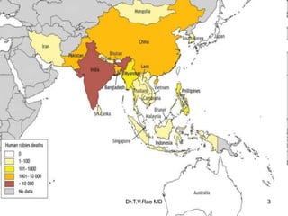
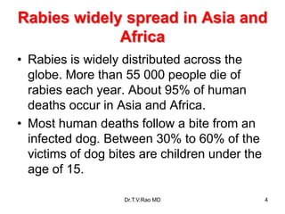
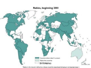
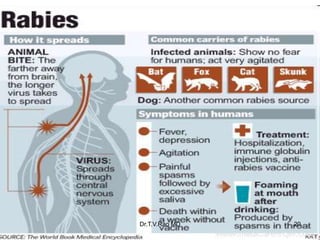
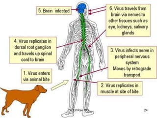
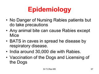
- Rabies is a fatal viral disease spread through animal bites that is endemic in India and parts of Asia/Africa.
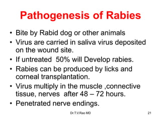
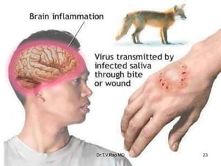
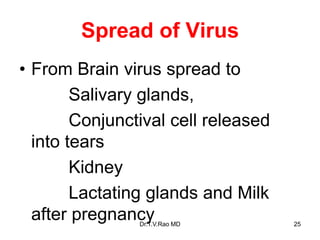
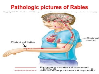
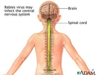
- It causes neurological symptoms like fear of water, paralysis, and death. The virus travels through nerves to the brain.
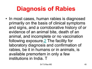
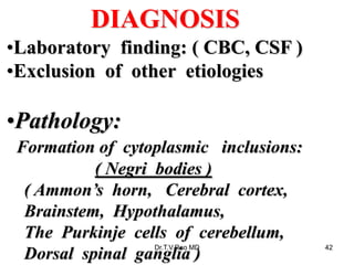
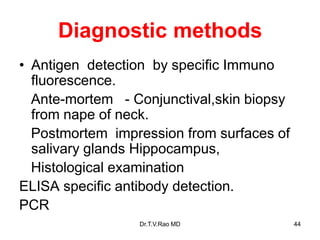
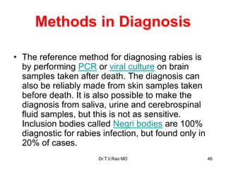
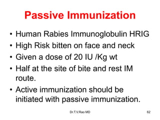
- Diagnosis involves identifying symptoms, exposure history, detecting viral antigens or Negri bodies post-mortem. Prevention relies on vaccination both before and after exposure. The first successful rabies vaccine was developed by Pasteur.