SlideShare a Scribd company logo
RETINOBLASTOMA
Dr. Abhishek Onkar
• Primary malignant neoplasm of the retina that arises from immature
retinal cells
• It is the most common primary intraocular malignancy of childhood
INHERITANCE
• 60–70% of retinoblastoma – unilateral
• 30–40% are bilateral.
• In unilateral cases, only a single tumor is usually present in the
affected eye.
• In bilateral cases, multifocal tumors in both eyes are the rule.
• Retinoblastoma is generally a sporadic condition (i.e., no previously
affected family members exist)
• Approximately 6% of newly diagnosed retinoblastoma cases are
familial and 94% are sporadic.
• Sporadic form of retinoblastoma are affected unilaterally.
• Familial cases have a autosomal dominant inheritance.
Classification
• Unifocal/multifocal.
• Unilateral (70%) or bilateral (30%).
• Sporadic (94%) or familial (6%).
• Non hereditary (50-60%) or hereditary (40-50%).
• It is recognized that bilateral and familial retinoblastoma are caused
by a germline mutation and are thus a heritable tumor.
• Bilateral retinoblastomas involve germinal mutations in all cases.
• Approximately 15% of unilateral sporadic retinoblastoma is caused by
germinal mutations affecting only one eye while the 85% are
sporadic.
• Knudson two-hit hypothesis
• The retinoblastoma gene (RB1) is located on the long arm of
chromosome 13 (13q14).
• Overexpression of MYCN (2p)
Histopathology of Retinoblastoma
• On low magnification, basophilic areas of tumor are seen along with
eosinophilic areas of necrosis and more basophilic areas of calcification
within the tumor.
• Poorly differentiated tumors consist of small to medium sized round cells
with large hyperchromatic nuclei and scanty cytoplasm with mitotic
figures.
• Well-differentiated tumors show the presence of rosettes and fleurettes.
• These can be of various types.:
• Flexner-Wintersteiner rosettes consist of columnar cells arranged around a
central lumen. This is highly characteristic of retinoblastoma and is also
seen in medulloepithelioma.
• Homer Wright rosettes consist of cells arranged around a central
neuromuscular tangle. This is also found in neuroblastomas,
medulloblastomas and medulloepitheliomas.
• Pseudorosette refers to the arrangement of tumor cells around blood
vessels. They are not signs of good differentiation.
• Fleurettes are eosinophilic structures composed of tumor cells with
pear shaped eosinophilic processes projecting through a fenestrated
membrane.
• Rosettes and fleurettes indicate that the tumor cells show
photoreceptor differentiation.
Life-Threatening Problems
Children with retinoblastoma are at risk for three important,
• life-threatening problems including metastasis from retinoblastoma,
• intracranial neuroblastic malignancy (trilateral retinoblastoma), and
• second primary tumors.
At Risk for Metastasis
• Retinoblastoma metastasis, when it occurs, generally develops within 1
year of the diagnosis of the intraocular tumor.
• Those at greatest risk for metastasis show features of:
1. retinoblastoma invasion beyond the lamina cribrosa in the optic nerve,
2. in the choroid (>2 mm dimension),
3. sclera,
4. Orbit
5. anterior chamber.
• Eyes with invasion of the optic nerve or choroid generally demonstrate
large retinoblastoma over 15 mm greatest dimension along with elevated
intraocular pressure and total retinal detachment.
At Risk for Neuroblastic Intracranial Malignancy
(Trilateral Retinoblastoma)
• There is an association of neuroblastic intracranial malignancy in
patients with the hereditary form of retinoblastoma, most often
manifesting as pineoblastoma.
At Risk for Second Primary Tumors
• child with retinoblastoma has approximately a 5% chance of
developing another malignancy during the first 10 years of follow-up,
18% during the first 20 years, and 26% within 30 years.
• Osteogenic sarcoma, often involving the femur, is most common
• other tumors such as spindle cell sarcoma, chondrosarcoma,
rhabdomyosarcoma, neuroblastoma, glioma, leukemia, sebaceous
cell carcinoma, squamous cell carcinoma, and malignant melanoma
have also been recognized.
• The mean latency period for the appearance of the second primary is
approximately 13 years.
Clinical Features of Retinoblastoma
• The clinical manifestations of retinoblastoma vary with the stage of the
disease at the time of recognition.
• In its earliest clinical stage, a small retinoblastoma, ie, less than 2 mm in
basal dimension, appears ophthalmoscopically as a subtle, transparent or
slightly translucent lesion in the sensory retina.
• Slightly larger tumors lead to dilated retinal blood vessels that feed and
drain the tumor.
• Some larger tumors show foci of chalk-like calcification that resemble
cottage cheese.
• Any retinoblastoma of any size can produce leukocoria (visible whiteness in
the pupil/ amaurotic "cat's eye reflex")
• Leucocoria is the most common presenting feature of retinoblastoma, followed by
strabismus, painful blind eye and loss of vision.
Common presenting features of retinoblastoma as reported in studies:
• 1. Leucocoria 56%
• 2. Strabismus 20%
• 3. Red painful eye 7%
• 4. Poor vision 5%
• 5. Asymptomatic 3%
• 6. Orbital cellulitis 3%
• 7. Unilateral mydriasis 2%
• 8. Heterochromia iridis 1%
• 9. Hyphema 1%
• As the tumor grows further, three patterns are usually seen
1. Endophytic, in which the tumor grows into the vitreous cavity. A yellow
white mass progressively fills the entire vitreous cavity and vitreous
seeds occur. The retinal vessels are not seen on the tumor surface.
2. Exophytic, in which the tumor grows towards the subretinal space.
Retinal detachment usually occurs and retinal vessels are seen over the
tumor
3. Diffuse infiltrating tumor, in which the tumor diffusely involves the retina
causing just a placoid thickness of the retina and not a mass. This is
generally seen in older children and usually there is a delay in the
diagnosis
Classification of Retinoblastoma
• Several classifications of retinoblastoma have been developed to
assist in prediction of globe salvage. The most popular grouping is the
Reese-Ellsworth classification
• A new International Classification of Retinoblastoma is currently being
developed to simplify the grouping scheme and allow a more
practical approach to judging results of chemoreduction.
Reese-Ellsworth Classification for
Conservative Treatment of Retinoblastoma*
Group I: Very favorable
• (a) Solitary tumor, less than 4 disc diameters in size, at or
• behind the equator
• (b) Multiple tumors, none over 4 disc diameters in size, all at
• or behind the equator
Group II: Favorable
• (a) Solitary tumor, 4 to 10 disc diameters in size, at or
• behind the equator
• (b) Multiple tumors, 4 to 10 disc diameters in size,
• behind the equator
Group III: Doubtful
• (a) Any lesion anterior to the equator
• (b) Solitary tumors larger than 10 disc diameters
• behind the equator
Group IV: Unfavorable
• (a) Multiple tumors, some larger than 10 disc diameters
• (b) Any lesion extending anteriorly to the ora serrata
Group V: Very unfavorable
• (a) Massive tumors involving over half the retina
• (b) Vitreous seeding
* Refers to chances of salvaging the affected eye and not systemic prognosis.
Differential Diagnosis
• The most common pseudoretinoblastomas include persistent
hyperplastic primary vitreous, Coats’ disease, and ocular toxocariasis
Conditions Closely Simulating Retinoblastoma
• Persistent hyperplastic primary vitreous 28
• Coats’ disease (exudative retinitis or retinal telangiectasis) 16
• Ocular toxocariasis 16
• Retinopathy of prematurity 5
• Combined hamartoma 4
• Coloboma 4
• Vitreous hemorrhage 4
• Astrocytic hamartoma 3
• Congenital cataract 2
• Toxoplasmic retinitis 1
• Idiopathic endophthalmitis 1
Diagnostic Testing
• Accurate diagnosis in a child with suspected retinoblastoma is
accomplished by taking a detailed history, physical evaluation, external
ocular examination, slit lamp biomicroscopy, and binocular indirect
ophthalmoscopy with scleral indentation.
• The diagnosis is established by the classic appearance of the retinal tumors
• Needle biopsy confirmation is rarely, if ever, necessary.
• Ancillary diagnostic studies can be helpful in confirming the diagnosis of
retinoblastoma.
• Fluorescein angiography shows early vascularity and late
hyperfluorescence of the tumor.
• Ultrasonography and computed tomography can demonstrate the
intraocular tumor and possibly detect calcium within the mass.
• Approximately 5% to 10% of retinoblastomas show no intrinsic
calcification
• Magnetic resonance imaging does not usually detect calcium but may
be of value in the assessment of the optic nerve, orbit, and brain.
Diagnosis of Retinoblastoma
Other Classifications of Retinoblastoma
International Classification of Intraocular Retinoblastoma
(Murphree)
International Classification of Retinoblastoma (Shields)
TNM Classification for Retinoblastoma
Pathologic Classification (pTNM)
International Staging System for Retinoblastoma
Stage 0 No enucleation (one or both eyes may have intraocular disease)
Stage I Enucleation, tumor completely resected
Stage II Enucleation with microscopic residual tumor
Stage III Regional extension
• A. Overt orbital disease
• B. Preauricular or cervical lymph node extension
Stage IV Metastatic disease
• A. Hematogenous metastasis
• 1. Single lesion
• 2. Multiple lesions
• B. CNS Extension
• 1. Prechiasmatic lesion
• 2. CNS mass
• 3. Leptomeningeal disease
Management of Retinoblastoma
• The most important objective in the management of a child with
retinoblastoma is survival of the patient, and the second most
important goal is preservation of the globe.
• The focus on visual acuity comes later, after safety of the patient and
globe is established.
• Therapy is tailored to each individual case and based on the overall
situation, including threat of metastatic disease, risks for second
cancers, systemic status, laterality of the disease, size and location.
Current Suggested Protocol
A. Intraocular tumor, International Classification Group A to C, Unilateral or Bilateral
1. Focal therapy (cryotherapy or transpupillary thermotherapy) alone for smaller
tumors (< 3mm in diameter and height) located in visually noncrucial areas
2. Standard 6 cycle chemoreduction and sequential aggressive focal therapy for
larger tumors and those located in visually crucial areas
3. Defer focal therapy until 6 cycles for tumors located in the macular and
juxtapapillary areas. Transpupillary thermotherapy or plaque brachytherapy for
residual tumor in the macular and juxtapapillary areas >6 cycles.
4. Focal therapy for small residual tumor, and plaque brachytherapy/external
beam radiotherapy (>12 months age) for large residual tumorif bilateral, and
enucleation if unilateral.
B. Intraocular tumor, International Classification Group D, Unilateral or
Bilateral
1. High dose chemotherapy and sequential aggressive focal therapy
2. Periocular carboplatin for vitreous seeds
3. Consider primary enucleation if unilateral, specially in eyes with no
visual prognosis
Intraocular tumor, International Classification Group E, Unilateral or Bilateral
1. Primary enucleation
2. Evaluate histopathology for high risk factors
• D. High risk factors on histopathology, International Staging, Stage 2
• 1. Baseline systemic evaluation for metastasis
• 2. Standard 6 cycle adjuvant chemotherapy
• 3. High dose adjuvant chemotherapy and orbital external beam
• radiotherapy in patients with scleral infiltration, extraocular extension, and
optic nerve extension to transection.
E. Extraocular tumor, International Staging, Stage 3A
1. Baseline systemic evaluation for metastasis
2. High dose chemotherapy for 3-6 cycles, followed by enucleation or
extended enucleation, external beam radiotherapy, and continued high
dose chemotherapy for 12 cycles
F. Regional Lymph Node Metastasis, International Staging, Stage 3B
1. Baseline evaluation for systemic metastasis
2. Neck dissection, high dose chemotherapy for 6 cycles, followed by
external beam radiotherapy, and continued high dose chemotherapy
for 12 cycles
G. Hematogenous or Central Nervous System Metastasis, International
Staging, Stage 4
• 1. Palliative therapy in discussion with the family
• 2. High dose chemotherapy with bone marrow rescue for
hematogenous metastasis
• 3. High dose chemotherapy with intrathecal chemotherapy for central
nervous system metastasis
Chemoreduction Regimen for Intraocular Retinoblastoma
Given for a Total of 6 Monthly Cycles
• Day Vincristine Etoposide Carboplatin
• 0 × × ×
• 1 ×
• Vincristine: 0.05 mg/kg
• Etoposide: 5 mg/kg
• Carboplatin: 18.6 mg/kg
Focal Therapies
Focal therapies include laser photocoagulation, thermotherapy,
cryotherapy, and plaque radiotherapy.
Plaque radiotherapy is generally reserved for tumors that fail other
focal therapies, even those that reach a moderate size, up to 8 or 10
mm in thickness.
The remainder of the focal therapies are reserved for small tumors,
generally those under 3 mm in greatest dimension.
• Laser photocoagulation is usually employed for small retinoblastomas
posterior to the equator of the eye.
• Thermotherapy is a method of tumor heating using a diode infrared
laser system. Thermotherapy coupled with chemoreduction is
especially suited for tumors adjacent to the fovea and optic nerve
where radiation or laser photocoagulation would possibly induce
more visual loss
• Cryotherapy is useful in the treatment of equatorial and peripheral
small retinoblastomas.
• Plaque radiotherapy is a method of brachytherapy in which a
radioactive implant is placed on the sclera over the base of a
retinoblastoma to irradiate the tumor transsclerally.
• Generally, it is limited to tumors less than 16 mm in base and 8 mm in
thickness.
• It requires an average of 2 to 4 days of treatment time to deliver the
total dose of 40 Gy to the apex of the tumor.
External Beam Radiotherapy
• Retinoblastoma is generally a radiosensitive tumor. External beam
radiotherapy is a method of delivering whole eye irradiation to treat
advanced retinoblastoma, particularly when there is diffuse vitreous
seeding.
Enucleation
• Enucleation is a frequently used and important method for managing
retinoblastoma. If there is advanced disease with no hope for useful
vision in the affected eye or if there is a concern of invasion of the
tumor into the optic nerve, choroid, or orbit, then enucleation is
appropriate.
• Those eyes with secondary glaucoma, pars plana seeding, or anterior
chamber invasion are also generally best managed with enucleation.
Primary enucleation continues to be the treatment of choice for
• advanced intraocular retinoblastoma with neovascularization of iris,
• secondary glaucoma,
• anterior chamber tumor invasion,
• tumors occupying >75% of the vitreous volume,
• necrotic tumors with secondary orbital inflammation, and
• tumors associated with hyphema or vitreous hemorrhage where the
tumor characteristics can not be visualized, especially when only one
eye is involved
Special considerations for enucleation in retinoblastoma
• a. Minimal manipulation
• b. Avoid perforation of the eye
• c. Harvest long (> 15 mm) optic nerve stump
• d. Inspect the enucleated eye for macroscopic extraocular extension and
• optic nerve involvement
• e. Harvest fresh tissue for genetic studies
• f. Place a primary implant
• g. Avoid biointegrated implant if postoperative radiotherapy is necessary
Histopathologic high-risk factors predictive of metastasis
• • Anterior chamber seeding
• • Iris infiltration
• • Ciliary body infiltration
• • Massive choroidal infiltration
• • Invasion of the optic nerve lamina cribrosa
• • Retrolaminar optic nerve invasion
• • Invasion of optic nerve transection
• • Scleral infiltration
• • Extrascleral extension

More Related Content

What's hot

ocular Toxoplasmosis
ocular Toxoplasmosisocular Toxoplasmosis
ocular Toxoplasmosis
Dinesh Madduri
 
Ectopia lentis edit
Ectopia lentis editEctopia lentis edit
Ectopia lentis edit
Laxmi Eye Institute
 
Ocular surface squamous neoplasia(ossn)
Ocular surface squamous neoplasia(ossn)Ocular surface squamous neoplasia(ossn)
Ocular surface squamous neoplasia(ossn)
SSSIHMS-PG
 
Chemical injuries of the eye
Chemical injuries of the eyeChemical injuries of the eye
Chemical injuries of the eye
Nitish Narang
 
Intraocular Tumours
Intraocular TumoursIntraocular Tumours
Intraocular Tumours
OphthalmicDocs Chiong
 
Keratoconus
KeratoconusKeratoconus
Thyroid eye disease
Thyroid eye disease Thyroid eye disease
Thyroid eye disease
Mohmmad Dmour , MD
 
Neurotrophic keratopathy
Neurotrophic keratopathyNeurotrophic keratopathy
Neurotrophic keratopathy
Dinesh Madduri
 
Congenital glaucoma
Congenital glaucomaCongenital glaucoma
Congenital glaucoma
Shubham Vasudeva
 
Conjunctival tumors
Conjunctival tumorsConjunctival tumors
Conjunctival tumors
Poonam Rawat
 
Vogt Koyanagi Harada Disease
Vogt Koyanagi Harada DiseaseVogt Koyanagi Harada Disease
Vogt Koyanagi Harada Disease
Gauree Gattani
 
DIABETES AND THE EYE
DIABETES AND THE EYE DIABETES AND THE EYE
DIABETES AND THE EYE
Abdulrazzak Serafi
 
Pediatric cataract
Pediatric cataractPediatric cataract
Pediatric cataract
Dr. Ajay Kumar Singh
 
Age related macular degeneration
Age  related  macular degenerationAge  related  macular degeneration
Age related macular degeneration
ikramdr01
 
Rhegmatogenous retinal detachment
Rhegmatogenous retinal detachmentRhegmatogenous retinal detachment
Rhegmatogenous retinal detachmentSamuel Ponraj
 
Herpetic eye disease
Herpetic eye diseaseHerpetic eye disease
Herpetic eye disease
Othman Al-Abbadi
 
Ocular Manifestation of Diabetes Mellitus
Ocular Manifestation of Diabetes MellitusOcular Manifestation of Diabetes Mellitus
Ocular Manifestation of Diabetes Mellitus
Joobin Khadamy . MD
 
Coats' Disease
Coats' DiseaseCoats' Disease
Coats' Disease
Dr. Shah Noor Hassan
 
Orbital tumours
Orbital tumoursOrbital tumours
Orbital tumours
airwave12
 
Diabetic macular edema
Diabetic macular edemaDiabetic macular edema
Diabetic macular edema
drkvasantha
 

What's hot (20)

ocular Toxoplasmosis
ocular Toxoplasmosisocular Toxoplasmosis
ocular Toxoplasmosis
 
Ectopia lentis edit
Ectopia lentis editEctopia lentis edit
Ectopia lentis edit
 
Ocular surface squamous neoplasia(ossn)
Ocular surface squamous neoplasia(ossn)Ocular surface squamous neoplasia(ossn)
Ocular surface squamous neoplasia(ossn)
 
Chemical injuries of the eye
Chemical injuries of the eyeChemical injuries of the eye
Chemical injuries of the eye
 
Intraocular Tumours
Intraocular TumoursIntraocular Tumours
Intraocular Tumours
 
Keratoconus
KeratoconusKeratoconus
Keratoconus
 
Thyroid eye disease
Thyroid eye disease Thyroid eye disease
Thyroid eye disease
 
Neurotrophic keratopathy
Neurotrophic keratopathyNeurotrophic keratopathy
Neurotrophic keratopathy
 
Congenital glaucoma
Congenital glaucomaCongenital glaucoma
Congenital glaucoma
 
Conjunctival tumors
Conjunctival tumorsConjunctival tumors
Conjunctival tumors
 
Vogt Koyanagi Harada Disease
Vogt Koyanagi Harada DiseaseVogt Koyanagi Harada Disease
Vogt Koyanagi Harada Disease
 
DIABETES AND THE EYE
DIABETES AND THE EYE DIABETES AND THE EYE
DIABETES AND THE EYE
 
Pediatric cataract
Pediatric cataractPediatric cataract
Pediatric cataract
 
Age related macular degeneration
Age  related  macular degenerationAge  related  macular degeneration
Age related macular degeneration
 
Rhegmatogenous retinal detachment
Rhegmatogenous retinal detachmentRhegmatogenous retinal detachment
Rhegmatogenous retinal detachment
 
Herpetic eye disease
Herpetic eye diseaseHerpetic eye disease
Herpetic eye disease
 
Ocular Manifestation of Diabetes Mellitus
Ocular Manifestation of Diabetes MellitusOcular Manifestation of Diabetes Mellitus
Ocular Manifestation of Diabetes Mellitus
 
Coats' Disease
Coats' DiseaseCoats' Disease
Coats' Disease
 
Orbital tumours
Orbital tumoursOrbital tumours
Orbital tumours
 
Diabetic macular edema
Diabetic macular edemaDiabetic macular edema
Diabetic macular edema
 

Similar to Retinoblastoma

retinoblastoma by dr george deogratias
 retinoblastoma by dr george deogratias  retinoblastoma by dr george deogratias
retinoblastoma by dr george deogratias
Deogratias George
 
Retinoblastoma, general veiw.. by DR.ZEINAB MEDANI
Retinoblastoma, general veiw.. by DR.ZEINAB MEDANIRetinoblastoma, general veiw.. by DR.ZEINAB MEDANI
Retinoblastoma, general veiw.. by DR.ZEINAB MEDANI
Omdurman Islamic University
 
Retinoblastoma
RetinoblastomaRetinoblastoma
Retinoblastoma
Othman Al-Abbadi
 
Retinoblastoma 7th
Retinoblastoma 7thRetinoblastoma 7th
Retinoblastoma 7th
Gowtham Manimaran
 
Retinoblastoma
RetinoblastomaRetinoblastoma
Retinoblastoma
Dr. Renesha Islam
 
Retinoblastoma
RetinoblastomaRetinoblastoma
Retinoblastoma
Griheet Reddy
 
Ocular and Orbital Tumours.pptx
Ocular and Orbital Tumours.pptxOcular and Orbital Tumours.pptx
Ocular and Orbital Tumours.pptx
Medinfopedia Blog
 
Retinoblastoma
RetinoblastomaRetinoblastoma
Retinoblastoma
Dr. Mahziba Rahman
 
Retinoblastoma .pptx
Retinoblastoma .pptxRetinoblastoma .pptx
Retinoblastoma .pptx
RituRMenon
 
Retinoblastoma: A Beginner's Guide....
Retinoblastoma: A Beginner's Guide....Retinoblastoma: A Beginner's Guide....
Retinoblastoma : Dr Shylesh B Dabke
Retinoblastoma : Dr Shylesh B DabkeRetinoblastoma : Dr Shylesh B Dabke
Retinoblastoma : Dr Shylesh B Dabke
Shylesh Dabke
 
Tumors of the eye
Tumors of the eyeTumors of the eye
Tumors of the eye
SOUMYA SUBRAMANI
 
CHOROIDAL MELANOMA.ppt
CHOROIDAL MELANOMA.pptCHOROIDAL MELANOMA.ppt
CHOROIDAL MELANOMA.ppt
Salman Khan
 
solid.pptx
solid.pptxsolid.pptx
solid.pptx
AmareDejene
 
Childhood ocular tumor
Childhood  ocular tumorChildhood  ocular tumor
Childhood ocular tumor
Dr Md Almas Hossain
 
Retinoblastoma
RetinoblastomaRetinoblastoma
Retinoblastoma
Brijesh Maheshwari
 
Retinoblastoma (Bsc. optometry 2nd year KUHS)Exam view point
Retinoblastoma (Bsc. optometry 2nd year KUHS)Exam view pointRetinoblastoma (Bsc. optometry 2nd year KUHS)Exam view point
Retinoblastoma (Bsc. optometry 2nd year KUHS)Exam view point
Navmii
 
Brain tumor in children
Brain tumor in childrenBrain tumor in children
Brain tumor in children
SharminNaharNabila
 
retinoblastoma 2.pptx
retinoblastoma 2.pptxretinoblastoma 2.pptx
retinoblastoma 2.pptx
Harshika Malik
 

Similar to Retinoblastoma (20)

retinoblastoma by dr george deogratias
 retinoblastoma by dr george deogratias  retinoblastoma by dr george deogratias
retinoblastoma by dr george deogratias
 
Retinoblastoma, general veiw.. by DR.ZEINAB MEDANI
Retinoblastoma, general veiw.. by DR.ZEINAB MEDANIRetinoblastoma, general veiw.. by DR.ZEINAB MEDANI
Retinoblastoma, general veiw.. by DR.ZEINAB MEDANI
 
Retinoblastoma
RetinoblastomaRetinoblastoma
Retinoblastoma
 
Retinoblastoma 7th
Retinoblastoma 7thRetinoblastoma 7th
Retinoblastoma 7th
 
Retinoblastoma
RetinoblastomaRetinoblastoma
Retinoblastoma
 
Retinoblastoma
RetinoblastomaRetinoblastoma
Retinoblastoma
 
Ocular and Orbital Tumours.pptx
Ocular and Orbital Tumours.pptxOcular and Orbital Tumours.pptx
Ocular and Orbital Tumours.pptx
 
Retinoblastoma
RetinoblastomaRetinoblastoma
Retinoblastoma
 
Retinoblastoma .pptx
Retinoblastoma .pptxRetinoblastoma .pptx
Retinoblastoma .pptx
 
Retinoblastoma: A Beginner's Guide....
Retinoblastoma: A Beginner's Guide....Retinoblastoma: A Beginner's Guide....
Retinoblastoma: A Beginner's Guide....
 
Retinoblastoma : Dr Shylesh B Dabke
Retinoblastoma : Dr Shylesh B DabkeRetinoblastoma : Dr Shylesh B Dabke
Retinoblastoma : Dr Shylesh B Dabke
 
Tumors of the eye
Tumors of the eyeTumors of the eye
Tumors of the eye
 
CHOROIDAL MELANOMA.ppt
CHOROIDAL MELANOMA.pptCHOROIDAL MELANOMA.ppt
CHOROIDAL MELANOMA.ppt
 
solid.pptx
solid.pptxsolid.pptx
solid.pptx
 
Childhood ocular tumor
Childhood  ocular tumorChildhood  ocular tumor
Childhood ocular tumor
 
Retinoblastoma
RetinoblastomaRetinoblastoma
Retinoblastoma
 
Retinoblastoma (Bsc. optometry 2nd year KUHS)Exam view point
Retinoblastoma (Bsc. optometry 2nd year KUHS)Exam view pointRetinoblastoma (Bsc. optometry 2nd year KUHS)Exam view point
Retinoblastoma (Bsc. optometry 2nd year KUHS)Exam view point
 
Brain tumor in children
Brain tumor in childrenBrain tumor in children
Brain tumor in children
 
retinoblastoma 2.pptx
retinoblastoma 2.pptxretinoblastoma 2.pptx
retinoblastoma 2.pptx
 
Retino
RetinoRetino
Retino
 

More from Abhishek Onkar

Hypertensive Retinopathy
Hypertensive RetinopathyHypertensive Retinopathy
Hypertensive Retinopathy
Abhishek Onkar
 
Diabetic Retinopathy
Diabetic RetinopathyDiabetic Retinopathy
Diabetic Retinopathy
Abhishek Onkar
 
CRAO
CRAOCRAO
CRVO
CRVOCRVO
BRVO
BRVOBRVO
Optic neuritis
Optic neuritisOptic neuritis
Optic neuritis
Abhishek Onkar
 
Optic atrophy
Optic atrophyOptic atrophy
Optic atrophy
Abhishek Onkar
 
Papilloedema
PapilloedemaPapilloedema
Papilloedema
Abhishek Onkar
 
Retinoscopy
RetinoscopyRetinoscopy
Retinoscopy
Abhishek Onkar
 
Sturm's Conoid
Sturm's ConoidSturm's Conoid
Sturm's Conoid
Abhishek Onkar
 
Aphakia
AphakiaAphakia
Cataract management
Cataract managementCataract management
Cataract management
Abhishek Onkar
 
Overview of Cataract
Overview of CataractOverview of Cataract
Overview of Cataract
Abhishek Onkar
 
Congenital cataract
Congenital cataractCongenital cataract
Congenital cataract
Abhishek Onkar
 
Sympathetic ophthalmitis
Sympathetic ophthalmitisSympathetic ophthalmitis
Sympathetic ophthalmitis
Abhishek Onkar
 
Uveitis basics for undergrads
Uveitis basics for undergradsUveitis basics for undergrads
Uveitis basics for undergrads
Abhishek Onkar
 

More from Abhishek Onkar (16)

Hypertensive Retinopathy
Hypertensive RetinopathyHypertensive Retinopathy
Hypertensive Retinopathy
 
Diabetic Retinopathy
Diabetic RetinopathyDiabetic Retinopathy
Diabetic Retinopathy
 
CRAO
CRAOCRAO
CRAO
 
CRVO
CRVOCRVO
CRVO
 
BRVO
BRVOBRVO
BRVO
 
Optic neuritis
Optic neuritisOptic neuritis
Optic neuritis
 
Optic atrophy
Optic atrophyOptic atrophy
Optic atrophy
 
Papilloedema
PapilloedemaPapilloedema
Papilloedema
 
Retinoscopy
RetinoscopyRetinoscopy
Retinoscopy
 
Sturm's Conoid
Sturm's ConoidSturm's Conoid
Sturm's Conoid
 
Aphakia
AphakiaAphakia
Aphakia
 
Cataract management
Cataract managementCataract management
Cataract management
 
Overview of Cataract
Overview of CataractOverview of Cataract
Overview of Cataract
 
Congenital cataract
Congenital cataractCongenital cataract
Congenital cataract
 
Sympathetic ophthalmitis
Sympathetic ophthalmitisSympathetic ophthalmitis
Sympathetic ophthalmitis
 
Uveitis basics for undergrads
Uveitis basics for undergradsUveitis basics for undergrads
Uveitis basics for undergrads
 

Recently uploaded

Flu Vaccine Alert in Bangalore Karnataka
Flu Vaccine Alert in Bangalore KarnatakaFlu Vaccine Alert in Bangalore Karnataka
Flu Vaccine Alert in Bangalore Karnataka
addon Scans
 
The POPPY STUDY (Preconception to post-partum cardiovascular function in prim...
The POPPY STUDY (Preconception to post-partum cardiovascular function in prim...The POPPY STUDY (Preconception to post-partum cardiovascular function in prim...
The POPPY STUDY (Preconception to post-partum cardiovascular function in prim...
Catherine Liao
 
Ophthalmology Clinical Tests for OSCE exam
Ophthalmology Clinical Tests for OSCE examOphthalmology Clinical Tests for OSCE exam
Ophthalmology Clinical Tests for OSCE exam
KafrELShiekh University
 
The hemodynamic and autonomic determinants of elevated blood pressure in obes...
The hemodynamic and autonomic determinants of elevated blood pressure in obes...The hemodynamic and autonomic determinants of elevated blood pressure in obes...
The hemodynamic and autonomic determinants of elevated blood pressure in obes...
Catherine Liao
 
24 Upakrama.pptx class ppt useful in all
24 Upakrama.pptx class ppt useful in all24 Upakrama.pptx class ppt useful in all
24 Upakrama.pptx class ppt useful in all
DrSathishMS1
 
micro teaching on communication m.sc nursing.pdf
micro teaching on communication m.sc nursing.pdfmicro teaching on communication m.sc nursing.pdf
micro teaching on communication m.sc nursing.pdf
Anurag Sharma
 
ARTIFICIAL INTELLIGENCE IN HEALTHCARE.pdf
ARTIFICIAL INTELLIGENCE IN  HEALTHCARE.pdfARTIFICIAL INTELLIGENCE IN  HEALTHCARE.pdf
ARTIFICIAL INTELLIGENCE IN HEALTHCARE.pdf
Anujkumaranit
 
heat stroke and heat exhaustion in children
heat stroke and heat exhaustion in childrenheat stroke and heat exhaustion in children
heat stroke and heat exhaustion in children
SumeraAhmad5
 
BRACHYTHERAPY OVERVIEW AND APPLICATORS
BRACHYTHERAPY OVERVIEW  AND  APPLICATORSBRACHYTHERAPY OVERVIEW  AND  APPLICATORS
BRACHYTHERAPY OVERVIEW AND APPLICATORS
Krishan Murari
 
Hemodialysis: Chapter 3, Dialysis Water Unit - Dr.Gawad
Hemodialysis: Chapter 3, Dialysis Water Unit - Dr.GawadHemodialysis: Chapter 3, Dialysis Water Unit - Dr.Gawad
Hemodialysis: Chapter 3, Dialysis Water Unit - Dr.Gawad
NephroTube - Dr.Gawad
 
Phone Us ❤85270-49040❤ #ℂall #gIRLS In Surat By Surat @ℂall @Girls Hotel With...
Phone Us ❤85270-49040❤ #ℂall #gIRLS In Surat By Surat @ℂall @Girls Hotel With...Phone Us ❤85270-49040❤ #ℂall #gIRLS In Surat By Surat @ℂall @Girls Hotel With...
Phone Us ❤85270-49040❤ #ℂall #gIRLS In Surat By Surat @ℂall @Girls Hotel With...
Savita Shen $i11
 
Physiology of Chemical Sensation of smell.pdf
Physiology of Chemical Sensation of smell.pdfPhysiology of Chemical Sensation of smell.pdf
Physiology of Chemical Sensation of smell.pdf
MedicoseAcademics
 
Triangles of Neck and Clinical Correlation by Dr. RIG.pptx
Triangles of Neck and Clinical Correlation by Dr. RIG.pptxTriangles of Neck and Clinical Correlation by Dr. RIG.pptx
Triangles of Neck and Clinical Correlation by Dr. RIG.pptx
Dr. Rabia Inam Gandapore
 
MANAGEMENT OF ATRIOVENTRICULAR CONDUCTION BLOCK.pdf
MANAGEMENT OF ATRIOVENTRICULAR CONDUCTION BLOCK.pdfMANAGEMENT OF ATRIOVENTRICULAR CONDUCTION BLOCK.pdf
MANAGEMENT OF ATRIOVENTRICULAR CONDUCTION BLOCK.pdf
Jim Jacob Roy
 
Lung Cancer: Artificial Intelligence, Synergetics, Complex System Analysis, S...
Lung Cancer: Artificial Intelligence, Synergetics, Complex System Analysis, S...Lung Cancer: Artificial Intelligence, Synergetics, Complex System Analysis, S...
Lung Cancer: Artificial Intelligence, Synergetics, Complex System Analysis, S...
Oleg Kshivets
 
Maxilla, Mandible & Hyoid Bone & Clinical Correlations by Dr. RIG.pptx
Maxilla, Mandible & Hyoid Bone & Clinical Correlations by Dr. RIG.pptxMaxilla, Mandible & Hyoid Bone & Clinical Correlations by Dr. RIG.pptx
Maxilla, Mandible & Hyoid Bone & Clinical Correlations by Dr. RIG.pptx
Dr. Rabia Inam Gandapore
 
ANATOMY AND PHYSIOLOGY OF URINARY SYSTEM.pptx
ANATOMY AND PHYSIOLOGY OF URINARY SYSTEM.pptxANATOMY AND PHYSIOLOGY OF URINARY SYSTEM.pptx
ANATOMY AND PHYSIOLOGY OF URINARY SYSTEM.pptx
Swetaba Besh
 
basicmodesofventilation2022-220313203758.pdf
basicmodesofventilation2022-220313203758.pdfbasicmodesofventilation2022-220313203758.pdf
basicmodesofventilation2022-220313203758.pdf
aljamhori teaching hospital
 
The Normal Electrocardiogram - Part I of II
The Normal Electrocardiogram - Part I of IIThe Normal Electrocardiogram - Part I of II
The Normal Electrocardiogram - Part I of II
MedicoseAcademics
 
For Better Surat #ℂall #Girl Service ❤85270-49040❤ Surat #ℂall #Girls
For Better Surat #ℂall #Girl Service ❤85270-49040❤ Surat #ℂall #GirlsFor Better Surat #ℂall #Girl Service ❤85270-49040❤ Surat #ℂall #Girls
For Better Surat #ℂall #Girl Service ❤85270-49040❤ Surat #ℂall #Girls
Savita Shen $i11
 

Recently uploaded (20)

Flu Vaccine Alert in Bangalore Karnataka
Flu Vaccine Alert in Bangalore KarnatakaFlu Vaccine Alert in Bangalore Karnataka
Flu Vaccine Alert in Bangalore Karnataka
 
The POPPY STUDY (Preconception to post-partum cardiovascular function in prim...
The POPPY STUDY (Preconception to post-partum cardiovascular function in prim...The POPPY STUDY (Preconception to post-partum cardiovascular function in prim...
The POPPY STUDY (Preconception to post-partum cardiovascular function in prim...
 
Ophthalmology Clinical Tests for OSCE exam
Ophthalmology Clinical Tests for OSCE examOphthalmology Clinical Tests for OSCE exam
Ophthalmology Clinical Tests for OSCE exam
 
The hemodynamic and autonomic determinants of elevated blood pressure in obes...
The hemodynamic and autonomic determinants of elevated blood pressure in obes...The hemodynamic and autonomic determinants of elevated blood pressure in obes...
The hemodynamic and autonomic determinants of elevated blood pressure in obes...
 
24 Upakrama.pptx class ppt useful in all
24 Upakrama.pptx class ppt useful in all24 Upakrama.pptx class ppt useful in all
24 Upakrama.pptx class ppt useful in all
 
micro teaching on communication m.sc nursing.pdf
micro teaching on communication m.sc nursing.pdfmicro teaching on communication m.sc nursing.pdf
micro teaching on communication m.sc nursing.pdf
 
ARTIFICIAL INTELLIGENCE IN HEALTHCARE.pdf
ARTIFICIAL INTELLIGENCE IN  HEALTHCARE.pdfARTIFICIAL INTELLIGENCE IN  HEALTHCARE.pdf
ARTIFICIAL INTELLIGENCE IN HEALTHCARE.pdf
 
heat stroke and heat exhaustion in children
heat stroke and heat exhaustion in childrenheat stroke and heat exhaustion in children
heat stroke and heat exhaustion in children
 
BRACHYTHERAPY OVERVIEW AND APPLICATORS
BRACHYTHERAPY OVERVIEW  AND  APPLICATORSBRACHYTHERAPY OVERVIEW  AND  APPLICATORS
BRACHYTHERAPY OVERVIEW AND APPLICATORS
 
Hemodialysis: Chapter 3, Dialysis Water Unit - Dr.Gawad
Hemodialysis: Chapter 3, Dialysis Water Unit - Dr.GawadHemodialysis: Chapter 3, Dialysis Water Unit - Dr.Gawad
Hemodialysis: Chapter 3, Dialysis Water Unit - Dr.Gawad
 
Phone Us ❤85270-49040❤ #ℂall #gIRLS In Surat By Surat @ℂall @Girls Hotel With...
Phone Us ❤85270-49040❤ #ℂall #gIRLS In Surat By Surat @ℂall @Girls Hotel With...Phone Us ❤85270-49040❤ #ℂall #gIRLS In Surat By Surat @ℂall @Girls Hotel With...
Phone Us ❤85270-49040❤ #ℂall #gIRLS In Surat By Surat @ℂall @Girls Hotel With...
 
Physiology of Chemical Sensation of smell.pdf
Physiology of Chemical Sensation of smell.pdfPhysiology of Chemical Sensation of smell.pdf
Physiology of Chemical Sensation of smell.pdf
 
Triangles of Neck and Clinical Correlation by Dr. RIG.pptx
Triangles of Neck and Clinical Correlation by Dr. RIG.pptxTriangles of Neck and Clinical Correlation by Dr. RIG.pptx
Triangles of Neck and Clinical Correlation by Dr. RIG.pptx
 
MANAGEMENT OF ATRIOVENTRICULAR CONDUCTION BLOCK.pdf
MANAGEMENT OF ATRIOVENTRICULAR CONDUCTION BLOCK.pdfMANAGEMENT OF ATRIOVENTRICULAR CONDUCTION BLOCK.pdf
MANAGEMENT OF ATRIOVENTRICULAR CONDUCTION BLOCK.pdf
 
Lung Cancer: Artificial Intelligence, Synergetics, Complex System Analysis, S...
Lung Cancer: Artificial Intelligence, Synergetics, Complex System Analysis, S...Lung Cancer: Artificial Intelligence, Synergetics, Complex System Analysis, S...
Lung Cancer: Artificial Intelligence, Synergetics, Complex System Analysis, S...
 
Maxilla, Mandible & Hyoid Bone & Clinical Correlations by Dr. RIG.pptx
Maxilla, Mandible & Hyoid Bone & Clinical Correlations by Dr. RIG.pptxMaxilla, Mandible & Hyoid Bone & Clinical Correlations by Dr. RIG.pptx
Maxilla, Mandible & Hyoid Bone & Clinical Correlations by Dr. RIG.pptx
 
ANATOMY AND PHYSIOLOGY OF URINARY SYSTEM.pptx
ANATOMY AND PHYSIOLOGY OF URINARY SYSTEM.pptxANATOMY AND PHYSIOLOGY OF URINARY SYSTEM.pptx
ANATOMY AND PHYSIOLOGY OF URINARY SYSTEM.pptx
 
basicmodesofventilation2022-220313203758.pdf
basicmodesofventilation2022-220313203758.pdfbasicmodesofventilation2022-220313203758.pdf
basicmodesofventilation2022-220313203758.pdf
 
The Normal Electrocardiogram - Part I of II
The Normal Electrocardiogram - Part I of IIThe Normal Electrocardiogram - Part I of II
The Normal Electrocardiogram - Part I of II
 
For Better Surat #ℂall #Girl Service ❤85270-49040❤ Surat #ℂall #Girls
For Better Surat #ℂall #Girl Service ❤85270-49040❤ Surat #ℂall #GirlsFor Better Surat #ℂall #Girl Service ❤85270-49040❤ Surat #ℂall #Girls
For Better Surat #ℂall #Girl Service ❤85270-49040❤ Surat #ℂall #Girls
 

Retinoblastoma

  • 2. • Primary malignant neoplasm of the retina that arises from immature retinal cells • It is the most common primary intraocular malignancy of childhood
  • 3. INHERITANCE • 60–70% of retinoblastoma – unilateral • 30–40% are bilateral. • In unilateral cases, only a single tumor is usually present in the affected eye. • In bilateral cases, multifocal tumors in both eyes are the rule. • Retinoblastoma is generally a sporadic condition (i.e., no previously affected family members exist) • Approximately 6% of newly diagnosed retinoblastoma cases are familial and 94% are sporadic. • Sporadic form of retinoblastoma are affected unilaterally. • Familial cases have a autosomal dominant inheritance.
  • 4. Classification • Unifocal/multifocal. • Unilateral (70%) or bilateral (30%). • Sporadic (94%) or familial (6%). • Non hereditary (50-60%) or hereditary (40-50%). • It is recognized that bilateral and familial retinoblastoma are caused by a germline mutation and are thus a heritable tumor.
  • 5. • Bilateral retinoblastomas involve germinal mutations in all cases. • Approximately 15% of unilateral sporadic retinoblastoma is caused by germinal mutations affecting only one eye while the 85% are sporadic. • Knudson two-hit hypothesis • The retinoblastoma gene (RB1) is located on the long arm of chromosome 13 (13q14). • Overexpression of MYCN (2p)
  • 6. Histopathology of Retinoblastoma • On low magnification, basophilic areas of tumor are seen along with eosinophilic areas of necrosis and more basophilic areas of calcification within the tumor. • Poorly differentiated tumors consist of small to medium sized round cells with large hyperchromatic nuclei and scanty cytoplasm with mitotic figures. • Well-differentiated tumors show the presence of rosettes and fleurettes. • These can be of various types.: • Flexner-Wintersteiner rosettes consist of columnar cells arranged around a central lumen. This is highly characteristic of retinoblastoma and is also seen in medulloepithelioma.
  • 7. • Homer Wright rosettes consist of cells arranged around a central neuromuscular tangle. This is also found in neuroblastomas, medulloblastomas and medulloepitheliomas. • Pseudorosette refers to the arrangement of tumor cells around blood vessels. They are not signs of good differentiation. • Fleurettes are eosinophilic structures composed of tumor cells with pear shaped eosinophilic processes projecting through a fenestrated membrane. • Rosettes and fleurettes indicate that the tumor cells show photoreceptor differentiation.
  • 8. Life-Threatening Problems Children with retinoblastoma are at risk for three important, • life-threatening problems including metastasis from retinoblastoma, • intracranial neuroblastic malignancy (trilateral retinoblastoma), and • second primary tumors.
  • 9. At Risk for Metastasis • Retinoblastoma metastasis, when it occurs, generally develops within 1 year of the diagnosis of the intraocular tumor. • Those at greatest risk for metastasis show features of: 1. retinoblastoma invasion beyond the lamina cribrosa in the optic nerve, 2. in the choroid (>2 mm dimension), 3. sclera, 4. Orbit 5. anterior chamber. • Eyes with invasion of the optic nerve or choroid generally demonstrate large retinoblastoma over 15 mm greatest dimension along with elevated intraocular pressure and total retinal detachment.
  • 10. At Risk for Neuroblastic Intracranial Malignancy (Trilateral Retinoblastoma) • There is an association of neuroblastic intracranial malignancy in patients with the hereditary form of retinoblastoma, most often manifesting as pineoblastoma.
  • 11. At Risk for Second Primary Tumors • child with retinoblastoma has approximately a 5% chance of developing another malignancy during the first 10 years of follow-up, 18% during the first 20 years, and 26% within 30 years. • Osteogenic sarcoma, often involving the femur, is most common • other tumors such as spindle cell sarcoma, chondrosarcoma, rhabdomyosarcoma, neuroblastoma, glioma, leukemia, sebaceous cell carcinoma, squamous cell carcinoma, and malignant melanoma have also been recognized. • The mean latency period for the appearance of the second primary is approximately 13 years.
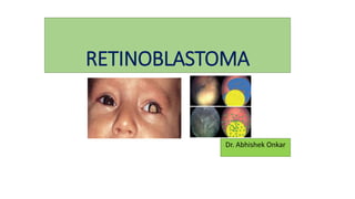
  • 12. Clinical Features of Retinoblastoma • The clinical manifestations of retinoblastoma vary with the stage of the disease at the time of recognition. • In its earliest clinical stage, a small retinoblastoma, ie, less than 2 mm in basal dimension, appears ophthalmoscopically as a subtle, transparent or slightly translucent lesion in the sensory retina. • Slightly larger tumors lead to dilated retinal blood vessels that feed and drain the tumor. • Some larger tumors show foci of chalk-like calcification that resemble cottage cheese. • Any retinoblastoma of any size can produce leukocoria (visible whiteness in the pupil/ amaurotic "cat's eye reflex")
  • 13. • Leucocoria is the most common presenting feature of retinoblastoma, followed by strabismus, painful blind eye and loss of vision. Common presenting features of retinoblastoma as reported in studies: • 1. Leucocoria 56% • 2. Strabismus 20% • 3. Red painful eye 7% • 4. Poor vision 5% • 5. Asymptomatic 3% • 6. Orbital cellulitis 3% • 7. Unilateral mydriasis 2% • 8. Heterochromia iridis 1% • 9. Hyphema 1%
  • 14.
  • 15.
  • 16.
  • 17. • As the tumor grows further, three patterns are usually seen 1. Endophytic, in which the tumor grows into the vitreous cavity. A yellow white mass progressively fills the entire vitreous cavity and vitreous seeds occur. The retinal vessels are not seen on the tumor surface. 2. Exophytic, in which the tumor grows towards the subretinal space. Retinal detachment usually occurs and retinal vessels are seen over the tumor 3. Diffuse infiltrating tumor, in which the tumor diffusely involves the retina causing just a placoid thickness of the retina and not a mass. This is generally seen in older children and usually there is a delay in the diagnosis
  • 18.
  • 19.
  • 20.
  • 21. Classification of Retinoblastoma • Several classifications of retinoblastoma have been developed to assist in prediction of globe salvage. The most popular grouping is the Reese-Ellsworth classification • A new International Classification of Retinoblastoma is currently being developed to simplify the grouping scheme and allow a more practical approach to judging results of chemoreduction.
  • 22. Reese-Ellsworth Classification for Conservative Treatment of Retinoblastoma* Group I: Very favorable • (a) Solitary tumor, less than 4 disc diameters in size, at or • behind the equator • (b) Multiple tumors, none over 4 disc diameters in size, all at • or behind the equator Group II: Favorable • (a) Solitary tumor, 4 to 10 disc diameters in size, at or • behind the equator • (b) Multiple tumors, 4 to 10 disc diameters in size, • behind the equator
  • 23. Group III: Doubtful • (a) Any lesion anterior to the equator • (b) Solitary tumors larger than 10 disc diameters • behind the equator Group IV: Unfavorable • (a) Multiple tumors, some larger than 10 disc diameters • (b) Any lesion extending anteriorly to the ora serrata Group V: Very unfavorable • (a) Massive tumors involving over half the retina • (b) Vitreous seeding * Refers to chances of salvaging the affected eye and not systemic prognosis.
  • 24. Differential Diagnosis • The most common pseudoretinoblastomas include persistent hyperplastic primary vitreous, Coats’ disease, and ocular toxocariasis
  • 25. Conditions Closely Simulating Retinoblastoma • Persistent hyperplastic primary vitreous 28 • Coats’ disease (exudative retinitis or retinal telangiectasis) 16 • Ocular toxocariasis 16 • Retinopathy of prematurity 5 • Combined hamartoma 4 • Coloboma 4 • Vitreous hemorrhage 4 • Astrocytic hamartoma 3 • Congenital cataract 2 • Toxoplasmic retinitis 1 • Idiopathic endophthalmitis 1
  • 26. Diagnostic Testing • Accurate diagnosis in a child with suspected retinoblastoma is accomplished by taking a detailed history, physical evaluation, external ocular examination, slit lamp biomicroscopy, and binocular indirect ophthalmoscopy with scleral indentation. • The diagnosis is established by the classic appearance of the retinal tumors • Needle biopsy confirmation is rarely, if ever, necessary. • Ancillary diagnostic studies can be helpful in confirming the diagnosis of retinoblastoma. • Fluorescein angiography shows early vascularity and late hyperfluorescence of the tumor.
  • 27. • Ultrasonography and computed tomography can demonstrate the intraocular tumor and possibly detect calcium within the mass. • Approximately 5% to 10% of retinoblastomas show no intrinsic calcification • Magnetic resonance imaging does not usually detect calcium but may be of value in the assessment of the optic nerve, orbit, and brain.
  • 29. Other Classifications of Retinoblastoma International Classification of Intraocular Retinoblastoma (Murphree) International Classification of Retinoblastoma (Shields) TNM Classification for Retinoblastoma Pathologic Classification (pTNM)
  • 30. International Staging System for Retinoblastoma Stage 0 No enucleation (one or both eyes may have intraocular disease) Stage I Enucleation, tumor completely resected Stage II Enucleation with microscopic residual tumor Stage III Regional extension • A. Overt orbital disease • B. Preauricular or cervical lymph node extension Stage IV Metastatic disease • A. Hematogenous metastasis • 1. Single lesion • 2. Multiple lesions • B. CNS Extension • 1. Prechiasmatic lesion • 2. CNS mass • 3. Leptomeningeal disease
  • 31. Management of Retinoblastoma • The most important objective in the management of a child with retinoblastoma is survival of the patient, and the second most important goal is preservation of the globe. • The focus on visual acuity comes later, after safety of the patient and globe is established. • Therapy is tailored to each individual case and based on the overall situation, including threat of metastatic disease, risks for second cancers, systemic status, laterality of the disease, size and location.
  • 32. Current Suggested Protocol A. Intraocular tumor, International Classification Group A to C, Unilateral or Bilateral 1. Focal therapy (cryotherapy or transpupillary thermotherapy) alone for smaller tumors (< 3mm in diameter and height) located in visually noncrucial areas 2. Standard 6 cycle chemoreduction and sequential aggressive focal therapy for larger tumors and those located in visually crucial areas 3. Defer focal therapy until 6 cycles for tumors located in the macular and juxtapapillary areas. Transpupillary thermotherapy or plaque brachytherapy for residual tumor in the macular and juxtapapillary areas >6 cycles. 4. Focal therapy for small residual tumor, and plaque brachytherapy/external beam radiotherapy (>12 months age) for large residual tumorif bilateral, and enucleation if unilateral.
  • 33. B. Intraocular tumor, International Classification Group D, Unilateral or Bilateral 1. High dose chemotherapy and sequential aggressive focal therapy 2. Periocular carboplatin for vitreous seeds 3. Consider primary enucleation if unilateral, specially in eyes with no visual prognosis
  • 34. Intraocular tumor, International Classification Group E, Unilateral or Bilateral 1. Primary enucleation 2. Evaluate histopathology for high risk factors • D. High risk factors on histopathology, International Staging, Stage 2 • 1. Baseline systemic evaluation for metastasis • 2. Standard 6 cycle adjuvant chemotherapy • 3. High dose adjuvant chemotherapy and orbital external beam • radiotherapy in patients with scleral infiltration, extraocular extension, and optic nerve extension to transection.
  • 35. E. Extraocular tumor, International Staging, Stage 3A 1. Baseline systemic evaluation for metastasis 2. High dose chemotherapy for 3-6 cycles, followed by enucleation or extended enucleation, external beam radiotherapy, and continued high dose chemotherapy for 12 cycles F. Regional Lymph Node Metastasis, International Staging, Stage 3B 1. Baseline evaluation for systemic metastasis 2. Neck dissection, high dose chemotherapy for 6 cycles, followed by external beam radiotherapy, and continued high dose chemotherapy for 12 cycles
  • 36. G. Hematogenous or Central Nervous System Metastasis, International Staging, Stage 4 • 1. Palliative therapy in discussion with the family • 2. High dose chemotherapy with bone marrow rescue for hematogenous metastasis • 3. High dose chemotherapy with intrathecal chemotherapy for central nervous system metastasis
  • 37. Chemoreduction Regimen for Intraocular Retinoblastoma Given for a Total of 6 Monthly Cycles • Day Vincristine Etoposide Carboplatin • 0 × × × • 1 × • Vincristine: 0.05 mg/kg • Etoposide: 5 mg/kg • Carboplatin: 18.6 mg/kg
  • 38. Focal Therapies Focal therapies include laser photocoagulation, thermotherapy, cryotherapy, and plaque radiotherapy. Plaque radiotherapy is generally reserved for tumors that fail other focal therapies, even those that reach a moderate size, up to 8 or 10 mm in thickness. The remainder of the focal therapies are reserved for small tumors, generally those under 3 mm in greatest dimension.
  • 39. • Laser photocoagulation is usually employed for small retinoblastomas posterior to the equator of the eye. • Thermotherapy is a method of tumor heating using a diode infrared laser system. Thermotherapy coupled with chemoreduction is especially suited for tumors adjacent to the fovea and optic nerve where radiation or laser photocoagulation would possibly induce more visual loss • Cryotherapy is useful in the treatment of equatorial and peripheral small retinoblastomas.
  • 40. • Plaque radiotherapy is a method of brachytherapy in which a radioactive implant is placed on the sclera over the base of a retinoblastoma to irradiate the tumor transsclerally. • Generally, it is limited to tumors less than 16 mm in base and 8 mm in thickness. • It requires an average of 2 to 4 days of treatment time to deliver the total dose of 40 Gy to the apex of the tumor.
  • 41.
  • 42. External Beam Radiotherapy • Retinoblastoma is generally a radiosensitive tumor. External beam radiotherapy is a method of delivering whole eye irradiation to treat advanced retinoblastoma, particularly when there is diffuse vitreous seeding.
  • 43. Enucleation • Enucleation is a frequently used and important method for managing retinoblastoma. If there is advanced disease with no hope for useful vision in the affected eye or if there is a concern of invasion of the tumor into the optic nerve, choroid, or orbit, then enucleation is appropriate. • Those eyes with secondary glaucoma, pars plana seeding, or anterior chamber invasion are also generally best managed with enucleation.
  • 44. Primary enucleation continues to be the treatment of choice for • advanced intraocular retinoblastoma with neovascularization of iris, • secondary glaucoma, • anterior chamber tumor invasion, • tumors occupying >75% of the vitreous volume, • necrotic tumors with secondary orbital inflammation, and • tumors associated with hyphema or vitreous hemorrhage where the tumor characteristics can not be visualized, especially when only one eye is involved
  • 45. Special considerations for enucleation in retinoblastoma • a. Minimal manipulation • b. Avoid perforation of the eye • c. Harvest long (> 15 mm) optic nerve stump • d. Inspect the enucleated eye for macroscopic extraocular extension and • optic nerve involvement • e. Harvest fresh tissue for genetic studies • f. Place a primary implant • g. Avoid biointegrated implant if postoperative radiotherapy is necessary
  • 46.
  • 47. Histopathologic high-risk factors predictive of metastasis • • Anterior chamber seeding • • Iris infiltration • • Ciliary body infiltration • • Massive choroidal infiltration • • Invasion of the optic nerve lamina cribrosa • • Retrolaminar optic nerve invasion • • Invasion of optic nerve transection • • Scleral infiltration • • Extrascleral extension