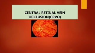
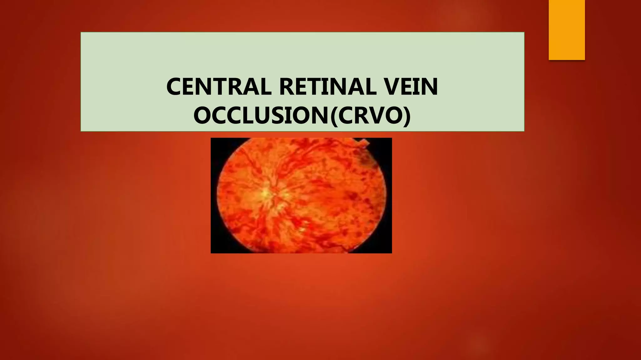
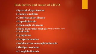
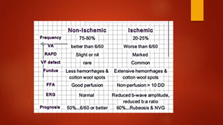
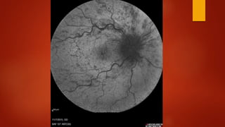
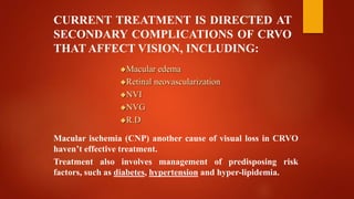
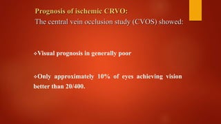
CENTRAL RETINAL VEIN
OCCLUSION(CRVO)
CRVO
CRVO IS AN IMPORTANT CAUSE OF VISUAL LOSS AMONG OLDER
ADULTS THROUGHOUT WORLD.
CRVO OCCURS DUE TO THROMBUS WITHIN THE CENTRAL RETINAL
VEIN AT THE LEVEL OF THE LAMINA CRIBROSA OF THE OPTIC
NERVE.
Risk factors and causes of CRVO
Systemic-hypertension
Diabetes mellitus
Cardiovascular disease
Hyperlipidemia
Open-angle claucoma
Blood dyscrasias such as:
Leukemia
Lymphoma
Paraproteinemias
Waldenstrom macroglobulinemia
Multiple myeloma
Cryoglobulinemia
•Polycythemia vera
Risk factors and causes of CRVO
Oral-contraceptive
Diuretics
Hyperhemocysteinemia
 protein S deficiency
Protein C deficiency
Thrombosis of the central retinal vein at and posterior to the
level of the lamina cribrosa.
In some cases, a thickened central retinal artery may impinge
on the central retinal vein turbulence Endothelial
damage thrombosis
Histologic studies:
Evaluation of CRVO: non ischemic- ischemic
Careful clinical examination
V.A
RAPD
SLE
Funduscopy (90-78 – indirect funduscopy)
Gonioscopy specially in F/U
Check IOP
IN CRVO VISION LOSS IS MOST COMMONLY
SUDDEN:
Mild vision loss nonischemic CRVO
Severe vision loss ischemic CRVO
Less commonly, patients may experience premonitory
symptoms of transient visual obscuration before overt
retinal manifestations appear.
NONISCHEMIC CRVO (MILD)
PARTIAL CRVO
VENOUS STASIS RETINOPATHY
Blurred –vision
No RAPD
Mild V-field defect
Mild dilation and tortuosity of all branches of central
retinal vein.
Dot and flame shaped hemorrhages in four quadrant
of retina.
Mild macular edema
Mild optic disc swelling
Ischemic CRVO or severe CRVO
Complete CRVO
Non perfused CRVO
Hemorrhagic retinopathy
Decrease V.A
RAPD: +
Marked venous dilation
Severe splinter and dot hemorrhage in four
quadrant
Cotton-wool spots
Macular edema
Hemi CRVO considered a variant of CRVO, is
associated with a congenital variation in
central vein anatomy; it may involve either the
superior or inferior half the retina.
Paraclinic ischemic CRVO
Prolonged retinovascular circulation times.
Widespread CNP (at least 10 disc areas)
OCT
Visual – field: dense central scotoma
ERG: decrease b-to a- wave amplitude ratio due to
inner retinal- ischemia
Ultra wide field angiography
F.A:
Differential diagnosis of CRVO:
Hyperviscosity retinopathy generally bilateral
Hypertensive retinopathy
Ocular ischemic syndrome
Ocular ischemic-syndrome
Conj-injection
Cataract
Rubeosis-iridis
Funduscopy:
Venous dilation
M.A
Mid peripheral intraretinal
hemorrhages
Attenuated arterioles
F.A in ocular ischemic syndrome:
Prolonged arm –to-retina circulation time
Patchy choroidal filling pattern.
Ophthalmodynamometer:
CRVO will have normal artery pressure.
Ocular ischemic syndrome will have low artery pressure.
Ocular ischemic syndrome
CURRENT TREATMENT IS DIRECTED AT
SECONDARY COMPLICATIONS OF CRVO
THAT AFFECT VISION, INCLUDING:
Macular edema
Retinal neovascularization
NVI
NVG
R.D
Macular ischemia (CNP) another cause of visual loss in CRVO
haven’t effective treatment.
Treatment also involves management of predisposing risk
factors, such as diabetes, hypertension and hyper-lipidemia.
Prognosis of ischemic CRVO:
The central vein occlusion study (CVOS) showed:
Visual prognosis in generally poor
Only approximately 10% of eyes achieving vision
better than 20/400.
VERY ISCHEMIC CRVO ANT-SEGMENT.
NEOVASCULARIZATION (IRIS AND ANGLE) 60%
OF CASES NVG
3-5 MONTHS AFTER THE ONSET OF SYMPTOMS
Risk factors for NVG
Iris-neovascularization in CRVO
Large areas of CNP
Massive intraretinal and preretinal hemorrhage
Cotton wool spots
In the absence of treatment, should be
monitored on a monthly for at least 6.0Ms
Follow up:
For the progression of CRVO
For the development of Ant-Seg-neovascularization
and NVG
In the patients treated with anti-VEGF agents should
be observed for a similar duration after D/C of the
drugs.
TREATMENT OF MACULAR EDEMA:
Intravitreal anti-VEGF agents is currently first line
therapy for macular edema
Intravitreal steroid therapy is considered an
alternative for patients with edema refractory to anti-
VEGF monotherapy.
Based on CRUISE study
GALILEO
COPERNICUS
Ranibizumab
Bevacizumab
aflibercept
Are effective for macular edema in CRVO
patients.
ANTERIOR SEGMENT NEOVASCULARIZATION:
patients with anterior segment neovascularization are
at risk of neovascular glaucoma.
Prophylactic scatter retinal photocoagulation is
recommended to reduce this risk in patients with NVI
( the effect of laser take 2 to 4 weeks to develop).
CVOS recommendation:
Waiting until undilated gonioscopic examination
revealed at least 2 clock-hours of Iris
neovascularization before performing PRP.
PRP is often performed at the first sign of NVI,
particularly when close F/U in not possible
CVOS the central vein occlusion study
Laser surgery for CRVO
Nonclearing VH after 3Ms
Vit-H +N.V.I or NV
Persistent NVG: glaucoma surgery (glaucoma valve)
PARS PLANA Vitrectomy Surgery

CRVO

  • 1.
  • 2.
    CRVO CRVO IS ANIMPORTANT CAUSE OF VISUAL LOSS AMONG OLDER ADULTS THROUGHOUT WORLD. CRVO OCCURS DUE TO THROMBUS WITHIN THE CENTRAL RETINAL VEIN AT THE LEVEL OF THE LAMINA CRIBROSA OF THE OPTIC NERVE.
  • 3.
    Risk factors andcauses of CRVO Systemic-hypertension Diabetes mellitus Cardiovascular disease Hyperlipidemia Open-angle claucoma Blood dyscrasias such as: Leukemia Lymphoma Paraproteinemias Waldenstrom macroglobulinemia Multiple myeloma Cryoglobulinemia •Polycythemia vera
  • 4.
    Risk factors andcauses of CRVO Oral-contraceptive Diuretics Hyperhemocysteinemia  protein S deficiency Protein C deficiency
  • 5.
    Thrombosis of thecentral retinal vein at and posterior to the level of the lamina cribrosa. In some cases, a thickened central retinal artery may impinge on the central retinal vein turbulence Endothelial damage thrombosis Histologic studies:
  • 6.
    Evaluation of CRVO:non ischemic- ischemic Careful clinical examination V.A RAPD SLE Funduscopy (90-78 – indirect funduscopy) Gonioscopy specially in F/U Check IOP
  • 7.
    IN CRVO VISIONLOSS IS MOST COMMONLY SUDDEN: Mild vision loss nonischemic CRVO Severe vision loss ischemic CRVO Less commonly, patients may experience premonitory symptoms of transient visual obscuration before overt retinal manifestations appear.
  • 8.
    NONISCHEMIC CRVO (MILD) PARTIALCRVO VENOUS STASIS RETINOPATHY Blurred –vision No RAPD Mild V-field defect Mild dilation and tortuosity of all branches of central retinal vein. Dot and flame shaped hemorrhages in four quadrant of retina. Mild macular edema Mild optic disc swelling
  • 9.
    Ischemic CRVO orsevere CRVO Complete CRVO Non perfused CRVO Hemorrhagic retinopathy Decrease V.A RAPD: + Marked venous dilation Severe splinter and dot hemorrhage in four quadrant Cotton-wool spots Macular edema
  • 12.
    Hemi CRVO considereda variant of CRVO, is associated with a congenital variation in central vein anatomy; it may involve either the superior or inferior half the retina.
  • 13.
    Paraclinic ischemic CRVO Prolongedretinovascular circulation times. Widespread CNP (at least 10 disc areas) OCT Visual – field: dense central scotoma ERG: decrease b-to a- wave amplitude ratio due to inner retinal- ischemia Ultra wide field angiography F.A:
  • 17.
    Differential diagnosis ofCRVO: Hyperviscosity retinopathy generally bilateral Hypertensive retinopathy Ocular ischemic syndrome
  • 18.
  • 19.
    F.A in ocularischemic syndrome: Prolonged arm –to-retina circulation time Patchy choroidal filling pattern. Ophthalmodynamometer: CRVO will have normal artery pressure. Ocular ischemic syndrome will have low artery pressure.
  • 20.
  • 21.
    CURRENT TREATMENT ISDIRECTED AT SECONDARY COMPLICATIONS OF CRVO THAT AFFECT VISION, INCLUDING: Macular edema Retinal neovascularization NVI NVG R.D Macular ischemia (CNP) another cause of visual loss in CRVO haven’t effective treatment. Treatment also involves management of predisposing risk factors, such as diabetes, hypertension and hyper-lipidemia.
  • 22.
    Prognosis of ischemicCRVO: The central vein occlusion study (CVOS) showed: Visual prognosis in generally poor Only approximately 10% of eyes achieving vision better than 20/400.
  • 23.
    VERY ISCHEMIC CRVOANT-SEGMENT. NEOVASCULARIZATION (IRIS AND ANGLE) 60% OF CASES NVG 3-5 MONTHS AFTER THE ONSET OF SYMPTOMS Risk factors for NVG Iris-neovascularization in CRVO Large areas of CNP Massive intraretinal and preretinal hemorrhage Cotton wool spots
  • 24.
    In the absenceof treatment, should be monitored on a monthly for at least 6.0Ms Follow up: For the progression of CRVO For the development of Ant-Seg-neovascularization and NVG In the patients treated with anti-VEGF agents should be observed for a similar duration after D/C of the drugs.
  • 25.
    TREATMENT OF MACULAREDEMA: Intravitreal anti-VEGF agents is currently first line therapy for macular edema Intravitreal steroid therapy is considered an alternative for patients with edema refractory to anti- VEGF monotherapy.
  • 26.
    Based on CRUISEstudy GALILEO COPERNICUS Ranibizumab Bevacizumab aflibercept Are effective for macular edema in CRVO patients.
  • 27.
    ANTERIOR SEGMENT NEOVASCULARIZATION: patientswith anterior segment neovascularization are at risk of neovascular glaucoma. Prophylactic scatter retinal photocoagulation is recommended to reduce this risk in patients with NVI ( the effect of laser take 2 to 4 weeks to develop).
  • 28.
    CVOS recommendation: Waiting untilundilated gonioscopic examination revealed at least 2 clock-hours of Iris neovascularization before performing PRP. PRP is often performed at the first sign of NVI, particularly when close F/U in not possible CVOS the central vein occlusion study Laser surgery for CRVO
  • 29.
    Nonclearing VH after3Ms Vit-H +N.V.I or NV Persistent NVG: glaucoma surgery (glaucoma valve) PARS PLANA Vitrectomy Surgery