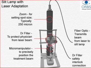
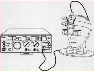
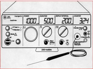
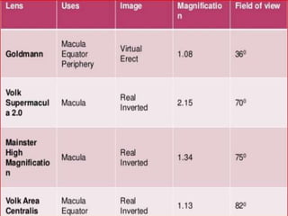
LASERS IN
OPHTHALMOLOGY
LASER
• Light Amplification by Stimulated Emission of
Radiation
• Term was coined by Gordon Gould.
Laser characteristics
• Monochromatic – One color
• Coherent – In-phase
• Collimated – Light waves alligned
• Highly energized
• Can be sharply focused
History of LASER
• 1917 - A. Einstein : Laser possible.
• 1960 - T. Maiman : Built first laser.
A pulsed ruby laser coupled
with a monocular DO delivery
system
• 1963 - C. Zweng : 1ST medical laser trial .
4 retinal breaks and PDR
• 1965 - W.Z. Yarn : 1ST clinical laser surgery.
• 1968-L’Esperance :developed the argon laser
LASER PHYSICS
• photons and atoms interact in 3 ways
Absorption
Spontaneous Emission
Stimulated Emission
• stimulated emission is the only method
known that produces coherent light.
How is LASER generated?
• BASIC LASER COMPONENTS
• Laser Tube (Laser medium + Resonating element)
• Pump or excitation source
• Power supply
• Cooling unit (water, air)
• Energy from LASER PUMP  substance in the
LASER MEDIUM (solid, gas, liquid with dissolved
organic dye,semiconductor) energy emission.
• Mirrors at either end of the laser tube reflect
photons parallel to tube axis strike other atoms
 stimulated emission of ↑photons of same λ .
• Photons moving in other directions
:absorbed/reflected tube sides.
• The remaining photonspartially reflective mirror
as a coherent beam.
• cooling unit :prevent heat damage to the laser
medium and pump.
• Pump :produce more electrons in high-energy
states population inversion.
can be current/heat/ionising radiation/light.
• Mirrors :maintain the chain reaction of stimulated
emissions, with an arrangement called a resonator.
DIFFERENT TYPES OF LASERS
Solid State : Ruby,Nd Yag,Erbium YAG
Gas :Ion,Argon,Krypton,He-Neon,CO2
Metal Vapour : Cu,Gold
Dye :Rhodamine
Excimer :Ar Fl, Kr Fl, Kr Cl
Diode : Gallium-AluminumArsenide (GaAlAs)
• krypton:metastable state  lower energy :647nm
(red).
• argon :2 metastable states, 2 λ :488 nm and 514 nm
(blue-green and green).
• no: of λ produced are increased by:
HARMONIC GENERATION
• laser light nonlinear crystal(2x wave Ỵ)exact
multiples of the laser’s frequency, called harmonics
Eg:“double-frequency” Nd:YAG laser.
USING ORGANIC DYES
• complex chemical structure of organic dyes  large
no:of metastable orbits differing in little
energydifferent λ
• rhodamine 6G λ 570 nm to 630 nm.
• drawbacks :least efficient producers of laser energy
:expensive to manufacture.
PROPERTIES
• Pulsed lasers :modest amounts of energy.
:energy concentrated into very brief time
:high power (energy/t).
:usually by intermittent pumping source.
:Nd YAG, excimer laser.
Q Switches Lasers : pulses 4 nano second
Mode-locked Lasers: pulses 4 picoseconds.
continuous laser :delivers energy in continuos manner
: relatively long time.
:power is lower
:pumping source constant
Gated pulse delivery: continuous wave laser used in
intermittent timed manner.
in between,laser output is gated
• Mode
longitudinal :photon packets of diff energy at diff
time diff frequency output.
transverse :across the beam’s c/s some areas will
have low intensity uneven intensity.
VARIABLES THAT DETERMINE THE LASER TISSUE
INTERACTION
• Power Density:
– Milliwatts to Kilowatts/cm²
-α power, inversly α ½ (spotsize )².
• Wavelength:
– Ultraviolet,Visible,Infrared.
-if <300nm & >1300 nm dont pass through tissue
• Duration of Pulse:
– Nanoseconds to Continuous
• Spot size
TISSUE FACTORS AFFECTING TISSUE LASER
INTERACTION
• Water content
• Transparency
• Pigmentation
Hb :Ar Green, Kr yellow absorbed.
to coagulate the blood vessels.
Xanthophyll :Maximum absorption is blue.
Argon blue not used in macular d/s.
Melanin :Ar Blue, Kr
PRP, and RPE destruction
TISSUE INTERACTIONS WITH LASER
PHOTOCHEMICAL DAMAGE/PHOTOABLATION:
• highly localised laser light of high energy breaks the
chemical bonds in a molecule clean edged incisions.
• excimer laser for Photorefractive surgeries of cornea.
• Disadv:malignancy risk.
PHOTOTHERMAL DAMAGE:
• laser Light causes temp rise in tissue by energy
absorption breaks weak hydrogen bonds in
molecules by heat.
• Divided into photocoagulation
• photovapourisation
• photodynamic therapy
PHOTOCOAGULATION :temp rise upto 10-20⁰c.
:by protein denaturation (PRP)
:by thrombus formation(focal pc)
:by collagen contraction(ALT)
:pigment dependant (melanin,Hb)
:Hb-Argon Green , Krypton yellow
absorbed to coagulate the bv.
PHOTOVAPOURISATION:pigment dependant
:Melanin-Argon Blue, Krypton
:temp rise upto 60-100⁰C 
vapourisation due to high power
:used in PI.
causes hemorhage from vessels as it
donot coagulate bv
PHOTODYNAMIC THERAPY: exogenous chromophore is injected IV.
: Commonly used photosensitizers
:Hematoporphyrin
Benzaporphyrin
: bind to specific tissuetemp riseO₂ free
radicallocal destruction of vessels
: 4 Treatment of ocular tumour and CNVM
PHOTODISRUPTION/PHOTO IONISATION DAMAGE:
– Lights of short duration and high energy  tissue ionisation (plasma)
Pigment independent
Usually 1015W/cm² over nano/ pico s used
Eg:YAG cap produces plasma cloud of 150 µm//PI
disadv:donot coagulate bv hemorhage.
PDT
Dye dose = 6 mg/m2 body surface area
Intravenous infusion over 10 min
Treatment at 15 min after start of dye infusion
wavelength of at 689 nm,irradiance of 600 mW/cm2 and
fluence of 100 J/cm2
•
DELIVERY SYSTEMS
• Most ophthalmic laser systems has:a laser medium,
laser pump, and cooling system coupled to a slit-
lamp by a flexible fiberoptic cable .
• Delivery Systems
• transpupillary: - Slit lamp
- Laser Indirect Ophthalmoscopy
Trans scleral : - Contact
- Non contact
Endophotocoagulation.
SLIT LAMP
• Most commonly used mode for a and p segment.
• ADVANTAGES:Binocular and stereoscopic view.
:Fixed distance.
:spot size standardisation is accurate.
:Aiming accuracy is good.
LASER INDIRECT OPHTHALMOSCOPY
• Advantages : Wider field(until periphery).
Better visualization in hazy medium.
Can treat in supine position.
• Disadvantage : difficulty in focusing.
Difficulty to standardize spot size.
Expensive.
Un co-operative patient.
Learning curve.
LASER USES
• THERAPEUTIC.
• DIAGNOSTIC
Anterior segment
• CORNEA :Keratorefractive Surgeries:
PRK,LASIK,LASEK,Epi Lasik
: Laser Thermal Keratoplasty .
:Corneal Neovascularization.
:Retrocorneal Pigmented Plaques.
GLAUCOMA : Laser Iridotomy.
Laser Trabeculoplasty (LT)
Selective Laser Trabeculoplasty
Trabecular ablation
Gonioplasty (Iridoplasty, Iridoretraction)
Pupilloplasty
Sphincterotomy
Iridolenticular Synechiolysis
Goniophotocoagulation & Goniotomy
PUPILLOPLASTY
• 2-3 rows of burns circumferentially 1mm away from
the pupillary margin.
• Innermost row:8spots,200micron size, 200-400mW.
• Outer row:10-12spots,400micronsize,300-500mW.
• STRETCHING THE UPDRAWN PUPIL
• Laser parameters are same for photomydriasis.
• Burns are placed along the inferior margin.
SCLEROSTOMY
• Ab Externosclerostomy (Holmium)
• Ab Internosclerostomy (Nd.YAG)
Contact /Non-contact
CYCLODESTRUCTIVE PROCEDURES
• Transscleral Cyclophotocoagulation
• Transpupillary Cyclophotocoagulation
• Diode Laser Endophotocoagulation
LENS :Posterior capsulotomy
Laser phacoemulsification
Phaco ablation.
LACRIMAL SURGERY : Laser DCR.
VITREOUS :Vitreous membranes
Vitreous traction bands
FUNDUS
Diabetic Retinopathy
Retinal Vascular Diseases
Choroidal Neovascularization (CNV)
Clinical Significant Macular Edema (CSME)
Central Serous Retinopathy (CSR)
Retinal Break/Detachment
Tumour
ARMD
Retinal Vein Occlusion
Eale’s Disease
Coats Disease
Peripheral Retinal Lesion
Retinopathy of prematurity.
Focal PC PRP
Retinal hemorrhage
RETINAL BREAK &TEAR
• λ :argon green, Nd YAG, dye yellow red , diode.
• Duration :0.1-0.2seconds.
• Retinal spot size: 200-500microns.
• Intensity : moderate retinal whitening
CHORIODAL MELANOMA
• Photocoagulation technique.
• Initial destruction of the surrounding choroidal blood
supply in 1-2rows -200-500 microns ,0.5-1sec-
intense burn.
• Direct tumour photocoagulation-low energy long
duration(5-30sec) burns.
RETINOBLASTOMA
• Diode (infrared)laser tumor surface in regions of
disease activity tumor cell death by raising the
temperature of tumor cells to above 45°C for
~1min(reduces blood supply , apoptosis).
PAttern SCAn Laser
• It is an integrated semi-automatic pattern scan laser
photocoagulation system designed to treat ocular
diseases using a single shot or predetermined
pattern array.
• Laser source :Nd:YAG laser.
• Delivery device: slit lamp or laser indirect
ophthalmoscope (LIO)
• Control system for selecting power,duration &
Method for selecting spot size.
SCANNING LASER OPHTHALMOSCOPY
• High-resolution, real-time motion images of the
macula without patient discomfort.
• SLO angiography: to study retinal and choroidal
blood flow.
• May be used to perform micro perimetry, an
extremely accurate mapping of the macula’svisual
field.
OCT
• Uses diode laser light in the near-infrared spectrum
(810 nm) high-resolution cross-sectional images
of the retina using coherence interferometry.
COMPLICATIONS
• General : Pain,Seizures.
• Anterior segment : Elevated IOP.
Corneal damage.
Iris burns.
Crystalline lens burns.
IOL and PC damage.
Internal ophthalmoplegia.
• POSTERIOR SEGMENT : Choroidal detachment and exudative RD.
Choroidal ,subretinal,vitreous hemorrhage.
Thermal induced retinal vascular damage.
Preretinal membranes.
Ischaemic papillitis.
Paracentral visual field loss and scotoma.
Photocoagulation scar enlargement.
Subretinal fibrosis.
Iatrogenic choroidal neovascularisation.
Accidental foveal burns.
LASER SAFETY
ANSI (American National Standard Institute)
• Class-I : Causing no biological damage.
• Class-II : Safe on momentary viewing but chronic
exposure may cause damage.
• Class-III: Not safe even in momentary view.
• Class-IV : Cause more hazardous than Class-III.
• Protective shutters built into the equipment, filters
incorporated into the slit-lamp biomicroscope, and
divergence of the beam at the exit optics risk to
clinicians during photocoagulation/disruption.
• Accessory lenses should have ARC reflected laser
light if bystanders are within nominal hazard zone
(the area where direct, reflected, or scattered radiation
exceeds safe exposure levels).
• When a handpiece is used in place of biomicroscopy,
precautions must be taken to minimize the chance of
specular reflection from instruments.
• Protective goggles should be used.
• warning signs listing the laser’s type and class should
be posted at all entrances to the laser suite.
EXCIMER(Excited dimer)
• High energy UV laser.
• Ar F(193nm),Kr F, Kr Cl, Xe Cl, Xe F (gas)
• Ar F :Low shock waves,thermal effects,ocular
penetration,max edge control.
• USE: corneal surgeries.
• Laser removes approximately 0.25microns of
corneal tissue with each pulse(12-15ns).
• Amount of tissue to be ablated ―munnerlyn
equation” Central ablation depth in microns=
diopters of myopia*(ablation zone diameter in mm)
FEMTOSECOND LASER
• ADVANTAGES:
• Flap are more accurate and uniform in thickness.
• Centration of flap is easier.
• Better adherence to underlying stroma.
• Patients comfortable.
• DISADVANTAGES:
• Suction break
• Costly
ARGON LASER
• 488 nm-blue green
• 514 nm-green
• As continuous,gated,true pulse wave.
• Adv :λ matches Hb absorption spectrum.
:RPE absorption 90-93%-good input output ratio.
• Disadv:blue end absorbed by xanthophyll of macula.
:poor optical electrical efficiency.
:water cooling system is a must.
DIODE LASER
• Temporally coherent & monochromatic .
• Doped with Al, λ-810 nm.
• Transmitted by cornea,AH,lens,VHno hindrance by
cataract/vitreous hemorrhage.
• Negligible xanthophyll absorption.
• Compact size,portable,no heat generation.
• Dis adv:wide cone anglespot is difficult .
• :20-25% absorbed by RPE4x Ar laser energy
:pain most common
Uses:transpupillary retinal PC,trans scleral
cycloPC,endoPC
Nd YAG laser
• Impregantion of Nd crystals with Yttr,Al.
• Delivery system used:binocular microscope with He-
Ne aiming beam & focussing beam.
• Aiming dot shud b round,shud not flicker.
Frequency doubled YAG(Kr YAG-532nm)
• Monochromatic green laser.
• Diode laser lampNd YAG crystalKr crystal532 nm
(1064 nm)
Lasers in ophthalmology

Lasers in ophthalmology

  • 1.
  • 2.
    LASER • Light Amplificationby Stimulated Emission of Radiation • Term was coined by Gordon Gould. Laser characteristics • Monochromatic – One color • Coherent – In-phase • Collimated – Light waves alligned • Highly energized • Can be sharply focused
  • 4.
    History of LASER •1917 - A. Einstein : Laser possible. • 1960 - T. Maiman : Built first laser. A pulsed ruby laser coupled with a monocular DO delivery system • 1963 - C. Zweng : 1ST medical laser trial . 4 retinal breaks and PDR • 1965 - W.Z. Yarn : 1ST clinical laser surgery. • 1968-L’Esperance :developed the argon laser
  • 5.
    LASER PHYSICS • photonsand atoms interact in 3 ways Absorption Spontaneous Emission Stimulated Emission • stimulated emission is the only method known that produces coherent light.
  • 11.
    How is LASERgenerated? • BASIC LASER COMPONENTS • Laser Tube (Laser medium + Resonating element) • Pump or excitation source • Power supply • Cooling unit (water, air)
  • 14.
    • Energy fromLASER PUMP  substance in the LASER MEDIUM (solid, gas, liquid with dissolved organic dye,semiconductor) energy emission. • Mirrors at either end of the laser tube reflect photons parallel to tube axis strike other atoms  stimulated emission of ↑photons of same λ . • Photons moving in other directions :absorbed/reflected tube sides. • The remaining photonspartially reflective mirror as a coherent beam.
  • 15.
    • cooling unit:prevent heat damage to the laser medium and pump. • Pump :produce more electrons in high-energy states population inversion. can be current/heat/ionising radiation/light. • Mirrors :maintain the chain reaction of stimulated emissions, with an arrangement called a resonator.
  • 16.
    DIFFERENT TYPES OFLASERS Solid State : Ruby,Nd Yag,Erbium YAG Gas :Ion,Argon,Krypton,He-Neon,CO2 Metal Vapour : Cu,Gold Dye :Rhodamine Excimer :Ar Fl, Kr Fl, Kr Cl Diode : Gallium-AluminumArsenide (GaAlAs)
  • 18.
    • krypton:metastable state lower energy :647nm (red). • argon :2 metastable states, 2 λ :488 nm and 514 nm (blue-green and green). • no: of λ produced are increased by: HARMONIC GENERATION • laser light nonlinear crystal(2x wave Ỵ)exact multiples of the laser’s frequency, called harmonics Eg:“double-frequency” Nd:YAG laser. USING ORGANIC DYES • complex chemical structure of organic dyes  large no:of metastable orbits differing in little energydifferent λ • rhodamine 6G λ 570 nm to 630 nm. • drawbacks :least efficient producers of laser energy :expensive to manufacture.
  • 19.
    PROPERTIES • Pulsed lasers:modest amounts of energy. :energy concentrated into very brief time :high power (energy/t). :usually by intermittent pumping source. :Nd YAG, excimer laser. Q Switches Lasers : pulses 4 nano second Mode-locked Lasers: pulses 4 picoseconds. continuous laser :delivers energy in continuos manner : relatively long time. :power is lower :pumping source constant Gated pulse delivery: continuous wave laser used in intermittent timed manner. in between,laser output is gated
  • 20.
    • Mode longitudinal :photonpackets of diff energy at diff time diff frequency output. transverse :across the beam’s c/s some areas will have low intensity uneven intensity.
  • 21.
    VARIABLES THAT DETERMINETHE LASER TISSUE INTERACTION • Power Density: – Milliwatts to Kilowatts/cm² -α power, inversly α ½ (spotsize )². • Wavelength: – Ultraviolet,Visible,Infrared. -if <300nm & >1300 nm dont pass through tissue • Duration of Pulse: – Nanoseconds to Continuous • Spot size
  • 22.
    TISSUE FACTORS AFFECTINGTISSUE LASER INTERACTION • Water content • Transparency • Pigmentation Hb :Ar Green, Kr yellow absorbed. to coagulate the blood vessels. Xanthophyll :Maximum absorption is blue. Argon blue not used in macular d/s. Melanin :Ar Blue, Kr PRP, and RPE destruction
  • 23.
    TISSUE INTERACTIONS WITHLASER PHOTOCHEMICAL DAMAGE/PHOTOABLATION: • highly localised laser light of high energy breaks the chemical bonds in a molecule clean edged incisions. • excimer laser for Photorefractive surgeries of cornea. • Disadv:malignancy risk. PHOTOTHERMAL DAMAGE: • laser Light causes temp rise in tissue by energy absorption breaks weak hydrogen bonds in molecules by heat. • Divided into photocoagulation • photovapourisation • photodynamic therapy
  • 24.
    PHOTOCOAGULATION :temp riseupto 10-20⁰c. :by protein denaturation (PRP) :by thrombus formation(focal pc) :by collagen contraction(ALT) :pigment dependant (melanin,Hb) :Hb-Argon Green , Krypton yellow absorbed to coagulate the bv. PHOTOVAPOURISATION:pigment dependant :Melanin-Argon Blue, Krypton :temp rise upto 60-100⁰C  vapourisation due to high power :used in PI. causes hemorhage from vessels as it donot coagulate bv
  • 25.
    PHOTODYNAMIC THERAPY: exogenouschromophore is injected IV. : Commonly used photosensitizers :Hematoporphyrin Benzaporphyrin : bind to specific tissuetemp riseO₂ free radicallocal destruction of vessels : 4 Treatment of ocular tumour and CNVM PHOTODISRUPTION/PHOTO IONISATION DAMAGE: – Lights of short duration and high energy  tissue ionisation (plasma) Pigment independent Usually 1015W/cm² over nano/ pico s used Eg:YAG cap produces plasma cloud of 150 µm//PI disadv:donot coagulate bv hemorhage.
  • 26.
    PDT Dye dose =6 mg/m2 body surface area Intravenous infusion over 10 min Treatment at 15 min after start of dye infusion wavelength of at 689 nm,irradiance of 600 mW/cm2 and fluence of 100 J/cm2
  • 27.
  • 28.
    DELIVERY SYSTEMS • Mostophthalmic laser systems has:a laser medium, laser pump, and cooling system coupled to a slit- lamp by a flexible fiberoptic cable . • Delivery Systems • transpupillary: - Slit lamp - Laser Indirect Ophthalmoscopy Trans scleral : - Contact - Non contact Endophotocoagulation.
  • 29.
    SLIT LAMP • Mostcommonly used mode for a and p segment. • ADVANTAGES:Binocular and stereoscopic view. :Fixed distance. :spot size standardisation is accurate. :Aiming accuracy is good. LASER INDIRECT OPHTHALMOSCOPY • Advantages : Wider field(until periphery). Better visualization in hazy medium. Can treat in supine position. • Disadvantage : difficulty in focusing. Difficulty to standardize spot size. Expensive. Un co-operative patient. Learning curve.
  • 35.
  • 36.
    Anterior segment • CORNEA:Keratorefractive Surgeries: PRK,LASIK,LASEK,Epi Lasik : Laser Thermal Keratoplasty . :Corneal Neovascularization. :Retrocorneal Pigmented Plaques. GLAUCOMA : Laser Iridotomy. Laser Trabeculoplasty (LT) Selective Laser Trabeculoplasty Trabecular ablation Gonioplasty (Iridoplasty, Iridoretraction) Pupilloplasty Sphincterotomy Iridolenticular Synechiolysis Goniophotocoagulation & Goniotomy
  • 38.
    PUPILLOPLASTY • 2-3 rowsof burns circumferentially 1mm away from the pupillary margin. • Innermost row:8spots,200micron size, 200-400mW. • Outer row:10-12spots,400micronsize,300-500mW. • STRETCHING THE UPDRAWN PUPIL • Laser parameters are same for photomydriasis. • Burns are placed along the inferior margin.
  • 40.
    SCLEROSTOMY • Ab Externosclerostomy(Holmium) • Ab Internosclerostomy (Nd.YAG) Contact /Non-contact CYCLODESTRUCTIVE PROCEDURES • Transscleral Cyclophotocoagulation • Transpupillary Cyclophotocoagulation • Diode Laser Endophotocoagulation
  • 43.
    LENS :Posterior capsulotomy Laserphacoemulsification Phaco ablation. LACRIMAL SURGERY : Laser DCR. VITREOUS :Vitreous membranes Vitreous traction bands
  • 44.
    FUNDUS Diabetic Retinopathy Retinal VascularDiseases Choroidal Neovascularization (CNV) Clinical Significant Macular Edema (CSME) Central Serous Retinopathy (CSR) Retinal Break/Detachment Tumour ARMD Retinal Vein Occlusion Eale’s Disease Coats Disease Peripheral Retinal Lesion Retinopathy of prematurity.
  • 48.
  • 49.
  • 50.
    RETINAL BREAK &TEAR •λ :argon green, Nd YAG, dye yellow red , diode. • Duration :0.1-0.2seconds. • Retinal spot size: 200-500microns. • Intensity : moderate retinal whitening
  • 51.
    CHORIODAL MELANOMA • Photocoagulationtechnique. • Initial destruction of the surrounding choroidal blood supply in 1-2rows -200-500 microns ,0.5-1sec- intense burn. • Direct tumour photocoagulation-low energy long duration(5-30sec) burns. RETINOBLASTOMA • Diode (infrared)laser tumor surface in regions of disease activity tumor cell death by raising the temperature of tumor cells to above 45°C for ~1min(reduces blood supply , apoptosis).
  • 52.
    PAttern SCAn Laser •It is an integrated semi-automatic pattern scan laser photocoagulation system designed to treat ocular diseases using a single shot or predetermined pattern array. • Laser source :Nd:YAG laser. • Delivery device: slit lamp or laser indirect ophthalmoscope (LIO) • Control system for selecting power,duration & Method for selecting spot size.
  • 54.
    SCANNING LASER OPHTHALMOSCOPY •High-resolution, real-time motion images of the macula without patient discomfort. • SLO angiography: to study retinal and choroidal blood flow. • May be used to perform micro perimetry, an extremely accurate mapping of the macula’svisual field. OCT • Uses diode laser light in the near-infrared spectrum (810 nm) high-resolution cross-sectional images of the retina using coherence interferometry.
  • 55.
    COMPLICATIONS • General :Pain,Seizures. • Anterior segment : Elevated IOP. Corneal damage. Iris burns. Crystalline lens burns. IOL and PC damage. Internal ophthalmoplegia. • POSTERIOR SEGMENT : Choroidal detachment and exudative RD. Choroidal ,subretinal,vitreous hemorrhage. Thermal induced retinal vascular damage. Preretinal membranes. Ischaemic papillitis. Paracentral visual field loss and scotoma. Photocoagulation scar enlargement. Subretinal fibrosis. Iatrogenic choroidal neovascularisation. Accidental foveal burns.
  • 56.
  • 57.
    ANSI (American NationalStandard Institute) • Class-I : Causing no biological damage. • Class-II : Safe on momentary viewing but chronic exposure may cause damage. • Class-III: Not safe even in momentary view. • Class-IV : Cause more hazardous than Class-III.
  • 58.
    • Protective shuttersbuilt into the equipment, filters incorporated into the slit-lamp biomicroscope, and divergence of the beam at the exit optics risk to clinicians during photocoagulation/disruption. • Accessory lenses should have ARC reflected laser light if bystanders are within nominal hazard zone (the area where direct, reflected, or scattered radiation exceeds safe exposure levels). • When a handpiece is used in place of biomicroscopy, precautions must be taken to minimize the chance of specular reflection from instruments. • Protective goggles should be used. • warning signs listing the laser’s type and class should be posted at all entrances to the laser suite.
  • 60.
    EXCIMER(Excited dimer) • Highenergy UV laser. • Ar F(193nm),Kr F, Kr Cl, Xe Cl, Xe F (gas) • Ar F :Low shock waves,thermal effects,ocular penetration,max edge control. • USE: corneal surgeries. • Laser removes approximately 0.25microns of corneal tissue with each pulse(12-15ns). • Amount of tissue to be ablated ―munnerlyn equation” Central ablation depth in microns= diopters of myopia*(ablation zone diameter in mm)
  • 61.
    FEMTOSECOND LASER • ADVANTAGES: •Flap are more accurate and uniform in thickness. • Centration of flap is easier. • Better adherence to underlying stroma. • Patients comfortable. • DISADVANTAGES: • Suction break • Costly
  • 63.
    ARGON LASER • 488nm-blue green • 514 nm-green • As continuous,gated,true pulse wave. • Adv :λ matches Hb absorption spectrum. :RPE absorption 90-93%-good input output ratio. • Disadv:blue end absorbed by xanthophyll of macula. :poor optical electrical efficiency. :water cooling system is a must.
  • 64.
    DIODE LASER • Temporallycoherent & monochromatic . • Doped with Al, λ-810 nm. • Transmitted by cornea,AH,lens,VHno hindrance by cataract/vitreous hemorrhage. • Negligible xanthophyll absorption. • Compact size,portable,no heat generation. • Dis adv:wide cone anglespot is difficult . • :20-25% absorbed by RPE4x Ar laser energy :pain most common Uses:transpupillary retinal PC,trans scleral cycloPC,endoPC
  • 65.
    Nd YAG laser •Impregantion of Nd crystals with Yttr,Al. • Delivery system used:binocular microscope with He- Ne aiming beam & focussing beam. • Aiming dot shud b round,shud not flicker. Frequency doubled YAG(Kr YAG-532nm) • Monochromatic green laser. • Diode laser lampNd YAG crystalKr crystal532 nm (1064 nm)